Abstract
Objectives:
Radiation toxicity of the dentition may present significant treatment-related morbidity in the paediatric head and neck cancer population. However, clear dose–effect relationships remain undetermined and must be predicated upon accurate structure delineation and dosimetry at the individual tooth level. Radiation oncologists generally have limited familiarity or experience with relevant dental anatomy.
Methods:
We therefore developed a detailed CT atlas of permanent and primary dentition. After studying this atlas, five radiation oncology clinicians delineated all teeth for each of eight different cases (selected for breadth of dental maturity and anatomical variability). They were asked to record confidence in their contours on a per-tooth basis as well as the duration of time required per case. Contour accuracy and interclinician variability were assessed by Hausdorff distance and Dice similarity coefficient. All analyses were performed using R v. 3.1.1 and the RadOnc v. 1.0.9 package.
Results:
Participating clinicians delineated teeth with varying degrees of completeness and accuracy, stratified primarily by the age of the subject. On a per-tooth basis, delineation of permanent dentition was feasible for incisors, canines, premolars and first molars among all subjects, even at the youngest ages. However, delineation of second and third molars was less consistent, commensurate with approximate timing of tooth development. Within each tooth contour, uncertainty was the greatest at the level of the dental roots.
Conclusions:
Delineation of individual teeth is feasible and serves as a necessary precursor for dental dose assessment and avoidance. Among the paediatric radiation oncology community in particular, this atlas may serve as a useful tool and reference.
Keywords: paediatrics, dentition, head and neck neoplasms, radiation oncology, tomography
Introduction
Therapeutic irradiation to the head and neck has been associated with a variety of toxicities, including xerostomia, osteoradionecrosis and trismus. In the adult population, prophylactic tooth extractions and long-term dental care are often emphasized owing to the unintended risks of radiation to the teeth and jaws. In the paediatric cohort, the risks of radiation to the developing dentition may be even more pronounced. Specific abnormalities have been reported throughout the literature and include tooth and root hypoplasia or involution, delayed eruption, local enamel defects and excessive caries of crowns and cementoenamel junctions.1–4
Despite this, there are no current paediatric protocols or international practice guidelines that treat the dentition as an organ at risk (OAR). While delineating the entire mandible or maxilla may serve as a crude surrogate to assess dental dosimetry, radiation dose to neighbouring teeth can vary markedly.5 Moreover, because each tooth develops and exists as an independent unit, toxicities may differ significantly between adjacent teeth as a function of dose.5
To prospectively assess dental dose and minimize subsequent toxicity in young patients receiving therapeutic radiation to the head and neck, individual teeth must be identified as discrete OARs. For this purpose, we have developed a dental contouring atlas, analogous to other routinely employed tools, as a reference for the radiation oncology community.
Methods and materials
Development of a CT-based contouring atlas for primary and permanent dentition
Basic background information, including tooth development, anatomy and nomenclature, were included along with axially segmented dentition for three patients as a reference/atlas for tooth delineation. These reference patients included both young and old individuals who were identified as representative examples of varying dentition (i.e. mixed, permanent and partial dentition). Note that all imaging data was retrospectively obtained from departmental databases and analysed as part of an institutional review board-approved effort to study the dentition. Reference patients were classified into four groups by age: Group A (younger than 2 years), Group B (2–5 years), Group C (5–13 years) and Group D (older than 13 years). The full atlas is provided as Supplementary material to this article.
Individual teeth were manually contoured on axial images (1.5-mm CT slice thickness) in a standard bone window (e.g. −100 to 1500 HU) by a single physician (RFT) in accordance with general dental anatomy,6 beginning with the central and lateral incisors, with progression through adjacent teeth in an anterior to posterior fashion. Each tooth was delineated as a single high-resolution structure, including both crown and roots and excluding identifiable gingival tissue and alveolar bone. All teeth were labelled according to the International Standard World Dental Federation (Federation Dentaire Internationale) two-digit tooth notation system.7 These “gold standard” contours were revised and verified by a paediatric oral and maxillofacial surgeon (LML) and independently by an oral and maxillofacial radiologist (MM). When fully calcified (e.g. primary teeth in all patients and permanent teeth in all but the youngest patients), delineated teeth had a median signal intensity of approximately 1300 HU, with each tooth exhibiting a 5–95% interquantile range of values from approximately 500–2600 HU. At the youngest age (15 months), permanent teeth were incompletely calcified with a per-tooth range of signal intensities from approximately 200–800 HU (5–95% interquantile range).
Implementation of CT-based atlas among independent clinicians
Five radiation oncology clinicians (two attending physicians with expertise in head/neck and paediatric cancers and three trainees) from the University of Pennsylvania, Philadelphia, PA, participated in this study. Each clinician was provided a copy of the dental atlas and was asked to independently delineate individual primary and permanent teeth for each of eight patients accordingly. Clinicians were also asked to document confidence scores on a per-tooth basis (0–100%) and overall degree of effort required (approximated as duration of each case in minutes). All contouring work was performed using Varian's Eclipse™ treatment planning system (Varian® Medical Systems, Palo Alto, CA).
The patient cohort consisted of eight individuals of different ages, chosen to represent different phases of dental development with varying degrees of anticipated challenge (Table 1). Additionally, the patient cohort contained a mix of individuals with and without third molars (“wisdom teeth”) as well as a several individuals with anatomical deviations (e.g. supernumerary teeth). None of the eight patients possessed dental implants, orthodontic appliances or other reconstructive work, thus all CT scans were free of dental artefacts. All images were acquired supine without contrast at 1.5-mm axial slice width and 512 × 512 pixel resolution.
Table 1.
Patient-specific information
| Patient identifier | Age (years) | Gender | Dentition |
Group | ||
|---|---|---|---|---|---|---|
| Primary (%) | Permanent (%) | Third molar (number) | ||||
| a | 1 | Female | 100 | 85 | 0 | A |
| b | 2 | Male | 100 | 85 | 0 | A |
| c | 4 | Male | 100 | 100 | 0 | B |
| d | 5 | Female | 105a | 100 | 0 | B |
| e | 7 | Female | 90 | 100 | 0 | C |
| f | 9 | Male | 60 | 100 | 2 | C |
| g | 14 | Female | 0 | 100 | 4 | D |
| h | 15 | Male | 0 | 100 | 4 | D |
Supernumerary incisor (#82).
Groups A−D, varying degrees of difficulty.
Contour analysis and evaluation
All CT images and clinician-delineated structures were exported from the treatment planning system in standard digital imaging and communications in medicine, radiotherapy extension format and were imported into and analysed in R v. 3.1.1 using the RadOnc package v. 1.0.9.8 Volumetric comparisons with gold standard contours and among clinician volumes were performed using pairwise Dice similarity coefficients (DSCs)9 and Hausdorff10 distances (dH). For subvolume analyses (e.g. root/crown comparisons), all tooth volumes were split manually along the axial plane. The importance of clinician, subject and tooth identity in predicting clinician confidence scores and contour accuracy was assessed using both ANOVA and Random Forest methods (R package ggRandomForests v. 1.1.011).
Results
Contour accuracy
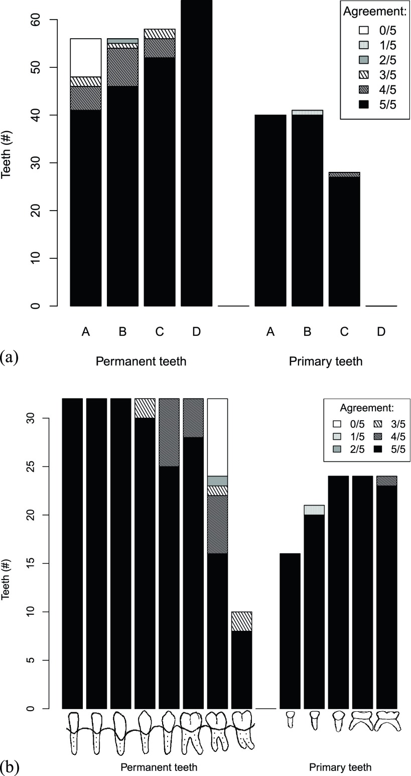
Participating clinicians delineated contours for primary and permanent teeth with varying degrees of completeness, stratified principally by the age of the subject. All clinicians successfully contoured all permanent teeth among the oldest cohort (Group D, age more than 13 years), whereas no clinician was able to identify eight permanent molars among the youngest cohort (Group A, age less than 2 years). Overall, clinicians were able to successfully identify 86% of permanent teeth among subjects in Group A and 100% among Groups B–D, with each tooth successfully contoured by all clinicians in 73/82/90/100% of cases (Groups A/B/C/D, respectively; Figure 1a).
Figure 1.
Groupwise distribution of tooth delineation success rates for permanent and primary teeth. For both panels, values depicted along the y-axis represent the number of potentially identifiable teeth, with greyscale colours and shading corresponding to the number of clinicians who correctly identified and contoured teeth in agreement with gold standard volumes. (a) Group A–D labels along the x-axis correspond to the analogous subjects in Table 1. (b) Grouping along the x-axis is stratified by permanent and primary tooth type (e.g. first incisors represent four teeth from bilateral mandibular and maxillary dentition). Among the permanent teeth, data corresponds to first incisors, second incisors, canines, first premolars, second premolars, first molars, second molars and third molars from left to right, respectively. The primary teeth correspond to the first incisors, second incisors, canines, first molars and second molars from left to right, respectively. #, number.
For all clinicians, complete delineation of primary dentition was more readily achieved (note that subjects in Group D had previously shed all primary dentition). However, one subject in Group B with a supernumerary primary tooth posed a significant challenge to four of five clinicians.
Measured on a per-tooth basis, delineation of permanent dentition was feasible for incisors, canines, premolars and primary molars among all groups, even at the youngest ages (Figure 1b). However, delineation of second molars was only possible commensurate with approximate timing of tooth development (Figure 1b, Supplementary materials). The presence/absence of third molars was unable to be assessed in Groups A and B, however, third molars that were present among the older cohorts (Groups C and D) were reliably identified by most clinicians (Figure 1b).
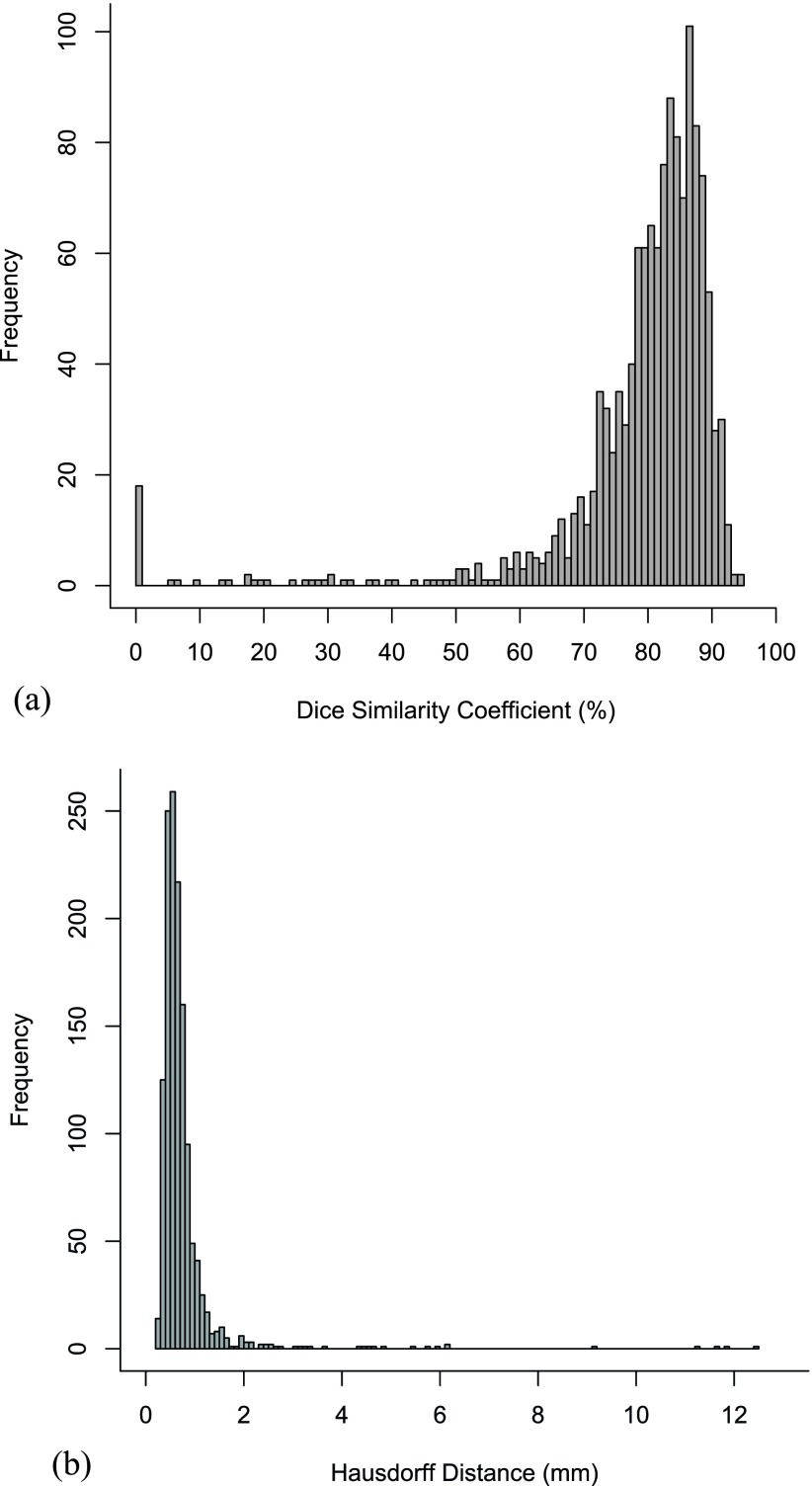
Next, clinician-delineated volumes were compared with gold standard contours for all teeth among all patients. Pairwise analysis of volumetric structure overlap was determined by DSC, with good volumetric agreement in the majority of cases (Figure 2a). Median DSC was 82%, with maximal agreement between independent clinician-delineated and gold standard contours as high as 95%. These findings are reproduced using dH as an independent volume–volume comparison metric, with dH < 1 mm in 88% of cases (Figure 2b).
Figure 2.
Histogram frequencies of the degree of contour agreement with gold standard volumes for all teeth. (a) Volumetric comparison was performed by pairwise Dice similarity coefficient (DSC). The y-axis represents the absolute number of clinician-delineated structures corresponding to the respective DSC along the x-axis. (b) Volumetric comparison was performed by pairwise Hausdorff distance (dH). The y-axis represents the absolute number of clinician-delineated structures corresponding to the respective dH along the x-axis.
However, in approximately 4% of cases, clinician-delineated volumes departed significantly from gold standard volumes (DSC <50%). In 17 specific instances (approximately 1% of cases), a clinician delineated a tooth volume that shared 0 overlap with the corresponding gold standard volume. Seven of these cases were permanent mandibular molars contoured too deeply within the mandible itself, while six separate cases consisted of permanent maxillary molars defined medial to the primary dentition instead of deep to it. Similarly, two mandibular molars were defined posteriorly to the primary teeth instead of underlying them as anatomically appropriate. Finally, one tooth was contoured in the space between two other teeth, while a separate tooth replaced the root of its neighbour. Note that 76% of these errors occurred among the youngest patient cohort (Group A), reflecting the relative challenge of accurately contouring the dentition in individuals less than 2 years of age.
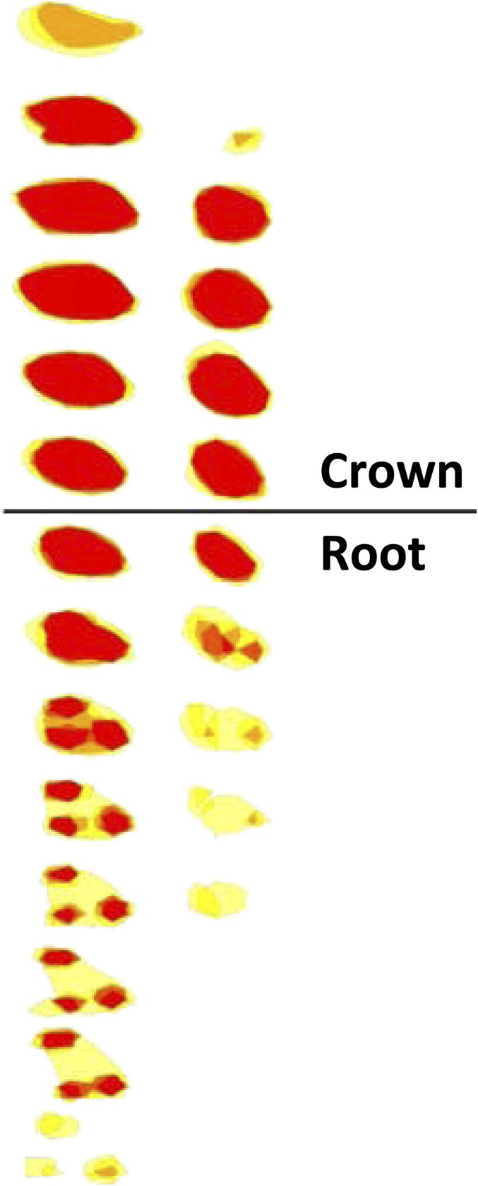
Within each tooth, the area of largest uncertainty (i.e. variability among clinician contours) was located at the level of the dental roots. Among the permanent dentition, the average dH was 2–5 mm larger in the roots than in the crowns for all clinician volumes. The same phenomenon was observed among the primary dentition, with 3–8 mm larger average dH at the level of the roots than at crowns for all clinician volumes. Figure 3 depicts the variability among clinician contours for representative permanent and primary teeth. As noted above, per-tooth contour variability was demonstrably larger in the roots than in the crowns.
Figure 3.
Variability among clinician contours for two representative teeth. Axial cross-sections are displayed along the y-axis for two representative teeth, a permanent maxillary first molar (left) and a primary maxillary first molar (right). Within each axial cross-section, the degree of structural overlap among clinician contours is depicted in shades of red (complete agreement between clinicians) to yellow (no agreement). The axial cross sections are divided at the approximate separation of tooth crown and root. For colour images see online.
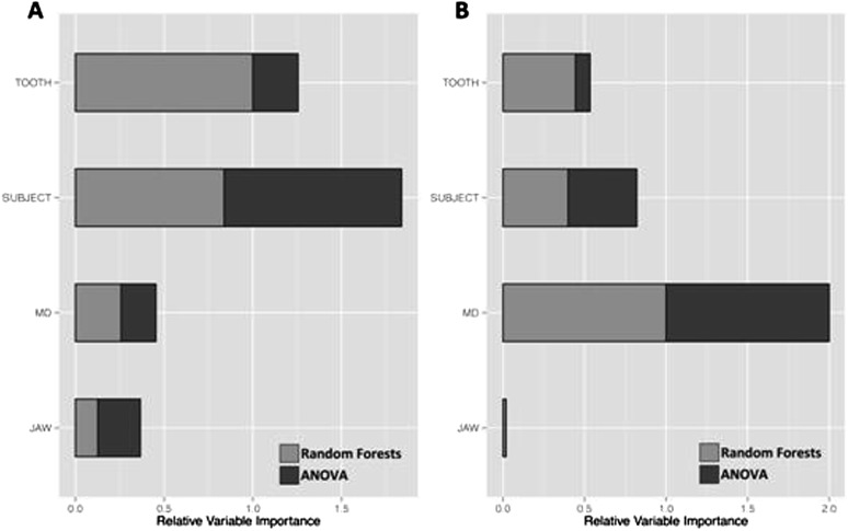
Overall, the age of the subject and the type of tooth (e.g. primary canine) were principal predictors of contour accuracy as assessed by dH (p < 0.001). To a lesser degree, contour accuracy varied between the mandible and the maxilla and between each clinician (p < 0.001). The relative importance of each of these variables as predictors of contour accuracy (assessed by both ANOVA and Random Forests methods) is shown in Figure 4a.
Figure 4.
Relative variable importance in predicting contour accuracy and clinician confidence. (a) The relative importance of four variables—“TOOTH” (e.g. primary canine, permanent first premolar); “SUBJECT” (subject age); “MD” (clinician identity); and “JAW” (mandible/maxilla)—is depicted along the x-axis with higher numbers indicating relatively larger importance. Relative variable importance was assessed by Random Forests (gray) and ANOVA (dark gray) methods, with respect to contour accuracy. (b) Analogous to Panel A, data are displayed as a function of relative variable importance in predicting clinician confidence scores.
Clinician effort
On average, clinicians spent 2.2 min delineating each tooth (range, 40 s to 4.5 min), equivalent to 93 min for the entire dentition for each subject (range, 30–200 min). Strikingly, the duration of effort allotted to each case was stratified principally by the clinician's level of training (average case duration was 46 and 127 min for attending physicians and trainees, respectively). The average duration per case increased linearly with the number of teeth present for each subject (R = 0.98), with the oldest patient cohort requiring the least overall effort owing to the absence of primary dentition.
Interobserver variability and clinician confidence
Agreement among independent clinician contours was generally good, with a median 0.7 mm dH between structures (range, 0.2 mm–1.3 cm). This degree of variability increased with the degree of structural complexity (average dH for premolars/molars was greater than that for incisors/canines, 1.1/1.0 mm vs 0.7/0.7 mm, respectively; p < 0.001) and was predicted, inversely, by clinician confidence (R from −0.17 to −0.55). The time allotted to each case was also inversely related to confidence scores (R = −0.33) but had no direct bearing on contour variability (note that per-contour effort was not assessed).
Clinician-reported confidence scores were found to be principally dependent upon the clinician him/herself (p < 0.001), with each physician applying a distinct grading scale to their own data. Secondarily, confidence scores were influenced by the age of the subject and the location of the tooth within the jaw (e.g. permanent canine, p < 0.001). However, confidence scores were not significantly different between maxillary and mandibular dentition as a whole. The relative importance of each of these variables as predictors of clinician confidence scores (assessed by both ANOVA and Random Forests methods) is shown in Figure 4b.
Discussion
This study describes the development of a detailed tooth contouring atlas and its implementation among five independent clinicians. We not only show feasibility of dental identification on CT scans in children and infants as young as 1 year of age but also describe the degree of interclinician variability and the areas of greatest uncertainty and challenge. Contouring accuracy generally increases with subject age (and dental maturity), correlating with expected developmental time points such as tooth calcification (Supplementary materials). On a per-tooth basis, we observe relatively decreased accuracy of root delineation as well as the added challenge of accounting for congenital anomalies such as supernumerary teeth, which may occur in approximately 1% of the population.12–14
The tooth-contouring atlas includes a subject with dental artefacts (i.e. fillings), however, the study data set excluded individuals with dental fillings, orthodontic appliances or implants to minimize confounding variables. As a consequence, we are unable to evaluate interclinician variability among dental contours in the setting of metallic artefacts. In the paediatric population, the prevalence of such obfuscating implants is relatively rare, however, the cumulative incidence of dental fillings increases with age, reaching an estimated 50% in the adult population.15 Given the frequency and potential impact, the scenario of dental artefacts may warrant future investigation in the adult population.
While tooth delineation is generally straightforward among older subjects with exclusively permanent dentition (e.g. teenagers and adults, excluding dental artefacts), it is the youngest cohort with the most challenging anatomy (e.g. infants) who may benefit the most from dental dose avoidance. As each tooth develops and exists as an independent organ, we assert that dental toxicity can and should be accounted for in the context of localized dosimetric variability (e.g. each tooth as an independent OAR). Tooth dose in excess of 20 Gy may be particularly detrimental among the youngest paediatric patients, and prospective efforts should avoid dose to the developing dentition where feasible and clinically appropriate.5
The purpose of this contouring atlas is to facilitate accurate and consistent dental contouring among the paediatric radiation oncology community. Nonetheless, based on our observations, contouring of the entire dentition may add an average of more than 90-min effort to a given case, potentially rendering this approach impractical in a busy clinical work environment. As a viable compromise in select cases, individual teeth or a single quadrant of the dentition may be identified with significantly reduced effort (approximately 2.2 min of effort per tooth). Alternatively, tooth delineation by an expert clinician (e.g. oral radiologist) could potentially reduce the time required per case, although this was not explored in the present study. Lastly, automated segmentation approaches may also be attempted, with the caveat of decreased accuracy of tooth delineation in adults (note that such tools are not currently applicable for dental contouring in the paediatric population).16
Of note, we did not formally assess clinician ease or success at tooth delineation prior to introduction of this atlas. Therefore, we are currently unable to report on the effectiveness of this atlas as a teaching tool or clinical resource. Anecdotally, the atlas was observed to be a helpful reference for anatomy and spatial orientation as well as tooth nomenclature. However, its use does not preclude the possibility of significant contouring errors (approximately 1% error rate in this study). Additional studies regarding effectiveness are therefore warranted.
We envision a potential important role for this atlas, particularly among the paediatric radiation oncology community, as a reference for accurately identifying and delineating the permanent and primary dentition, on an individual tooth basis or in its entirety. We anticipate that more widespread adoption and implementation of this approach will enable further evaluation of the teeth as OARs and may ultimately reduce treatment-related morbidity.
Conclusions
Delineation of individual teeth was feasible in all cases, albeit with varying degrees of completeness and accuracy, dependent upon the age of the subject. We conclude that this dental contouring atlas may serve as a useful tool and reference, in particular for the paediatric radiation oncology community.
References
- 1.Fromm M, Littman P, Raney RB, Nelson L, Handler S, Diamond G, et al. Late effects after treatment of twenty children with soft tissue sarcomas of the head and neck. Experience at a single institution with a review of the literature. Cancer 1986; 57: 2070–6. doi: 10.1002/1097-0142(19860515)57:103.0.co;2-g [DOI] [PubMed] [Google Scholar]
- 2.Kaste SC, Hopkins KP, Jenkins JJ, 3rd. Abnormal odontogenesis in children treated with radiation and chemotherapy: imaging findings. AJR Am J Roentgenol 1994; 162: 1407–11. doi: 10.2214/ajr.162.6.8192008 [DOI] [PubMed] [Google Scholar]
- 3.Weyman J. The effect of irradiation on developing teeth. Oral Surg Oral Med Oral Pathol 1968; 25: 623–9. doi: 10.1016/0030-4220(68)90310-1 [DOI] [PubMed] [Google Scholar]
- 4.Jaffe N, Toth BB, Hoar RE, Ried HL, Sullivan MP, McNeese MD. Dental and maxillofacial abnormalities in long-term survivors of childhood cancer: effects of treatment with chemotherapy and radiation to the head and neck. Pediatrics 1984; 73: 816–23. [PubMed] [Google Scholar]
- 5.Thompson RF, Schneider RA, Albertini F, Lomax AJ, Ares C, Goitein G, et al. Dose to the developing dentition during therapeutic irradiation: organ at risk determination and clinical implications. Int J Radiat Oncol Biol Phys 2013; 86: 108–13. doi: 10.1016/j.ijrobp.2012.11.041 [DOI] [PubMed] [Google Scholar]
- 6.Nelson SJ, Ash MM. Wheeler's dental anatomy, physiology and occlusion. 9th edn. St Louis, MO: Saunders Elsevier; 2009. [Google Scholar]
- 7.Turp JC, Alt KW. Designating teeth: the advantages of the FDI's two-digit system. Quintessence Int 1995; 26: 501–4. [PubMed] [Google Scholar]
- 8.Thompson RF. RadOnc: an R package for analysis of dose-volume histogram and three-dimensional structural data. J Radiat Oncol Inform 2014; 6: 71–83. [Google Scholar]
- 9.Dice LR. Measures of the amount of ecologic association between species. Ecology 1945; 26: 297–302. doi: 10.2307/1932409 [DOI] [Google Scholar]
- 10.Hausdorff F. Basics of Set Theory. [German.] Leipzig, Germany: Veit & Company Inc.; 1914. [Google Scholar]
- 11.Ehrlinger J. ggRandomForests: graphical exploration of random forests. 1.1.0 ed. 2014. [Published 5 December 2014.] Available from: http://cran.r-project.org/web/packages/ggRandomForests/index.html [Google Scholar]
- 12.Celikoglu M, Kamak H, Oktay H. Prevalence and characteristics of supernumerary teeth in a non-syndrome Turkish population: associated pathologies and proposed treatment. Med Oral Patol Oral Cir Bucal 2010; 15: e575–8. doi: 10.4317/medoral.15.e575 [DOI] [PubMed] [Google Scholar]
- 13.Delli K, Livas C, Bornstein MM. Lateral incisor agenesis, canine impaction and characteristics of supernumerary teeth in a South European male population. Eur J Dent 2013; 7: 278–83. doi: 10.4103/1305-7456.115410 [DOI] [PMC free article] [PubMed] [Google Scholar]
- 14.Anegundi RT, Tegginmani VS, Battepati P, Tavargeri A, Patil S, Trasad V, et al. Prevalence and characteristics of supernumerary teeth in a non-syndromic South Indian pediatric population. J Indian Soc Pedod Prev Dent 2014; 32: 9–12. doi: 10.4103/0970-4388.127041 [DOI] [PubMed] [Google Scholar]
- 15.Kirkevang LL, Vaeth M, Wenzel A. Prevalence and incidence of caries lesions in relation to placement and replacement of fillings: a longitudinal observational radiographic study of an adult Danish population. Caries Res 2009; 43: 286–93. doi: 10.1159/000217861 [DOI] [PubMed] [Google Scholar]
- 16.Thariat J, Ramus L, Maingon P, Odin G, Gregoire V, Darcourt V, et al. Dentalmaps: automatic dental delineation for radiotherapy planning in head-and-neck cancer. Int J Radiat Oncol Biol Phys 2012; 82: 1858–65. doi: 10.1016/j.ijrobp.2011.03.035 [DOI] [PubMed] [Google Scholar]


