Abstract
Objectives:
To evaluate a new method for assessing the radio-opacity of endodontic sealers and to compare radio-opacity values with a well-established standard method.
Methods:
The sealers evaluated in this study were AH Plus® (Dentsply DeTrey GmbH, Konstanz, Germany), Endo CPM Sealer (EGEO SRL, Buenos Aires, Argentina) and MTA Fillapex® (Angelus Dental Products Industry S/A, Londrina, Parana, Brazil). Two methods were used to evaluate radio-opacity: (D) standard discs and (S) a tissue simulator. For (D), ten standard discs were prepared for each sealer and were radiographed using Digora® phosphor storage plates (Soredex; Orion Corporation, Helsinki, Finland), alongside an aluminium stepwedge. For (S), polyethylene tubes filled with sealer (n = 10 for each) were radiographed inside the simulator as described. The digital images were analysed using Adobe Photoshop® software v. 10.0 (Adobe Systems, San Jose, CA). To compare the radio-opacity among the sealers, the data were analysed by ANOVA and Tukey's test, and to compare methods, they were analysed by the Mann–Whitney U test. To compare the data obtained from dentin and sealers in method (S), Student's paired t-test was used (=0.05).
Results:
In both methods, the sealers showed significant differences, according to the following decreasing order: AH Plus, MTA Fillapex and Endo CPM. In (D), MTA Fillapex and Endo CPM showed less radio-opacity than aluminium. For all of the materials, the radio-opacity was higher in (S) than in (D). Compared with dentin, all of the materials were more radio-opaque.
Conclusions:
The comparison of the two assessment methods for sealer radio-opacity testing validated the use of a tissue simulator block.
Keywords: dental materials, root canal therapy, radiography, dental
Introduction
Radio-opacity has been widely acknowledged as an important property of endodontic sealers. Among other physical/chemical properties, it has been stated that an ideal root canal filling material should have a certain degree of radio-opacity that allows for a clear distinction between the material and the surrounding anatomical structures, to facilitate the evaluation of the quality of root fillings.1,2
According to the American National Standards Institute/American Dental Association (ANSI/ADA), to determine the minimal requirement of radio-opacity for root canal filling materials, it has been established that they should have radio-opacity values of at least 3 mm of aluminium at a thickness of 1 mm.3,4 The radiographic images must be obtained by chemical processing of the radiographic film, and radio-opacity must be evaluated by an optical densitometer.3–6
By contrast, the advantages of digital radiography systems have motivated researchers, and a number of studies have compared the radio-opacity of restorative materials measured from conventional and digital radiographic images.7–20
Furthermore, in the standard method, the samples are radiographed with no tissue association. The absence of tooth, bone and soft-tissue constitutes an important differential compared with clinical situations in which radio-opacity is investigated, and it could alter the perception of radio-opacity of dental materials. Aiming to simulate clinical conditions, Gegler and Fontanella21 developed a “tissue simulator block”. This experimental model has already been successfully used in studies on the diagnosis of external apical root resorption, but it has not been used to evaluate the radio-opacity of endodontic sealers.
Thus, the present study aimed to evaluate in vitro a new method for assessing the radio-opacity of three endodontic sealers [AH Plus® (Dentsply DeTrey GmbH, Konstanz, Germany); Endo CPM Sealer (EGEO SRL, Buenos Aires, Argentina) and MTA Fillapex® (Angelus Dental Products Industry S/A, Londrina, Parana, Brazil)] in digital images (phosphor storage plates) and to compare these radio-opacity values with a well-established standard method.
Methods and materials
To evaluate the radio-opacity of three root canal sealers—AH Plus, Endo CPM sealer and MTA Fillapex —the following methods were used:
(D) Materials in standard discs
Cylindrical samples were fabricated according to their manufacturers' instructions by pouring the manipulated sealers into plastic rings measuring 4 mm in diameter by 1.5 mm in thickness. Ten specimens were prepared from each sealer. The filled rings were stored at 37 °C (±1) in 95% (±5) humidity, until the material was completely set.
Each sample was then radiographed using Digora® phosphor storage plates (Soredex; Orion Corporation, Helsinki, Finland), alongside an aluminium stepwedge that was used as a reference. The radiographs were obtained using a radiographic unit (Dabi Atlante Spectro 70X, São Paulo, Brazil) operating at 70 kV and 10 mA, with a 0.3-s exposure time and a 30-cm focal distance set.
(S) Materials in a tissue simulator
The endodontic sealers were prepared according to their manufacturers' instructions. The freshly mixed sealer was introduced into polyethylene tubes (10 mm × 1.5 mm; Abbott Lab do Brasil, São Paulo, Brazil) with a syringe to avoid bubbles. The filled tubes were stored at 37 °C (±1) in 95% (±5) humidity, until the material was completely set.
The tubes with sealers (n = 10 for each) were individually placed in the root canals of teeth positioned in the tissue simulator, as described previously by Gegler and Fontanella.21 Briefly, the maxillary anterior region of a human skull was used, divided by sagittal osteotomy into two segments fixed with wax (Wilson, São Paulo, Brazil) in a plastic container (length = 6 cm; width = 2.5 cm; depth = 3.5 cm). Distances of 1 cm were established between the external surfaces of the buccal and palatal segments and the container's walls, with this latter space filled with pored self-curing acrylic (Artigos Odontológicos Clássico, São Paulo, Brazil) that could simulate the soft tissues.
A distance of 0.5 cm was established between the internal surface of the buccal bone and the internal surface of the palatal bone. The space was filled with wax and was used to fix a human canine root with the root canal previously prepared. The root was inserted up to the point at which the cementum–enamel junction coincided with the level of the alveolar crest.
The set (tubes with sealers in the root canals of the teeth positioned in the tissue simulator) was radiographed as previously described.
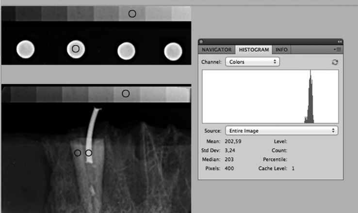
A 24-inch liquid crystal display monitor at 1920 × 1980 resolution was used to display the images in a dimmed light room. One observer, a dental radiologist with several years experience in digital radiography, evaluated the images at a 50-cm distance from the monitor. The digital images were analysed in Adobe Photoshop® software v. 10.0 (Adobe Systems, San Jose, CA). For the materials in (D), a standard-size circle (400 pixels) was drawn in the centre of the standard disc, and another circle was drawn in the sixth step of the aluminium stepwedge, equivalent to 3 mm of aluminium. For the materials in (S), three standard-size circles (400 pixels) were drawn: one under the tube and another under the dentin, both in the cervical third, and the third in the sixth step of the aluminium stepwedge (Figure 1).
Figure 1.
Analysis of the images in Adobe Photoshop® software v. 10.0 (Adobe Systems, San Jose, CA). Std Dev, standard deviation.
The average and standard deviation of the greyscale pixel values of the area selected were measured using the histogram tool and were recorded. The pixel values obtained for the materials were subtracted from the pixel values obtained in the 3-mm aluminium stepwedge.
To compare the radio-opacity among the sealers, the data were subjected to statistical analysis using ANOVA and Tukey's test. To compare methods, the data were evaluated by the Mann–Whitney U test. To compare the data obtained from dentin and sealers in method (S), the Student's paired t-test was used. The significance level was set at 5%, and the data were processed using SPSS® software, v. 10.0 (SPSS Inc., Chicago, IL).
Results
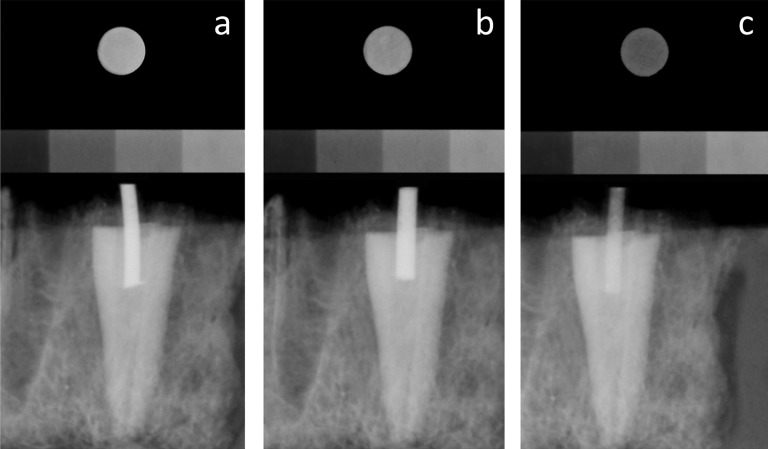
When considering the difference in pixel density between the material and 3 mm of aluminium, it was observed that the materials in both methods showed significant differences (ANOVA and Tukey post hoc test; p < 0.05) according to the following order of decreasing radio-opacity: AH Plus, MTA Fillapex and Endo CPM. It is worth mentioning that in (D), MTA Fillapex and Endo CPM showed less radio-opacity than aluminium (negative values), which was not observed in (S) (Table 1, Figure 2).
Table 1.
Comparison between sealers and methods, considering the difference in pixel density obtained from the materials and from 3 mm of aluminium
| Groups | n | Disc |
Tissue simulator |
p-value | ||
|---|---|---|---|---|---|---|
| Mean | SD | Mean | SD | |||
| AH Plus® | 10 | 7.6aA | 28.2 | 50.4aB | 5.2 | 0.0001 |
| MTA Fillapex® | 10 | −18.6bA | 19.7 | 34.0bB | 5.2 | |
| Endo CPM | 10 | −124.5cA | 4.5 | 14.1cB | 2.8 | |
SD, standard deviation.
Different lower case letters indicate significant difference among sealers in the same column (ANOVA and Tukey post hoc test).
Different capital letters indicate significant difference among methods in the same row (Mann–Whitney U test).
AH Plus was obtained from Dentsply DeTrey GmbH, Konstanz, Germany; Endo CPM was obtained from EGEO SRL, Buenos Aires, Argentina; MTA Fillapex was obtained from Angelus Dental Products Industry S/A, Londrina, Parana, Brazil.
Figure 2.
Image illustrating the differences between methods considering each material, showing higher radio-opacity in the tissue simulator than in standard discs: (a) AH Plus® (Dentsply DeTrey GmbH, Konstanz, Germany); (b) MTA Fillapex® (Angelus Dental Products Industry S/A, Londrina, Parana, Brazil); and (c) Endo CPM (EGEO SRL, Buenos Aires, Argentina).
When comparing both methods, regardless of the material used, the radio-opacity was higher in (S) than in (D) (Mann–Whitney U test; p < 0.05) (Table 1, Figure 2). Compared with dentin, in (S), all of the materials were significantly more radio-opaque (<0.05) (Table 2).
Table 2.
Comparison of pixel density between the materials and dentin with the simulator method
| Groups | n | Material |
Dentin |
p-value | ||
|---|---|---|---|---|---|---|
| Mean | SD | Mean | SD | |||
| AH Plus® | 10 | 205.8a | 9.0 | 126.8b | 9.8 | 0.0001a |
| MTA Fillapex® | 10 | 201.5a | 16.1 | 125.6b | 15.5 | |
| Endo CPM | 10 | 187.7a | 6.7 | 142.8b | 9.8 | |
SD, standard deviation.
Different lower case letters on the same row indicate significant difference.
Paired Student's t-test.
AH Plus was obtained from Dentsply DeTrey GmbH, Konstanz, Germany; Endo CPM was obtained from EGEO SRL, Buenos Aires, Argentina; MTA Fillapex was obtained from Angelus Dental Products Industry S/A, Londrina, Parana, Brazil.
Discussion
The radio-opacity of root canal sealers has particular relevance for assessing the quality of endodontic treatment.14,17 To evaluate this property, the ANSI/ADA standards have traditionally been employed. However, more recently, proposals to simplify this method have been presented, both to reduce the number of steps with the aluminium stepwedge18 and to use digital radiographs and software to replace optical densitometry.7–17,19,20 These changes have occurred not only for endodontic sealers but also for other categories of dental materials.22
Additionally, further progress towards the improvement of in vitro testing is expected to simulate clinical conditions more closely.23 The inclusion of materials in the tooth structure was proposed in a radio-opacity study.24
In effect, our results showed differences in the relative radio-opacity of the sealers between methods, with increased radio-opacity of endodontic sealers when they were radiographed inside the simulator. The overlapping of soft tissues, bone and dental structures was intrinsic in the clinical situation and was an important issue when radio-opacity was investigated.16,20 It is interesting to observe that the differences in radio-opacity among materials found in the standard method were reduced in the simulator method, owing to the overlapping of tissues, which resulted in a certain degree of radio-opacity that allowed for the distinctions between the materials and the surrounding anatomical structures.
With the standard method, certain sealers could present lower radio-opacity values than those recommended by ANSI/ADA, requiring the addition of radio-opaque substances to their compositions. These substances could negatively influence the other properties of the sealers.25 The present study suggested that when radio-opacity was evaluated by the simulator method, the same sealer that was considered only slightly radio-opaque by the standard method could be considered sufficiently radio-opaque to be used clinically with the original composition.
Furthermore, higher grey values were found in the sealers than in dentin in the simulator method, proving that the tested materials presented sufficient radio-opacity to be identified under clinical conditions. AH Plus, a two-component paste/paste sealer, has been continuously used in comparative studies of the physicochemical, biological and antimicrobial properties of root canal sealers.26–28 This sealer contains zirconium and iron oxide, which contribute to its greater radio-opacity. Agreeing with the findings of this investigation, its adequate radio-opacity has been demonstrated in several studies that have used the standard method.10–14,17,20
By contrast, for the two MTA-based filling materials tested (MTA Fillapex and Endo CPM), which were introduced to the market with the promise of improving clinical performance, the findings demonstrated herein should be confirmed in future investigations.
According to the ANSI/ADA specifications, to evaluate the radio-opacity of endodontic filling materials, discs that are 1-mm thick should be imaged and compared with 3 mm of aluminium. Because the objective of this study was to compare methods, the material thickness of 1.5 mm was used to standardize this parameter in both methods. This fact did not allow for quantitative comparison of the data obtained regarding the radio-opacity of the endodontic sealers in the standard method with the data from other investigations that used the same method.
In addition, it is important to consider that the teeth used in the simulator were superior canines, owing to their large diameters in the cervical third of the root canal. This anatomy allowed the insertion of polyethylene tube within the canal. The interference of the tube in material radio-opacity was evaluated by Salles et al.29 They compared the tooth radio-opacity with and without the tube within the root canal and found no significant differences. Based on these findings, it can be inferred that, in this study, the tube did not affect the sealers' radio-opacity.
Future studies to develop methodologies that allow for the use of other dental groups, with different root diameters and bone cortical thicknesses, should be conducted to investigate whether these anatomical variations have any influence on the radio-opacity of the sealers inside the simulator.
In conclusion, the comparison of the two assessment methods for sealer radio-opacity testing, considering clinical reality, validated the use of a tissue simulator block.
References
- 1.McComb D, Smith DC. Comparison of physical properties of polycarboxylate-based and conventional root canal sealers. J Endod 1976; 2: 228–35. [DOI] [PubMed] [Google Scholar]
- 2.Borges AH, Pedro FL, Semanoff-Segundo A, Miranda CE, Pécora JD, Cruz Filho AM. Radiopacity evaluation of Portland and MTA-based cements by digital radiographic system. J Appl Oral Sci 2011; 19: 228–32. [DOI] [PMC free article] [PubMed] [Google Scholar]
- 3.International Organization for Standardization ISO 6876: dental root canal sealing materials. 2nd edn. Geneva, Switzerland: ISO; 2001. [Google Scholar]
- 4.American Dental Association. ANSI/ADA specification no. 57—endodontic sealing material. Chicago, IL: ADA; 2000. [Google Scholar]
- 5.Beyer-Olsen EM, Orstavik D. Radiopacity of root canal sealers. Oral Surg Oral Med Oral Pathol 1981; 51: 320–8. [DOI] [PubMed] [Google Scholar]
- 6.Bodrumlu E, Sumer AP, Gungor K. Radiopacity of a new root canal sealer, Epiphany. Oral Surg Oral Med Oral Pathol Oral Radiol Endod 2007; 104: e59–61. [DOI] [PubMed] [Google Scholar]
- 7.Akcay I, Ilhan B, Dundar N. Comparison of conventional and digital radiography systems with regard to radiopacity of root canal filling materials. Int Endod J 2012; 45: 730–6. doi: 10.1111/j.1365-2591.2012.02026.x [DOI] [PubMed] [Google Scholar]
- 8.Baksi Akdeniz BG, Eyüboglu TF, Sen BH, Erdilek N. The effect of three different sealers on the radiopacity of root fillings in simulated canals. Oral Surg Oral Med Oral Pathol Oral Radiol Endod 2007; 103: 138–41. [DOI] [PubMed] [Google Scholar]
- 9.Baldi JV, Bernardes RA, Duarte MA, Ordinola-Zapata R, Cavenago BC, Moraes JC, et al. Variability of physicochemical properties of an epoxy resin sealer taken from different parts of the same tube. Int Endod J 2012; 45: 915–20. doi: 10.1111/j.1365-2591.2012.02049.x [DOI] [PubMed] [Google Scholar]
- 10.Bodanezi A, Pereira AL, Munhoz EA, Bernardineli N, de Moraes IG, Bramante CM. Digital radiopacity measurement of different resin- and zinc oxide-based root canal sealers. Rev Odonto Ciênc 2010; 25: 74–7. [Google Scholar]
- 11.Candeiro GT, Correia FC, Duarte MA, Ribeiro-Siqueira DC, Gavini G. Evaluation of radiopacity, pH, release of calcium ions, and flow of a bioceramic root canal sealer. J Endod 2012; 38: 842–5. doi: 10.1016/j.joen.2012.02.029 [DOI] [PubMed] [Google Scholar]
- 12.Carvalho-Junior JR, Correr-Sobrinho L, Correr AB, Sinhoreti MA, Consani S, Sousa-Neto MD. Radiopacity of root filling materials using digital radiography. Int Endod J 2007; 40: 514–20. [DOI] [PubMed] [Google Scholar]
- 13.Duarte MA, Ordinola-Zapata R, Bernardes RA, Bramante CM, Bernardineli N, Garcia RB, et al. Influence of calcium hydroxide association on the physical properties of AH Plus. J Endod 2010; 36: 1048–51. doi: 10.1016/j.joen.2010.02.007 [DOI] [PubMed] [Google Scholar]
- 14.Garrido AD, Lia RC, França SC, da Silva JF, Astolfi-Filho S, Sousa-Neto MD. Laboratory evaluation of the physicochemical properties of a new root canal sealer based on Copaifera multijuga oil-resin. Int Endod J 2010; 43: 283–91. doi: 10.1111/j.1365-2591.2009.01678.x [DOI] [PubMed] [Google Scholar]
- 15.Gu S, Rasimick BJ, Deutsch AS, Musikant BL. Radiopacity of dental materials using a digital X-ray system. Dent Mater 2006; 22: 765–70. [DOI] [PubMed] [Google Scholar]
- 16.Guerreiro-Tanomaru JM, Duarte MA, Gonçalves M, Tanomaru-Filho M. Radiopacity evaluation of root canal sealers containing calcium hydroxide and MTA. Braz Oral Res 2009; 23: 119–23. [DOI] [PubMed] [Google Scholar]
- 17.Marin-Bauza GA, Rached-Junior FJ, Souza-Gabriel AE, Sousa-Neto MD, Miranda CE, Silva-Sousa YT. Physicochemical properties of methacrylate resin-based root canal sealers. J Endod 2010; 36: 1531–6. doi: 10.1016/j.joen.2010.05.002 [DOI] [PubMed] [Google Scholar]
- 18.Poorsattar Bejeh Mir A, Poorsattar Bejeh Mir M. Assessment of radiopacity of restorative composite resins with various target distances and exposure times and a modified aluminum step wedge. Imaging Sci Dent 2012; 42: 163–7. doi: 10.5624/isd.2012.42.3.163 [DOI] [PMC free article] [PubMed] [Google Scholar]
- 19.Tagger M, Katz A. Radiopacity of endodontic sealers: development of a new method for direct measurement. J Endod 2003; 29: 751–5. [DOI] [PubMed] [Google Scholar]
- 20.Tanomaru-Filho M, Jorge EG, Guerreiro-Tanomaru JM, Gonçalves M. Radiopacity evaluation of new root canal filling materials by digitalization of images. J Endod 2007; 33: 249–51. [DOI] [PubMed] [Google Scholar]
- 21.Gegler A, Fontanella V. In vitro evaluation of a method for obtaining periapical radiographs for diagnosis of external apical root resorption. Eur J Orthod 2008; 30: 315–19. doi: 10.1093/ejo/cjm125 [DOI] [PubMed] [Google Scholar]
- 22.Kurşun Ş, Dinç G, Oztaş B, Yüksel S, Kamburoğlu K. The visibility of secondary caries under bonding agents with two different imaging modalities. Dent Mater J 2012; 31: 975–9. [DOI] [PubMed] [Google Scholar]
- 23.Kelly JR, Benetti P, Rungruanganunt P, Bona AD. The slippery slope: critical perspectives on in vitro research methodologies. Dent Mater 2012; 28: 41–51. doi: 10.1016/j.dental.2011.09.001 [DOI] [PubMed] [Google Scholar]
- 24.Goracci C, Juloski J, Schiavetti R, Mainieri P, Giovannetti A, Vichi A, et al. The influence of cement filler load on the radiopacity of various fibre posts ex vivo. Int Endod J 2015; 48: 60–7. doi: 10.1111/iej.12275 [DOI] [PubMed] [Google Scholar]
- 25.Kuga MC, Faria G, Só MV, Keine KC, Santos AD, Duarte MA, et al. The impact of the addition of iodoform on the physicochemical properties of an epoxy-based endodontic sealer. J Appl Oral Sci 2014; 22: 125–30. [DOI] [PMC free article] [PubMed] [Google Scholar]
- 26.Sousa CJ, Montes CR, Pascon EA, Loyola AM, Versiani MA. Comparison of the intraosseous biocompatibility of AH Plus, EndoREZ, and Epiphany root canal sealers. J Endod 2006; 32: 656–62. [DOI] [PubMed] [Google Scholar]
- 27.Versiani MA, Carvalho-Junior JR, Padilha MI, Lacey S, Pascon EA, Sousa-Neto MD. A comparative study of physicochemical properties of AH Plus and Epiphany root canal sealants. Int Endod J 2006; 39: 464–71. [DOI] [PubMed] [Google Scholar]
- 28.Tavares CO, Böttcher DE, Assmann E, Kopper PM, de Figueiredo JA, Grecca FS, et al. Tissue reactions to a new mineral trioxide aggregate-containing endodontic sealer. J Endod 2013; 39: 653–7. doi: 10.1016/j.joen.2012.10.009 [DOI] [PubMed] [Google Scholar]
- 29.Salles AA, Hauschild FM, Paranhos L, Fontanella VRC. Evaluation of three calcium hydroxide pastes' optical density. Rev Fac Odontol P Alegre 2007; 48: 17–21. [Google Scholar]


