

Abstract
Resorption of tooth structures can occur as a result of physiological, pathological, and idiopathic factors. Early diagnosis and appropriate treatment can prevent its serious complications. This case report presents surgical endodontic management of a trauma-induced perforating external root resorption, which was diagnosed with the help of cone beam computed tomography. Following root canal treatment, intentional replantation of the tooth was performed so as to expose the opening of the resorption defect to allow for complete debridement and closure. Eighteen months follow-up showed arrest of root resorption, and progressive healing of the defect.
Keywords: Biodentine, cone-beam computerized tomography, root resorption, sodium trichloroacetate, surgical replantation
INTRODUCTION
The etiology of root resorption involves complex processes. Classification of root resorption involves the categorization of different types on the basis of designated morphologic characteristics, as well as etiologic factors, for predicting potential biologic behavior; so that appropriate treatment may be delivered.[1] Heithersay had classified dental resorptions into three groups: Induced by trauma, induced by infection and hyperplasic invasive.[2] Among the various studies, Andreasen's classification is widely accepted, who divided resorptions into internal (replacement and inflammatory) and external (superficial, replacement, and inflammatory).[3]
Whether resorption has extended from an external inflammatory lesion to involve the internal surface or vice versa, a perforating lesion is created. External inflammatory resorption is a progressive inflammatory process, and the main factor accounting for its maintenance is necrotic pulp.[2] The presence of either necrotic pulp rests or bacteria within dentinal tubules attracts large number of osteoclasts to the area. Consequently, resorption progresses continuously up to root canal that is ultimately exposed.[4]
In cases without a perforation, the removal of the granulation tissue and the blood supply to the resorbing cells followed by repair of the perforation site with a suitable sealing material should be sufficient.[2] However, in cases in which a pathway between the pulp canal space and the periodontal tissues is present, root canal treatment should also be done.
Successful management of the root resorption involves an accurate diagnosis and insight knowledge of the various etiological factors, and the pathological processes involved.[3] Conventional intraoral radiograph produces images that have objects superimposed upon each other. The observer has to make three-dimensional decisions on the basis of a two-dimensional image. Cone beam computed tomography (CBCT) is a new and innovative technology that provides us with the relevant information that cannot be obtained from conventional radiographs. It offers the clinician ability to assess the area of interest in three different planes, and by doing so, it eliminates the superimposition that is inherent in conventional radiographic imaging. CBCT has high sensitivity and specificity in the detection of resorption lesions, and it performs significantly better than that of digital intraoral radiographs.[5]
This case report presents the 18 months follow-up of a case of trauma induced perforating root resorption in maxillary central incisor diagnosed with the help of CBCT, in which Biodentine was used to seal the resorption defect after endodontic therapy.
CASE REPORT
A 28-year-old male patient reported to the Conservative Department of the Maulana Azad Institute of Dental Sciences, New Delhi, with a chief complaint of pain in right upper front central tooth for that he had already seen a fewer private clinics. The tooth was opened and debrided. However, the symptoms and signs including spontaneous pain and pain on percussion did not subside, and he was referred to our hospital.
Medical history was noncontributory, but there was a history of trauma to maxillary anterior region 5 years ago. Intraoral periapical radiographic examination showed an oval radiolucent lesion in the middle third of the root canal of right maxillary central incisor, and the lateral incisor was already root canal treated [Figure 1]. The central incisor had a temporary restoration, and the root canal treatment had already been started in a private clinic. To confirm the diagnosis, a CBCT scan in the axial and sagittal plane was advised. The scan showed a communicating resorption defect in the middle third of the root canal [Figure 2]. The defect was present at the palatal aspect approaching toward the center of the canal [Figure 3]. Such defects start at the cementum adjacent to the periodontal ligament, and neither the periodontal nor the bone breakdown develops at the site of the portal.[4]
Figure 1.

Pre-operative periapical radiograph showing irregular radiolucent lesions within the maxillary right central incisor's root.
Figure 2.

CBCT Sagittal section showing the extent and location of the resorption defect in sagittal plane.
Figure 3.

CBCT Axial section showing the extent and location of the resorption defect in axial plane.
Successful treatment relies on the complete removal or inactivation of a resorptive tissue as well as debridement and obliteration of the root canal system.[2] As the portal of entry was well within the bone, and could not be located through the root canal system, extraction of the involved tooth after root canal therapy, followed by debridement and obturation of the resorption defect, and replantation of the tooth was planned. Tooth replantation carries its own risks,[6] but due to the inaccessibility of the resorption defect, it had to be included in the treatment plan.
Access to the canal system was reestablished, and the working length was confirmed with apex locator ROOT ZX II (J Morita, Irvine, CA, USA). The root canal was instrumented with stainless steel hand K-files (Dentsply Malliefer, Switzerland), until an apical stop of ISO #60 could be created. The root canal was repeatedly irrigated with 2.5% sodium hypochlorite followed by normal saline, and calcium hydroxide dressing was placed. The patient was recalled after 2 weeks. However, in case of pain/swelling, the patient was advised to report immediately.
On the subsequent visit, the root canal was obturated using Gutta-percha cones (Dentsply Malliefer, Switzerland) and AH-Plus (Dentsply Detrey, Konstanz) endodontic sealer by lateral condensation technique [Figure 4]. After anesthesia, the tooth was extracted with only dental forceps to minimize the damage of the periodontal ligament as undue trauma to the ligament can hamper the replantation process [Figure 5]. The resorptive defect was cleaned of the granulation tissue and 90% aqueous solution of trichloroacetic acid was applied for approximately 2-3 min using a micro applicator tip so that the area of acid application could be confined to the resorption defect only. The irregular borders of the defect were smoothed with a small round bur [Figure 6]. Biodentine (Septodont, St. Maurdes Fossés, France) was mixed according to the manufacturer's instructions and was firmly condensed in the cavity [Figure 7]. During the time (15 min) that the tooth was treated extra-orally, the tooth was held in a wet gauze piece. The best prognosis of a replanted tooth is indirectly related to the amount of time the tooth is maintained extraorally during the procedure. It has been stated that the potential for resorption in replanted teeth increases if they remain outside the mouth for more than 30 min.[7]
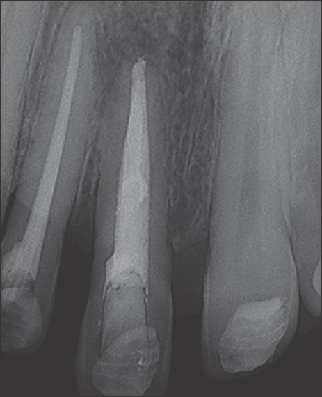
Figure 4.

Post-obturation radiograph with resorption areas still visible.
Figure 5.
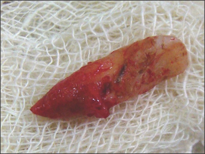
Photograph immediately after extraction showing the resorption defect.
Figure 6.

Photograph after complete debridement of the resorptive defect.
Figure 7.
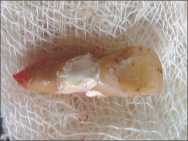
Resorptive defect restored with Biodentine.
The tooth was replanted back to its socket, and splinted with adjacent teeth using fiber-splint and composite. A periapical radiograph was taken for future comparison [Figure 8]. The patient was recalled after 2 weeks when the splint was removed.
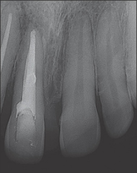
Figure 8.
Radiograph after replantation and splinting with resorption areas seen filled with biodentine which has the radio-opacity similar to dentine.
The case was followed up clinically and radiographically, which showed satisfactory results at 3, 6, 12 and 18 months post-operatively [Figures 9-12]. There was no pain on percussion, percussion sound was normal; and radiographically there was no sign of replacement resorption.
Figure 9.

Radiograph at 3 months.
Figure 12.

Radiograph at 18 months.
Figure 10.
Radiograph at 6 months.
Figure 11.

Radiograph at 12 months showing progressive healing.
DISCUSSION
Root resorption is either a physiologic (tooth eruption) or pathological (trauma induced) event mainly occurring due to the action of activated clastic cells. It is a serious dental complication, which can even lead to extraction of the tooth. Early diagnosis and appropriate treatment minimizes the sequelae related to this problem.[8] As the conventional intraoral radiograph does not reveal the true nature and exact location of the lesion, CBCT scans are recommended for this purpose. These images are displayed on the three orthogonal planes axial, sagittal and coronal simultaneously, allowing the clinician to gain a truly three-dimensional view of the area of interest. CBCT images are geometrically accurate, and the problems of anatomical noise seen with periapical radiographs can be eliminated.[9]
For the management of such cases, it is essential to trace and inactivate the resorptive tissue prior to the internal placement of a restorative material. The rationale for using trichloroacetic acid in order to remove or inactivate the resorbing tissue lies on the fact that it renders the tissue avascular by a process of coagulation necrosis. The self-limiting necrosis zone is well demarcated from the adjacent tissue, and therefore an uncomplicated repair of the surrounding periodontal tissue can be expected.[10]
The use of calcium hydroxide as an inter-appointment intra-canal medicament was aimed at dissolving remaining pulpal debris and alkalinizing the environment[11] to decrease the bacterial growth.
The insertion of a suitable restoration into the resorption cavity is also of paramount importance. Biodentine, a new calcium silicate-based material, has been proposed as a favorable perforation repair material as it can be placed in permanent and close contact with periradicular tissue due to its bioactivity and biocompatibility.[12] It has dentin-like mechanical properties, and can be used as a dentin substitute on crowns and roots.[13] Compared with other materials such as mineral trioxide aggregate (MTA), Biodentine handles easily and needs much less time for setting. Moreover, discoloration of marginal gingiva after perforation repair with gray MTA has been reported, which is not seen with Biodentine.[14]
Various studies advocate the filling of the root canal after repair of the perforation defect. On the other hand, Hsien et al. had repaired an iatrogenic root perforation with MTA after root canal filling had been completed.[15] In the present case, we also preferred filling the root canal before placement of Biodentine. Previously, placed gutta-percha acts as a barrier material allowing successful condensation of Biodentine. Moreover, it eliminates the risk of displacing Biodentine from the perforation site during condensation of gutta-percha.[16]
In the present case, after treating the defect, intentional replantation of the tooth was planned as the resorption was on the palatal aspect, well beneath the bone and surgery was not possible. Intentional replantation is defined as the purposeful extraction of a tooth in order to repair a defect or cause of treatment failure, and thereafter the return of the tooth to its original socket.[17] This treatment modality is considered as a last resort and should only be attempted only when no other treatment is feasible, as it is associated with the risk of root fracture, replacement resorption and ankylosis.[18,19]
Filippi et al. investigated the outcome of intentional replantation using resection of the ankylosed sites and observed that such treatment following intentional replantation prevents or delays the recurrence of ankylosis in 7 of 15 teeth.[20] Tang et al. successfully, treated a case of intentional replantation of a mandibular molar that had severe periodontal destruction resulting from iatrogenic perforation of the furcation and suggested the possibility of a successful surgical technique for the otherwise hopeless condition.[21] In another case report, Filho et al. concluded that intentional replantation could be indicated correctly as an alternative treatment for cases, in which conservative endodontic therapy or surgical technique cannot be performed.[22]
Intentional replantation has been found to be associated with the risk of root fracture, replacement resorption and ankylosis.[19] Although it is suggested only for limited cases, this procedure has been reported with a success rate of up to 95%, if done by following the guidelines.[23] The current case presented with inaccessibility of the lesion being the major challenge that indicated this treatment modality.
CONCLUSION
The success of intentional replantation is likely dependent upon a minimally traumatic extraction, short extraoral time with copious irrigation and meticulous instrumentation as well as carefully controlled postoperative patient compliance. This case presented some characteristics that required intentional replantation and the follow-up result suggested that the technique applied represented a viable treatment option.
Financial support and sponsorship
Nil.
Conflicts of interest
The authors of this manuscript declare that they have no conflicts of interest, real or perceived, financial or non-financial in this article.
REFERENCES
- 1.Santos BO, Mendoca DS, Sousa DL, Neto JJ, Araujo RB. Root resorption after dental traumas: Classification and clinical, radiographic and histologic aspects. Rev Bras Odontol. 2011;8:439–45. [Google Scholar]
- 2.Heithersay GS. Management of tooth resorption. Aust Dent J. 2007;52:S105–21. doi: 10.1111/j.1834-7819.2007.tb00519.x. [DOI] [PubMed] [Google Scholar]
- 3.Andreasen JO. Luxation of permanent teeth due to trauma. A clinical and radiographic follow-up study of 189 injured teeth. Scand J Dent Res. 1970;78:273–86. doi: 10.1111/j.1600-0722.1970.tb02074.x. [DOI] [PubMed] [Google Scholar]
- 4.Fuss Z, Tsesis I, Lin S. Root resorption: Diagnosis, classification and treatment choices based on stimulation factors. Dent Traumatol. 2003;19:175–82. doi: 10.1034/j.1600-9657.2003.00192.x. [DOI] [PubMed] [Google Scholar]
- 5.Leung SF. Cone beam computed tomography in endodontics. Dent Bull. 2010;15:3. [The Hong Kong Medical diary] [Google Scholar]
- 6.Pohl Y, Filippi A, Kirschner H. Results after replantation of avulsed permanent teeth. I. Endodontic considerations. Dent Traumatol. 2005;21:80–92. doi: 10.1111/j.1600-9657.2004.00297.x. [DOI] [PubMed] [Google Scholar]
- 7.Peñarrocha M, García B, Martí E, Palop M, von Arx T. Intentional replantation for the management of maxillary sinusitis. Int Endod J. 2007;40:891–9. doi: 10.1111/j.1365-2591.2007.01278.x. [DOI] [PubMed] [Google Scholar]
- 8.Heithersay GS. Clinical endodontic and surgical management of tooth and associated bone resorption. Int Endod J. 1985;18:72–92. doi: 10.1111/j.1365-2591.1985.tb00425.x. [DOI] [PubMed] [Google Scholar]
- 9.Patel S. New dimensions in endodontic imaging: Part 2. Cone beam computed tomography. Int Endod J. 2009;42:463–75. doi: 10.1111/j.1365-2591.2008.01531.x. [DOI] [PubMed] [Google Scholar]
- 10.Heithersay GS. Treatment of invasive cervical resorption: An analysis of results using topical application of trichloracetic acid, curettage, and restoration. Quintessence Int. 1999;30:96–110. [PubMed] [Google Scholar]
- 11.Siqueira JF, Jr, Lopes HP. Medicac¸ a˜o intracanal. In: Siqueira JF Jr, Lopes HP, editors. Endodontia: Biologia e te´ Cnica. 2nd ed. Rio de Janeiro: Guanabara Koogan; 2004. pp. 81–618. [Google Scholar]
- 12.Nikhil V, Arora V, Jha P, Verma M. Non surgical management of trauma induced external root resorption at two different sites in a single tooth with Biodentine: A case report. Endodontology. 2012;24:150–5. [Google Scholar]
- 13.Wang X, Sun H, Chang J. Characterization of Ca3SiO5/CaCl2 composite cement for dental application. Dent Mater. 2008;24:74–82. doi: 10.1016/j.dental.2007.02.006. [DOI] [PubMed] [Google Scholar]
- 14.Bortoluzzi EA, Araújo GS, Guerreiro Tanomaru JM, Tanomaru-Filho M. Marginal gingiva discoloration by gray MTA: A case report. J Endod. 2007;33:325–7. doi: 10.1016/j.joen.2006.09.012. [DOI] [PubMed] [Google Scholar]
- 15.Hsien HC, Cheng YA, Lee YL, Lan WH, Lin CP. Repair of perforating internal resorption with mineral trioxide aggregate: A case report. J Endod. 2003;29:538–9. doi: 10.1097/00004770-200308000-00011. [DOI] [PubMed] [Google Scholar]
- 16.Meire M, De Moor R. Mineral trioxide aggregate repair of a perforating internal resorption in a mandibular molar. J Endod. 2008;34:220–3. doi: 10.1016/j.joen.2007.11.011. [DOI] [PubMed] [Google Scholar]
- 17.Kingsbury BC, Jr, Wiesenbaugh JM., Jr Intentional replantation of mandibular premolars and molars. J Am Dent Assoc. 1971;83:1053–7. doi: 10.14219/jada.archive.1971.0425. [DOI] [PubMed] [Google Scholar]
- 18.Frank AL, Torabinejad M. Diagnosis and treatment of extracanal invasive resorption. J Endod. 1998;24:500–4. doi: 10.1016/S0099-2399(98)80056-3. [DOI] [PubMed] [Google Scholar]
- 19.Peer M. Intentional replantation: A ‘last resort’ treatment or a conventional treatment procedure?. Nine case reports. Dent Traumatol. 2004;20:48–55. doi: 10.1046/j.1600-4469.2003.00218.x. [DOI] [PubMed] [Google Scholar]
- 20.Filippi A, Pohl Y, von Arx T. Treatment of replacement resorption by intentional replantation, resection of the ankylosed sites, and Emdogain: Results of a 6-year survey. Dent Traumatol. 2006;22:307–11. doi: 10.1111/j.1600-9657.2005.00363.x. [DOI] [PubMed] [Google Scholar]
- 21.Tang PM, Chan CP, Huang SK, Huang CC. Intentional replantation for iatrogenic perforation of the furcation: A case report. Quintessence Int. 1996;27:691–6. [PubMed] [Google Scholar]
- 22.Filho FB, Vanni JR, Limongi O, Fariniuk LF, Travassos R, Albuquerque DS. Intentional replantation: Case report of an alternative treatment for endodontic therapy failure. Rev Bras Odontol. 2004;1:36–40. [Google Scholar]
- 23.Messkoub M. Intentional replantation: A successful alternative for hopeless teeth. Oral Surg Oral Med Oral Pathol. 1991;71:743–7. doi: 10.1016/0030-4220(91)90286-l. [DOI] [PubMed] [Google Scholar]