

Abstract
Sialolithiasis is a common disease of the major salivary glands, characterized by the obstruction of a salivary gland or its excretory duct due to the formation of calcareous concretions. Sialoliths usually measure from 1 mm to <10 mm. They rarely measure more than 15 mm, and infrequently giant salivary gland calculi >15 mm have been reported in the literature. The submandibular gland and its duct appear to be the most susceptible sites for this disease. In this article, we report two unique cases, including a giant bilateral case, measuring 50 mm in length and 5 mm in width on the right side and one, 30 mm in length, and 5 mm in width on the left side; and another case, measuring 83 mm in length. The diagnostic and therapeutic approaches consisted of transocclusal radiography with the conservative transoral surgical technique in both cases. The follow-up showed the normal function of the relevant salivary glands. To the best of our knowledge and belief, similar cases have not been reported in the literature.
Keywords: Sialoliths, Submandibular Glands, Wharton duct, Salivary duct
INTRODUCTION
Sialolithiasis is the second most common disease of salivary glands after mumps and also the most common disease of the submandibular gland.[1,2,3] It is more common in middle-aged males.[4] In the orofacial region, sialoliths are recognized as calcified masses with possible development through a mineralization nucleus consisting of debris, bacterial colonies, ductal epithelial cell remnants, mucus plugs, and foreign bodies.[5,6] They are usually small in size, varying from <1 mm up to 3 mm. Most of them are <10 mm in size whereas only 7.6% are larger than 15 mm.[7] Submandibular sialolith formation is more common because its saliva is more alkaline, has an increased concentration of calcium and phosphate, and has a higher mucous content than saliva from the parotid or sublingual glands. Furthermore, the submandibular duct is longer than the ducts of the other major glands, and the saliva flows against the gravity. Submandibular sialoliths tend to be single and form within the duct.[8] The exact cause of sialolith formation is still unknown, but a decrease in saliva production due to medications may contribute to a higher incidence rate of calculi.[8,9] The most common clinical symptoms are swelling and pain during food intake.[8] Sialoliths may be detected on palpation at an area superior to the mylohyoid muscle insertion. In many cases, the diagnosis is easy due to obvious clinical features, but imaging techniques are always necessary for therapeutic approaches.[10] Most sialoliths have enough calcium content to be readily identified on conventional dental radiographs, especially on occlusal and panoramic views.[4] Other effective techniques include computed tomography (CT), ultrasound, sialography, and magnetic resonance imaging; and the treatment is aimed at sialolith removal.[7,10] The present article reports two unusually large submandibular sialoliths; similar cases have not been reported in the literature to the best of our knowledge.
CASE REPORT
Case A
A healthy 25-year-old smoking male presented to the Department of Oral and Maxillofacial Surgery, Taleghani Hospital, Tehran, Iran. The patient complained of a tender, small swelling, and white creamy discharge in the right sublingual area.
The presence of a giant salivary calculus of the Wharton duct was confirmed by intraoral radiography and CT scan that showed two radiopaque oval formations, one about 50 mm in length and 5 mm in width on the right side and the other 30 mm in length and 5 mm in width on the left side [Figure 1].
Figure 1.

Transocclusal endoral radiography showed two radiopacities within the Wharton duct on the right and left sides.
Under intravenous sedation and local anesthesia, the sialolith was removed through an incision parallel to the submandibular gland duct. The incision was sutured, and antibiotic therapy with amoxicillin 500 mg and nonsteroidal anti-inflammatory drug (NSAID) analgesics were administrated. The patient was discharged from the hospital 2 days after the operation. Two weeks after the operation, the patient was recalled and examined. The operation site exhibited normal healing process, and secretion of saliva was observed.
Case B
A healthy 30-year-old male was referred by his dentist and presented with lower jaw pain. He also reported difficulty breathing and a foul-tasting mouth since 21 days previously and also a history of right submandibular swelling episodes occurring with meals. These symptoms disappeared within a relatively short period, never lasting more than 2 h. In fact, a panoramic radiograph, taken 3 months before, showed a radiopaque area appearing like an impacted lower right canine. Intraoral examination revealed a localized swelling at the right side of the floor of the mouth, solid on the touch and not adherent to any deeper structures. The diagnosis of a giant salivary calculus of the Wharton duct was confirmed by intraoral radiography and CT scan that showed a radiopaque oval formation approximately 83 mm × 12 mm located within the Wharton duct on the right side [Figures 2-4].
Figure 2.
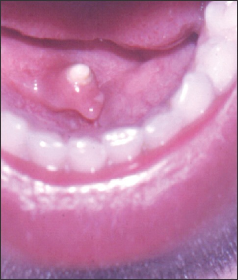
Huge right submandibular sialolith, exposed in the orifice of Wharton duct.
Figure 4.

Giant submandibular sialolith.
Figure 3.

Transocclusal endoral radiography showed a radiopaque stone within the right Wharton duct.
Once the symptoms were controlled, the surgical removal of the calculus was planned. Under local anesthesia, once the sialolith had been located, it was distally fixed with a suture in order to prevent any movement along the duct during the surgical procedure. The orifice of the salivary duct was surgically enlarged with an approximately 8-mm-long incision. Light pressure exerted at the level of the distal ligature provoked the discharge of the sialolith through the incision. The incised mucosa was rotated outward with an everted suture in order to keep the ductal orifice open. The salivary flow immediately became regular. NSAIDs and antibiotics (ibuprofen 600 mg, and amoxicillin 500 mg) were prescribed for a period of 7 days. After 2 days, the patient was symptom-free and discharged from the hospital.
DISCUSSION
Sialolithiasis is one of the most frequently encountered lesions of the major salivary glands, affecting men more than women in their middle age. Sialolithiasis can cause salivary gland dysfunction.[1,2] Sialoliths may be intraductal or lie within the gland parenchyma. The most common cause might be acute and chronic bacterial infections of salivary glands, resulting in the slowing of the salivary flow. Varying degrees of atrophy of the glandular parenchyma with ductal ectasia/expansion and fibrosis of the interstitium might also occur. This depends upon the position of the stone within the ductal system, hilum or gland parenchyma and the duration and degree of obstruction and the resultant retrograde pressure.[3,4,5] Sialoliths have varying sizes. In this article, two large submandibular sialoliths were reported, including a giant bilateral case, one approximately 50 mm in length and 5 mm in width on the right side and the other, 30 mm and 5 mm in length and width, respectively on the left side, and the other case, approximately 83 mm in length. Previous similar reports are summarized in Table 1.
Table 1.
The summarized information about previous reports of large sialoliths

In many cases, the diagnosis is easy due to obvious clinical features. Careful history and examination are important in the diagnosis of sialolithiasis. Sialoliths can occasionally be palpated. Pain and swelling of the affected gland at mealtimes are especially important. However, imaging studies are very often necessary for treatment.[3,4,5] Submandibular gland sialoliths have been reported to be radiopaque in 80-94.7% of cases and can be seen on plain films.[11,12] The most common radiographic techniques used to diagnose sialolithiasis are panoramic or occlusal views. Ultrasound, scintigraphy, or sialography (radiographic examination of the salivary glands and ducts after the introduction of a radiopaque material into the ducts) can be useful for arriving at a diagnosis.[13,14,15,16,17,18,19] Magnetic resonance sialography is a newer method of diagnosing sialolithiasis. It provides two- or three-dimensional images of the salivary gland without contrast medium and excessive exposure to radiation.[20] Depending on the site and size of salivary gland calculi, diagnosis, and various treatment modalities are available.[6] Conservative management is the treatment of choice, with plenty of fluids to keep the patient hydrated, followed by “milking” the duct to release the stone.[14,15,16] Stimulated salivation with lemon drops or the like should also help release the stone.[18] If the gland is infected, antibiotics should be prescribed.[19] If the conservative approach is not helpful, then the patient may need to undergo ductal dilatation or a minor incision of the duct to help remove the stone. Removal of an entire lobe or gland is reserved for the stone that is deeply embedded in the salivary gland parenchyma as sialolithotomy and sialodochoplasty.[15,16,17,20] However, interventional sialendoscopy for giant stones has resulted in improved gland preservation rates (86% vs. 57%), independent of stone location and with preservation of salivary function.[21,22,23,24]
CONCLUSION
Giant salivary gland calculi (>15 mm) and bilateral cases are considered rare and according to the best of our knowledge, this report included unique cases of very large sialoliths and also bilateral ones.
Financial support and sponsorship
Nil.
Conflicts of interest
The authors of this manuscript declare that they have no conflicts of interest, real or perceived, financial or non-financial in this article.
REFERENCES
- 1.Royce J, Biddle RJ, Arora S. Giant sialolith of the submandibular salivary gland. Radiol Case Rep. 2008;3:101. doi: 10.2484/rcr.2007.v3i2.101. [DOI] [PMC free article] [PubMed] [Google Scholar]
- 2.Ledesma-Montes C, Garcés-Ortíz M, Salcido-García JF, Hernández-Flores F, Hernández-Guerrero JC. Giant sialolith: Case report and review of the literature. J Oral Maxillofac Surg. 2007;65:128–30. doi: 10.1016/j.joms.2005.10.053. [DOI] [PubMed] [Google Scholar]
- 3.Brusati R, Fiamminghi L. Large calculus of the submandibular gland: Report of case. J Oral Surg. 1973;31:710–1. [PubMed] [Google Scholar]
- 4.Marchal F, Dulguerov P. Sialolithiasis management: The state of the art. Arch Otolaryngol Head Neck Surg. 2003;129:951–6. doi: 10.1001/archotol.129.9.951. [DOI] [PubMed] [Google Scholar]
- 5.Angiero F, Benedicenti S, Romanos GE, Crippa R. Sialolithiasis of the submandibular salivary gland treated with the 810- to 830-nm diode laser. Photomed Laser Surg. 2008;26:517–21. doi: 10.1089/pho.2007.2226. [DOI] [PubMed] [Google Scholar]
- 6.Rai M, Burman R. Giant submandibular sialolith of remarkable size in the comma area of Wharton's duct: A case report. J Oral Maxillofac Surg. 2009;67:1329–32. doi: 10.1016/j.joms.2008.11.014. [DOI] [PubMed] [Google Scholar]
- 7.Rontal M, Rontal E. The use of sialodochoplasty in the treatment of benign inflammatory obstructive submandibular gland disease. Laryngoscope. 1987;97:1417–21. doi: 10.1288/00005537-198712000-00008. [DOI] [PubMed] [Google Scholar]
- 8.Baurmash HD. Submandibular salivary stones: Current management modalities. J Oral Maxillofac Surg. 2004;62:369–78. doi: 10.1016/j.joms.2003.05.011. [DOI] [PubMed] [Google Scholar]
- 9.Grases F, Santiago C, Simonet BM, Costa-Bauzá A. Sialolithiasis: Mechanism of calculi formation and etiologic factors. Clin Chim Acta. 2003;334:131–6. doi: 10.1016/s0009-8981(03)00227-4. [DOI] [PubMed] [Google Scholar]
- 10.Williams MF. Sialolithiasis. Otolaryngol Clin North Am. 1999;32:819–34. doi: 10.1016/s0030-6665(05)70175-4. [DOI] [PubMed] [Google Scholar]
- 11.Udagatti V, Chandra S. Giant sialolith (megalith) of submandibular salivary gland. J Laryngol Otol. 1997;109:767–9. [Google Scholar]
- 12.Raksin SZ, Gould SM, Williams AC. Submandibular duct sialolith of unusual size and shape. J Oral Surg. 1975;33:142–5. [PubMed] [Google Scholar]
- 13.McGurk M, Makdissi J, Brown JE. Intra-oral removal of stones from the hilum of the submandibular gland: Report of technique and morbidity. Int J Oral Maxillofac Surg. 2004;33:683–6. doi: 10.1016/j.ijom.2004.01.024. [DOI] [PubMed] [Google Scholar]
- 14.Oteri G, Procopio RM, Cicciù M. Giant Salivary Gland Calculi (GSGC): Report of two cases. Open Dent J. 2011;5:90–5. doi: 10.2174/1874210601105010090. [DOI] [PMC free article] [PubMed] [Google Scholar]
- 15.Mason DK, Chisholm DM. 1st ed. Philadelphia: WB Saunders; 1975. Salivary Glands in Health and Disease; pp. 118–9. [Google Scholar]
- 16.Yoshimura Y, Inoue Y, Odagawa T. Sonographic examination of sialolithiasis. J Oral Maxillofac Surg. 1989;47:907–12. doi: 10.1016/0278-2391(89)90372-8. [DOI] [PubMed] [Google Scholar]
- 17.Capaccio P, Torretta S, Ottavian F, Sambataro G, Pignataro L. Modern management of obstructive salivary diseases. Acta Otorhinolaryngol Ital. 2007;27:161–72. [PMC free article] [PubMed] [Google Scholar]
- 18.Alkurt MT, Peker I. Unusually large submandibular sialoliths: Report of two cases. Eur J Dent. 2009;3:135–9. [PMC free article] [PubMed] [Google Scholar]
- 19.Cottrell D, Courtney M, Bhatia I, Gallagher G, Sundararajan D. Intraoral removal of a giant submandibular sialolith obstructing Wharton's duct: A case report. J Mass Dent Soc. 2011;60:14–6. [PubMed] [Google Scholar]
- 20.Markiewicz MR, Margarone JE, 3rd, Tapia JL, Aguirre A. Sialolithiasis in a residual Wharton's duct after excision of a submandibular salivary gland. J Laryngol Otol. 2007;121:182–5. doi: 10.1017/S0022215106003525. [DOI] [PubMed] [Google Scholar]
- 21.Wallace E, Tauzin M, Hagan J, Schaitkin B, Walvekar RR. Management of giant sialoliths: Review of the literature and preliminary experience with interventional sialendoscopy. Laryngoscope. 2010;120:1974–8. doi: 10.1002/lary.21082. [DOI] [PubMed] [Google Scholar]
- 22.Bodner L. Giant salivary gland calculi: Diagnostic imaging and surgical management. Oral Surg Oral Med Oral Pathol Oral Radiol Endod. 2002;94:320–3. [PubMed] [Google Scholar]
- 23.Boffano P, Gallesio C. Surgical treatment of a giant sialolith of the Wharton duct. 2010;21:134–5. doi: 10.1097/SCS.0b013e3181c46bc4. [DOI] [PubMed] [Google Scholar]
- 24.El Gehani R, Krishnan B, Shehoumi MI. Submandibular giant sialoliths: Report of two cases and review of the literature. Ear Nose Throat J. 2010;89:E1–4. [PubMed] [Google Scholar]