

Abstract
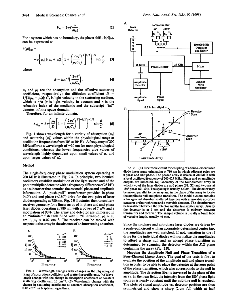
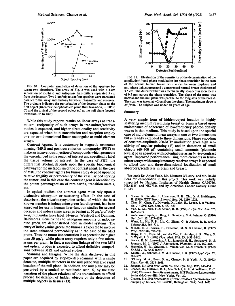
Based upon previous observations of low-frequency photon diffusion waves within highly scattering tissue, this paper explores the "near-field" phenomena of such waves of approximately 10-cm wavelength with 200-MHz phase modulation equipment. Multiple-element source arrays consist of laser diode sources modulated at 180 degrees out of phase with respect to the other sources. The diffusing waves originating from the out-of-phase sources give, in the midplane, an amplitude null and a sharp phase transition. These may be observed in a highly scattering intralipid medium simulating the breast or brain (0.5% intralipid), 3-5 cm from the transmitting laser diodes. In the plane containing the array, there is a high sensitivity for a small volume of a hidden absorber (indocyanine green) deep within a highly scattering medium; 20 pmol in a volume of 70 microliters can be detected. Two-dimensional arrays consisting of four or more elements in two orthogonal planes give sensitivity on both axes similar to the one-dimensional array. Measurements show that in the presence of a light-absorbing object, the amplitude null and the interference plane becomes a curved surface which is deflected toward the heterogeneity. The degree of deflection is related to the volume and the absorption characteristics of the heterogeneity and provides detection of the heterogeneity, and thereby may provide localization information for the detection of small tumors within the human breast, or stroke volumes, aneurysms, and tumors in the human brain.
Full text
PDF
Selected References
These references are in PubMed. This may not be the complete list of references from this article.
- Delpy D. T., Cope M., van der Zee P., Arridge S., Wray S., Wyatt J. Estimation of optical pathlength through tissue from direct time of flight measurement. Phys Med Biol. 1988 Dec;33(12):1433–1442. doi: 10.1088/0031-9155/33/12/008. [DOI] [PubMed] [Google Scholar]
- O'Leary MA, Boas DA, Chance B, Yodh AG. Refraction of diffuse photon density waves. Phys Rev Lett. 1992 Nov 2;69(18):2658–2661. doi: 10.1103/PhysRevLett.69.2658. [DOI] [PubMed] [Google Scholar]
- Sevick E. M., Lakowicz J. R., Szmacinski H., Nowaczyk K., Johnson M. L. Frequency domain imaging of absorbers obscured by scattering. J Photochem Photobiol B. 1992 Oct 30;16(2):169–185. doi: 10.1016/1011-1344(92)80007-i. [DOI] [PubMed] [Google Scholar]
- Spears K. G., Serafin J., Abramson N. H., Zhu X. M., Bjelkhagen H. Chrono-coherent imaging for medicine. IEEE Trans Biomed Eng. 1989 Dec;36(12):1210–1221. doi: 10.1109/10.42116. [DOI] [PubMed] [Google Scholar]
- Wang L., Ho P. P., Liu C., Zhang G., Alfano R. R. Ballistic 2-d imaging through scattering walls using an ultrafast optical kerr gate. Science. 1991 Aug 16;253(5021):769–771. doi: 10.1126/science.253.5021.769. [DOI] [PubMed] [Google Scholar]