Fig. 5.

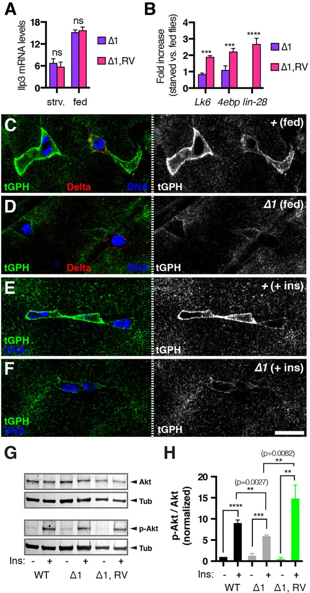
Lin-28 promotes insulin signaling in intestinal progenitors. (A) Ilp3 mRNA levels in lin-28Δ1 (Δ1) and rescued lin-28Δ1 (Δ1,RV) adults under starved (strv.) and fed conditions as determined by qRT-PCR. (B) Fold change in Lk6, 4e-bp and lin-28 mRNA levels in the intestines of starved versus fed lin-28Δ1 (Δ1) and rescued lin-28Δ1 (Δ1,RV) adults determined by qRT-PCR. ***P<0.001 for Δ1 versus Δ1,RV. ns, not significant. (C,D) Fed control (C) and lin-28Δ1 mutant (D) intestines stained for Delta (red), tGPH (green) and DNA (blue). (E,F) Insulin-treated control (E) and lin-28Δ1 mutant (F) intestines stained for tGPH (green) and DNA (blue). Scale bar: 7.5 μm. (G) Western blot analysis of control (WT), lin-28Δ1 mutant (Δ1) and rescued lin-28Δ1 mutant (Δ1,RV) intestines treated with (+) or without (−) insulin and probed with anti-Akt (Akt), anti-p-Akt (p-Akt) and anti-tubulin (Tub, loading control) antibodies. (H) Quantification of average ratio of p-Akt to total Akt levels detected in three independent western blots. **P<0.01, ***P<0.001, ****P<0.0001. Data are mean±s.e.m.
