eLife logoLink to eLife
. 2015 Nov 4;4:e08483. doi: 10.7554/eLife.08483

Re-examining the role of Drosophila Sas-4 in centrosome assembly using two-colour-3D-SIM FRAP

Paul T Conduit 1,2,*,, Alan Wainman 2,, Zsofia A Novak 2,, Timothy T Weil 1, Jordan W Raff 2,*
Editor: Tony Hunter3
PMCID: PMC4631861  PMID: 26530814

Abstract

Centrosomes have many important functions and comprise a ‘mother’ and ‘daughter’ centriole surrounded by pericentriolar material (PCM). The mother centriole recruits and organises the PCM and templates the formation of the daughter centriole. It has been reported that several important Drosophila PCM-organising proteins are recruited to centrioles from the cytosol as part of large cytoplasmic ‘S-CAP’ complexes that contain the centriole protein Sas-4. In a previous paper (Conduit et al., 2014b) we showed that one of these proteins, Cnn, and another key PCM-organising protein, Spd-2, are recruited around the mother centriole before spreading outwards to form a scaffold that supports mitotic PCM assembly; the recruitment of Cnn and Spd-2 is dependent on another S-CAP protein, Asl. We show here, however, that Cnn, Spd-2 and Asl are not recruited to the mother centriole as part of a complex with Sas-4. Thus, PCM recruitment in fly embryos does not appear to require cytosolic S-CAP complexes.

DOI: http://dx.doi.org/10.7554/eLife.08483.001

Research organism: D. melanogaster

Results

The centrosomal recruitment of Sas-4, Cnn and Spd-2 differs in space and time

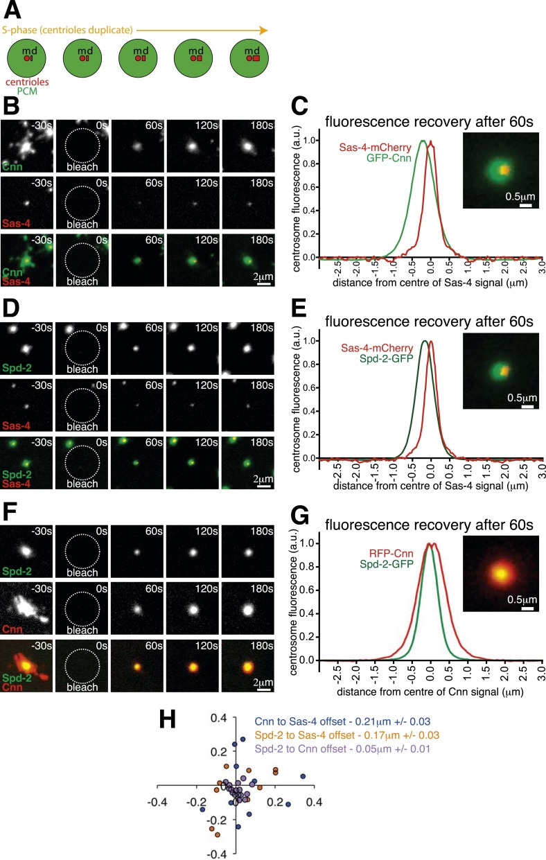
Centrosomes are crucial cell organisers (Nigg and Raff, 2009; Arquint et al., 2014; Chavali et al., 2014; Reina and Gonzalez, 2014; Stinchcombe and Griffiths, 2014). We previously showed that Cnn and Spd-2 are initially recruited around mother centrioles and then spread outward to form an extended pericentriolar material (PCM) scaffold (Conduit et al., 2010, 2014a, 2014b) (Note that we define ‘recruitment’ here as when a new protein molecule is added into the centrosome from the cytosol, irrespective of whether this molecule replaces an existing molecule or adds to the existing pool of molecules). Cnn has previously been identified as part of a multi-protein ‘S-CAP’ complex, which pre-assembles in the cytosol with the centriole protein Sas-4 before being recruited into the centrosome via a Sas-4–centriole interaction (Gopalakrishnan et al., 2011, 2012; Zheng et al., 2014). We reasoned, therefore, that Cnn and Spd-2 molecules might initially be recruited to the centrioles in S-CAP complexes, but could then be released from the centriolar-Sas-4 to spread outwards through the PCM. To test this possibility we compared the spatiotemporal centrosomal recruitment of Sas-4 to Cnn or Spd-2 in living Drosophila syncytial embryos, where S-CAP complexes were initially identified (Gopalakrishnan et al., 2011). These embryos cycle rapidly between S- and M-phases with no gap phases, and the mother centrioles organise large amounts of PCM throughout both S- and M-phases; during S-phase, each mother centriole also assembles a new daughter centriole (Figure 1A).

Figure 1. The centrosomal recruitment of Sas-4, Cnn and Spd-2 differ in space and time.

Figure 1.

(A) A schematic illustration of the centrosomal events that occur during S-phase in Drosophila syncytial embryos. The mother centriole (‘m’) constantly organises pericentriolar material (PCM, green) and also templates the formation of a new daughter centriole (‘d’) that grows throughout S-phase. (B) Images show how GFP-Cnn (top row, green in bottom row) and Sas-4-mCherry (middle row, red in bottom row) fluorescence signals recover after photobleaching. Time in seconds before and after photobleaching (t = 0 s) is shown in the top right of each panel. (C) The graph shows the normalised average recovery profiles of GFP-Cnn (green) and Sas-4-mCherry (red) 60 s after bleaching (n = 10 centrosomes from 10 embryos). Each profile is normalised so its maximum signal equals one and plotted taking into account the average spatial offset between the two signals—∼0.21 μm—see (H). The inset image shows the average fluorescent signals of GFP-Cnn (green) and Sas-4-mCherry (red) overlaid taking into account their average spatial offset. (DG) Images (D, F) and graphs (E, G) depict the same data as in (B) and (C) but for either Spd-2-GFP (green) and Sas-4-mCherry (red) (D, E) or for Spd-2-GFP (green) and RFP-Cnn (red) (F, G). (H) The graph shows the position of each GFP signal at 60 s post bleaching relative to the position of the mCherry/RFP signal (always positioned at 0; 0) for each combination of proteins, as indicated. Each dot represents a single centrosome.

DOI: http://dx.doi.org/10.7554/eLife.08483.002

Figure 1—source data 1. Measuring the spatial offset between recovering GFP-Cnn and Sas-4-mCherry, Spd-2-GFP and Sas-4-mCherry, and Spd-2-GFP and RFP-Cnn during S-phase.
(S1) This sheet includes the raw data used to calculate the offset between the centroids of the green and red fluorescent signals of sub-resolution beads. (S2) This sheet includes the raw data used to calculate the offset between the centroids of the GFP-Cnn and Sas-4-mCherry fluorescent signals shown in Figure 1H, and the raw data used to plot and offset the centrosomal profiles of GFP-Cnn and Sas-4-mCherry shown in Figure 1C. (S3) This sheet includes the raw data used to calculate the offset between the centroids of the Spd-2-GFP and Sas-4-mCherry fluorescent signals shown in Figure 1H, and the raw data used to plot and offset the centrosomal profiles of Spd-2-GFP and Sas-4-mCherry shown in Figure 1E. (S4) This sheet includes the raw data used to calculate the offset between the centroids of the Spd-2-GFP and RFP-Cnn fluorescent signals shown in Figure 1H, and the raw data used to plot and offset the centrosomal profiles of Spd-2-GFP and RFP-Cnn shown in Figure 1G. (S5) This sheet collates the offset data calculated in S2, S3 and S4 in order to plot the graph seen in Figure 1H.
elife08483s001.xlsx (594.6KB, xlsx)
DOI: 10.7554/eLife.08483.003

We co-expressed Sas-4-mCherry with either GFP-Cnn or Spd-2-GFP and performed two-colour Fluorescence Recovery After Photobleaching (two-colour FRAP) on a spinning disk confocal microscope. We first examined the centrosomal recruitment of these proteins from the cytosol during S-phase (Figure 1B,D; Videos 1, 2). Prior to photobleaching, Sas-4-mCherry appeared as a single tight spot, presumably localising to the two centrioles (Gopalakrishnan et al., 2011; Fu and Glover, 2012; Mennella et al., 2012), which cannot be resolved with a standard confocal microscope; GFP-Cnn and Spd-2-GFP occupied a relatively broad area around the centrioles (Figure 1B,D; t = −30 s), consistent with their known PCM localisation (Conduit et al., 2014b). The centrosomal fluorescence of all three proteins recovered after photobleaching, but we noticed that the recovering GFP and mCherry signals were not aligned in the X-Y plane (Figure 1B,D; t = 60–180 s). We plotted the relative positions of the recovering signals after using fluorescent beads to adjust for any systemic shift between the green and red channels (Figure 1H; Figure 1—source data 1; see ‘Materials and methods’), and calculated the average distance between the recovering signals: at 60 s post bleaching, recovering Sas-4-mCherry was offset from recovering GFP-Cnn by an average of ∼0.21 μm (blue dots, Figure 1H) and from recovering Spd-2-GFP by an average of ∼0.17 μm (orange dots, Figure 1H). In contrast, the recovering Spd-2-GFP signal was offset from the recovering RFP-Cnn signal by an average of only ∼0.053 μm (purple dots, Figure 1H). We illustrate these differences by displaying the average fluorescence profiles of each pair of markers offset by the average distance between each marker at 60 s post-bleaching (Figure 1C,E,G).

Video 1. Recovery dynamics of GFP-Cnn and Sas-4-mCherry during S-phase.

Download video file (2.1MB, avi)
DOI: 10.7554/eLife.08483.004

This video shows the fluorescent signals of GFP-Cnn (left panel) and Sas-4-mCherry (right panel) recovering during S-phase after photobleaching at t = 0 s. Both signals are detectable 30 s after photobleaching and continue to increase in intensity thereafter. The GFP-Cnn signal initially recovers centrally and then spreads outwards, as described previously (Conduit et al., 2010, 2014a, 2014b), whereas the Sas-4-mCherry signal recovers as a single tight focus and does not spread outwards.

DOI: http://dx.doi.org/10.7554/eLife.08483.004

Video 2. Recovery dynamics of Spd-2GFP and Sas-4-mCherry during S-phase.

Download video file (1.7MB, avi)
DOI: 10.7554/eLife.08483.005

This video shows the fluorescent signals of Spd-2-GFP (left panel) and Sas-4-mCherry (right panel) recovering during S-phase after photobleaching at t = 0 s. Both signals are detectable 30 s after photobleaching and continue to increase in intensity thereafter. The Spd-2-GFP signal initially recovers centrally and then spreads outwards, as described previously (Conduit et al., 2014b), whereas the Sas-4-mCherry signal recovers as a single tight focus and does not spread outwards.

DOI: http://dx.doi.org/10.7554/eLife.08483.005

During S-phase, an excess of Sas-4 is recruited to centrioles, and a large fraction of these molecules become irreversibly incorporated into newly forming daughter centrioles, while the remainder is later shed from the centrioles during mitosis (Novak et al., 2014). We wondered, therefore, if the offset between the Sas-4-mCherry and GFP-Cnn or Spd-2-GFP recovering signals was a result of Sas-4-mCherry being largely recruited to daughter centrioles and GFP-Cnn and Spd-2-GFP being largely recruited around mother centrioles.

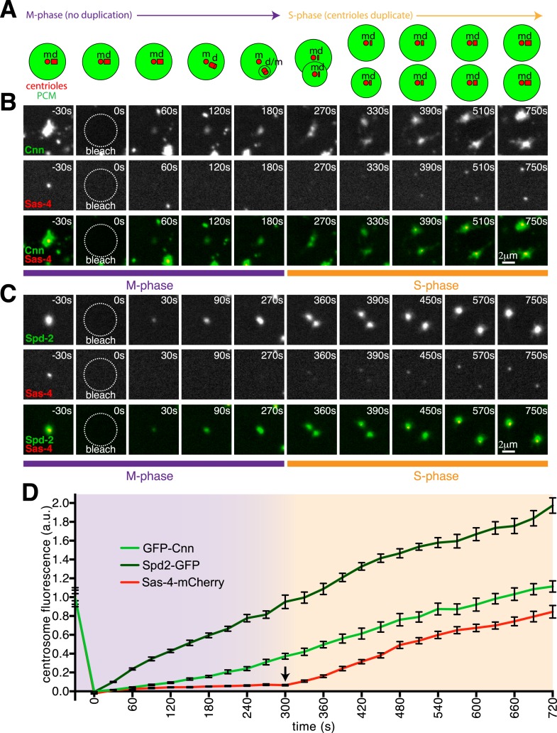
As an initial test of this hypothesis, we performed two-colour FRAP experiments in M-phase, when centriole duplication has been completed. After photobleaching, the centrosomal GFP-Cnn and Spd-2-GFP signals recovered immediately, while the Sas-4-mCherry signal only began to recover robustly after the centrosomes separated at the end of mitosis—when a new round of centriole duplication begins (Figure 2; Videos 3, 4; Figure 2—source data 1). These findings are consistent with our hypothesis that Sas-4 molecules are only recruited to growing daughter centrioles, and they strongly suggest that the Cnn and Spd-2 molecules that are recruited around mother centrioles during M-phase are not recruited there as part of a complex with Sas-4.

Video 3. Recovery dynamics of GFP-Cnn and Sas-4-mCherry during M-phase/S-phase.

Download video file (4.1MB, avi)
DOI: 10.7554/eLife.08483.008

This video shows the fluorescent signals of GFP-Cnn (left panel) and Sas-4-mCherry (right panel) recovering during M-phase and then during the following S-phase; the centrosome was bleached at t = 0 s in M-phase. The GFP-Cnn signal is detectable 30 s after photobleaching and continues to increase during M-phase and the following S-phase, when the centrosome divides into two. The Sas-4-mCherry signal, however, only becomes detectable 270 s after photobleaching, once the embryo has transitioned from M-phase into the following S-phase.

DOI: http://dx.doi.org/10.7554/eLife.08483.008

Video 4. Recovery dynamics of Spd-2-GFP and Sas-4-mCherry during M-phase/S-phase.

Download video file (4MB, avi)
DOI: 10.7554/eLife.08483.009

This video shows the fluorescent signals of Spd-2-GFP (left panel) and Sas-4-mCherry (right panel) recovering during M-phase and then during the following S-phase; the centrosome was bleached at t = 0 s in M-phase. The Spd-2-GFP signal is detectable 30 s after photobleaching and continues to increase during M-phase and the following S-phase, when the centrosome divides into two. The Sas-4-mCherry signal, however, only becomes detectable 330 s after photobleaching, once the embryo has transitioned from M-phase into the following S-phase.

DOI: http://dx.doi.org/10.7554/eLife.08483.009

Figure 2. Cnn and Spd-2, but not Sas-4, are recruited to centrosomes during M-phase.

Figure 2.

(A) A schematic illustration of the centrosomal events that occur during M-phase (purple arrow) and the following S-phase (orange arrow) in Drosophila syncytial embryos. The mother centriole (‘m’) organises the PCM (green) and remains ‘engaged’ to its fully formed daughter centriole (‘d’) until late M-phase. During late M-phase the centrioles disengage and the daughter centriole matures into a mother and starts to organise its own domain of PCM. At the start of the following S-phase, both the old and new mother centrioles template the formation of a new daughter centriole. (B, C) Images show how GFP-Cnn (top row in B, green in bottom row), Spd-2-GFP (top row in C, green in bottom row) and Sas-4-mCherry (middle rows in B and C, red in bottom rows) fluorescence signals recover after photobleaching during M-phase and the following S-phase. Time in seconds before and after photobleaching (t = 0 s) is shown in the top right of each panel. Note how the GFP-Cnn (B) and Spd-2-GFP (C) signals recover immediately after photobleaching, but that a recovering Sas-4-mCherry signal can only be detected once the embryos enter the following S-phase (and initiate a new round of centriole duplication). (D) A graph showing the fluorescence recovery over time of Sas-4-mCherry (red), GFP-Cnn (light green) and Spd-2-GFP (dark green) relative to their initial fluorescence values. Measurements were taken in the region of the centrosome where the fluorescent signals overlapped. The arrow indicates the sudden change in the recovery dynamics of the Sas-4-mCherry signal.

DOI: http://dx.doi.org/10.7554/eLife.08483.006

Figure 2—source data 1. A comparison of the centrosomal fluorescence recovery after photobleaching of GFP-Cnn, Spd-2-GFP and Sas-4-mCherry during M-phase and the following S-phase.
(S1) This sheet includes the raw data used to calculate the fluorescence recovery of GFP-Cnn and Sas-4-mCherry during M-phase and the following S-phase. (S2) This sheet includes the raw data used to calculate the fluorescence recovery of Spd-2-GFP and Sas-4-mCherry during M-phase and the following S-phase. (S3) This sheet combines the Sas-4-mCherry recovery data from S1 and S2. (S4) This sheet collates the data from S1, S2 and S3 in order to plot the final recovery graph seen in Figure 2D.
elife08483s002.xlsx (249.6KB, xlsx)
DOI: 10.7554/eLife.08483.007

Super-resolution microscopy confirms that Sas-4 molecules are recruited exclusively to growing daughter centrioles

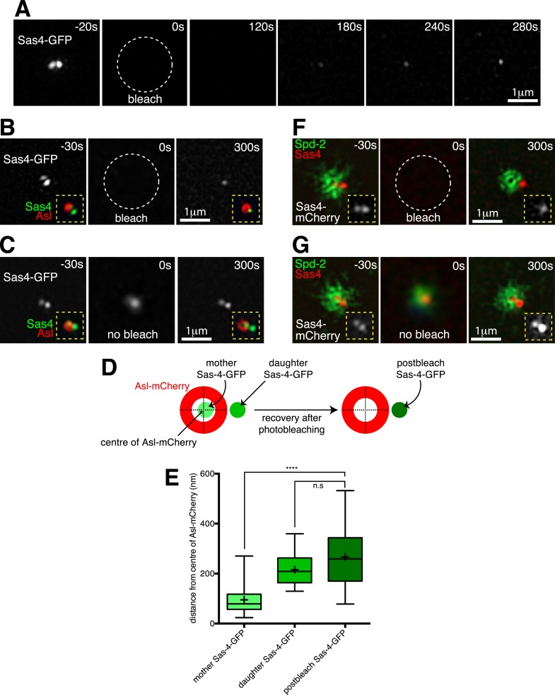
In order to directly test if Sas-4 molecules are only recruited to growing daughter centrioles, we turned to 3D-Structured Illumination Microscopy (3D-SIM), which has approximately twice the spatial resolution of standard confocal microscopy. Using 3D-SIM in living embryos, we could clearly distinguish two adjacent Sas-4-GFP foci at individual centrosomes during S-phase (Figure 3A, t = −20 s), presumably representing mother-daughter centriole pairs. We combined 3D-SIM with FRAP (Conduit et al., 2014b) and found that the Sas-4-GFP signal only recovered at a single foci (Figure 3A, t = 120 s to t = 280 s). We confirmed that this recovery occurred at the daughter centriole by performing a two-colour-3D-SIM-FRAP experiment in embryos co-expressing Sas-4-GFP and Asl-mCherry, as Asl forms a toroid around only the mother centrioles (Figure 3B, t = −30 s) (Novak et al., 2014). Strikingly, the recovering Sas-4-GFP fluorescence always lay outside of the Asl-mCherry toroid (Figure 3B, t = 300 s), whereas control unbleached centrosomes still contained two Sas-4-GFP foci, one of which lay inside the Asl-mCherry toroid (Figure 3C, t = 300 s). This suggested that new Sas-4 molecules are recruited only to the daughter centrioles. However, during acquisition centrosomes move in the x-y plane, and given that the green and red channels are acquired sequentially on this particular imaging system it is possible for the green and red signals to become misaligned. To be sure the recovering Sas-4-GFP signal represented the daughter centriole, rather than a mis-positioned mother centriole, we therefore measured the distance between the centre of the Asl-mCherry signal and the centres of the pre- and post-bleached Sas-4-GFP signals (Figure 3D; Figure 3—source data 1). This revealed that the average position of the post-bleached Sas-4-GFP signal closely matched the position of the daughter Sas-4-GFP signal, but not the mother Sas-4-GFP signal (Figure 3E), confirming that Sas-4 molecules are recruited only to daughter centrioles. As Asl molecules are known to turn over at the mother centriole at this stage in the cycle (Novak et al., 2014), their recruitment cannot occur as part of Sas-4 dependent S-CAP complexes.

Figure 3. Two-colour-3D-SIM FRAP reveals that Sas-4-mCherry is recruited only to growing daughter centrioles, while PCM proteins are recruited only around mother centrioles.

Figure 3.

(A) 3D-SIM images show how during S-phase two adjacent Sas-4-GFP foci can be resolved at an individual centrosome (t = −20 s), and how the Sas-4-GFP fluorescence signal recovers only as a single foci after photobleaching (t = 120 s to t = 280 s). Time in seconds before and after photobleaching (t = 0 s) is shown in the top right of each panel. (B) Two-colour 3D-SIM images show how Sas-4-GFP fluorescence recovers relative to Asl-mCherry fluorescence (which surrounds the mother centriole). The Sas-4-GFP is shown in greyscale; insets (yellow dashed lines) display the overlay of Sas-4-GFP (green) and Asl-mCherry (red). Note how after photobleaching the Sas-4-GFP fluorescence recovers outside of the Asl-mCherry toroid. (C) Complementary images of a control centrosome adjacent to the one shown in (B) where the Sas-4-GFP signal was not photobleached. The t = 0 s panel is a widefield image (see ‘Materials and methods’). (D, E) Schematic (D) and box-plot (E) show how the average position of the post-bleached Sas-4-GFP signal is similar to the position of the daughter, but not the mother, centriole's prebleached Sas-4-GFP signal, relative to the Asl-mCherry signal. Boxes in E extend from the 25th-75th percentiles, whiskers extend from min to max values, lines in boxes are the median values, ‘+’ in boxes are the mean values; n = 25 centrosomes from 4 embryos. **** indicates where p < 0.0001; n.s. indicates where p = 0.09, and is therefore not significant. (F) 3D-SIM images show how Sas-4-mCherry (red) recovers relative to recovering Spd-2-GFP (green). An overlay of Sas-4-mCherry and Spd-2-GFP fluorescence is shown in the main panels; insets (yellow dashed lines) display the Sas-4-mCherry signal (greyscale). Note how after photobleaching the Sas-4-mCherry recovers outside of the hollow created by the recovering Spd-2-GFP (t = 300 s). (G) Complementary images of a control centrosome adjacent to the one in (F) where the Sas-4-mCherry and Spd-2-GFP signals were not photobleached. The t = 0 s image is a widefield image, as in (C).

DOI: http://dx.doi.org/10.7554/eLife.08483.010

Figure 3—source data 1. Measuring the spatial offset between recovering Sas-4-GFP and Asl-mCherry at super resolution.
This flie includes the raw data used to calculate the offset between the centroids of the two Sas-4-GFP foci and the single Asl-mCherry foci prior to photobleaching and the offset between the centroids of the single Sas-4-GFP foci and the single Asl-mCherry foci 5 min post-photobleaching. The data is used to plot the graph shown in Figure 3E.
elife08483s003.xlsx (53.9KB, xlsx)
DOI: 10.7554/eLife.08483.011

We next performed a similar two-colour 3D-SIM FRAP experiment in embryos expressing Spd-2-GFP and Sas-4-mCherry. Here, the recovering Sas-4-mCherry foci lay adjacent to the recovering Spd-2-GFP signal (Figure 3F, t = 300 s), which is known to initially recover as a toroid around the mother centriole before spreading outwards in a fibrous network (Conduit et al., 2014b). Unbleached centrosomes still contained two Sas-4-mCherry foci, one of which lay at the centre of the Spd-2-GFP network (Figure 3G, t = 300 s). Together, these observations demonstrate that in these embryos Sas-4 is only recruited to growing daughter centrioles, while Asl and Spd-2 are recruited only around mother centrioles.

S-CAP complexes are of low abundance in the early embryonic cytosol

We next analysed the abundance of potential S-CAP complexes in syncytial embryos. We expressed a Sas-4-GFP construct at near endogenous levels in embryos lacking endogenous Sas-4—this construct is functional as it rescues the Sas-4 mutant phenotype (Novak et al., 2014). From these embryos we produced extracts where the centrosomes had been removed by centrifugation, immunoprecipitated Sas-4-GFP using anti-GFP coated beads and then examined the relative proportion of bound and unbound S-CAP complex proteins. This approach had two advantages over immunoprecipitating endogenous Sas-4 with anti-Sas-4 antibodies: (1) anti-GFP antibodies are less likely to perturb S-CAP complex assemblies; (2) we could perform negative controls using wild-type extracts that did not contain any Sas-4-GFP protein.

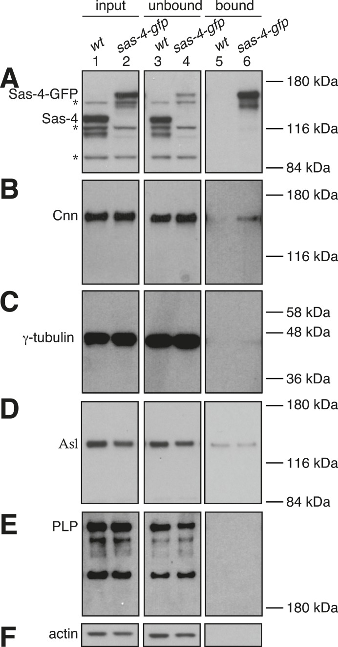
As expected, we saw a strong depletion of Sas-4-GFP from the Sas-4-GFP extract (compare lanes 2 and 4, Figure 4A) and a strong enrichment of Sas-4-GFP in the Sas-4-GFP bound sample (lane 6, Figure 4A). In contrast, the negative control showed no depletion of endogenous Sas-4 from the wild-type extract (compare lanes 1 and 3, Figure 4A), or enrichment of endogenous Sas-4 in the wild-type bound sample (lane 5, Figure 4A). Cnn, γ-tubulin, pericentrin-like-protein (PLP) and Asl (the other characterised components of the S-CAP complexes), were not obviously co-depleted with Sas-4-GFP from the extract (compare lanes 2 and 4, Figure 4B–E), although we reliably detected small amounts of Cnn and γ-tubulin in the Sas-4-GFP bound samples (compare lanes 5 and 6, Figure 4B,C). A small amount of Asl was also detectable in the Sas-4-GFP bound sample, but this was also seen in the control bound sample (compare lanes 5 and 6 in Figure 4D). We conclude that only a very small fraction of Cnn and γ-tubulin molecules form cytosolic complexes with Sas-4 in these embryo extracts, while any interaction between Sas-4 and Asl or PLP is undetectable with these methods. Thus, the previously described S-CAP complexes are either absent or present at very low levels in these embryo extracts.

Figure 4. Potential S-CAP complexes are of low abundance in Drosophila syncytial embryo extracts.

Figure 4.

Panels show western blots of anti-GFP immunoprecipitation experiments from WT embryos (lanes 1, 3, 5) or embryos expressing Sas-4-GFP in the absence of endogenous Sas-4 (lanes 2, 3, 6). The membranes were probed for Sas-4 (A), Cnn (B), γ-tubulin (C), Asl (D), pericentrin-like-protein (PLP) (E) or Actin (as a loading control) (F). Lanes 1 and 2 are taken from the initial embryo extracts (‘input’); lanes 3 and 4 are ‘unbound’ samples taken from the extracts after the beads had been removed; lanes 5 and 6 are ‘bound’ samples taken from the beads after incubation with extract. The * symbols in (A) highlight non-specific bands. Note that the signal intensities can only be directly compared between the ‘input’ and ‘unbound’ lanes (see ‘Materials and methods’).

DOI: http://dx.doi.org/10.7554/eLife.08483.012

Concluding remarks

Our results suggest that S-CAP complexes do not play a significant role in mitotic PCM assembly in Drosophila syncytial embryos. In support of this conclusion, previous reports have shown that centrioles in Drosophila cells lacking cytosolic Sas-4 can recruit PCM during mitosis (Stevens et al., 2007; Riparbelli and Callaini, 2011), and this also appears to be true in Caenorhabditis elegans embryos (Kirkham et al., 2003; Leidel and Gönczy, 2003) and in HeLa cells (Kitagawa et al., 2011). Moreover, SPD-2 and SPD-5, the likely functional homologues of Spd-2 and Cnn, exist mostly as monomers in the cytosol of C. elegans embryos and do not detectably interact with Sas-4 (Wueseke et al., 2014). Thus, the mechanism of mitotic PCM assembly in flies, worms and human cells does not appear to involve the pre-assembly of Sas-4-dependent cytosolic PCM complexes. Importantly, Sas-4 may have a more indirect role in mitotic PCM assembly in fly embryos as centriolar Sas-4 (as opposed to cytoplasmic Sas-4) is required to efficiently recruit Asl molecules around maturing mother centrioles (Dzhindzhev et al., 2010; Novak et al., 2014), and Asl has an important role in recruiting Spd-2 and Cnn around mother centrioles (Conduit et al., 2010, 2014a).

Materials and methods

Transgenic Drosophila lines

Sas-4-mCherry and Asl-mCherry P-element-mediated transformation vectors were made by introducing a full length Sas-4 or Asl cDNA into a mCherry C-terminal Gateway vector (Basto et al., 2008) downstream of a 2 kb predicted promoter region. Transgenic lines were generated by the Fly Facility in the Department of Genetics, Cambridge, United Kingdom. The other GFP and RFP fusions have been described previously: pUbq-GFP-Cnn (Lucas and Raff, 2007), pUbq-Spd-2-GFP (Dix and Raff, 2007), Sas-4-GFP (under control of endogenous promoter) (Novak et al., 2014) and pUbq-RFP-Cnn (Conduit et al., 2010).

Fly Stocks for live cell microscopy

To examine the dynamics of GFP-Cnn and Sas-4-mCherry at centrosomes, we analysed embryos from mothers expressing GFP-Cnn under the control of the pUbq promoter and Sas-4-mCherry under the control of its endogenous promoter in a cnnf04547/cnnHK21 and sas-42214/sas-42214 mutant background. To examine Spd-2-GFP and Sas-4-mCherry, we analysed embryos from mothers expressing Spd-2-GFP under the control of the pUbq promoter and Sas-4-mCherry under the control of its endogenous promoter in a sas-42214/sas-42214 mutant background. To examine Spd-2-GFP and RFP-Cnn, we analysed embryos from mothers expressing Spd-2-GFP and RFP-Cnn both under the control of the pUbq promoter in a cnnf04547/cnnf04547 mutant background. To examine Sas-4-GFP at super-resolution, we analysed embryos from mothers expressing Sas-4-GFP under the control of its endogenous promoter in a sas-42214/sas-42214 mutant background. To examine Sas-4-GFP and Asl-mCherry at super-resolution, we analysed embryos from mothers expressing Sas-4-GFP and Asl-mCherry both under the control of their respective endogenous promoters in a sas-42214/sas-42214 mutant background. To examine Spd-2-GFP and Sas-4-mCherry at super-resolution, we analysed embryos from mothers expressing Spd-2-GFP under the control of the pUbq promoter and Sas-4-mCherry under the control of its endogenous promoter in a sas-42214/sas-42214 mutant background.

FRAP experiments at standard resolution

Imaging was carried out on a Perkin Elmer Spinning Disk confocal system running Volocity software mounted on a Zeiss Axiovert microscope using a 60×/1.4 NA oil objective. Images shown are maximum intensity projections of 5 z-slices taken 0.5 μm apart. Photobleaching of individual centrosomes was carried out using a combination of a focussed 440 nm laser and a focussed 568 nm laser. ImageJ was used to calculate the distance between the centre points of the recovering green and red signals at 60 s post photobleaching. The images were first scaled up fivefold so that each pixel was divided into 5 × 5 pixels—this allowed a more accurate analysis. The X; Y location of the centre of mass of each signal was calculated by thresholding the image and running the ‘analyze particles’ (centre of mass) macro on the most central Z plane of the centrosome. To adjust for any residual shift in the green and red channels, we calculated the average distance and direction between the ‘green’ and ‘red’ signals coming from subresolution TetraSpeck beads (Life Technologies, United Kingdom) (total of 977 beads analysed from 12 images) and used this to correct the centrosome data for any microscope-induced channel misalignment; the green and red signals from the beads were offset by a distance of 0.058 μm ± 0.005 μm. Once corrected, we calculated the distance between the centres of the green and red signals from each centrosome, and then calculated an average distance from all the centrosomes.

ImageJ was used to calculate the 60 s fluorescence recovery profiles of GFP-Cnn, Spd-2-GFP and Sas-4-mCherry from the scaled (5 × 5) images described above. Using the previously calculated centre of mass of each signal, concentric rings (spaced at 0.028 μm and spanning across 3.02 μm) were centred and the average fluorescence around each ring was measured (radial profiling). After subtracting the average cytosolic signal and normalising so the peak intensity of the image was equal to 1, we mirrored the profiles to show a full symmetric centrosomal profile. For each profile, an average distribution from at least 10 centrosomes was calculated. The green and red profiles were plotted on the same graph after manually taking into account the previously calculated average distance between the centre of each signal.

To produce the images that represent the average fluorescent signals at 60 s post bleaching (inset into each graph described above), average projections of green and red images were initially generated separately (after being aligned using the centre of mass coordinates) and then overlaid manually after taking into account the previously calculated average distance between the centre of each signal.

To examine the rate of fluorescence recovery of GFP-Cnn, Spd-2-GFP and Sas-4-mCherry, we measured the green and red fluorescence signals at each timepoint in the pixels where the fluorescent signals overlapped. An ROI was drawn that included all Sas-4-mCherry pixels that had a value above 2 standard deviations from the mean image value, and the total value of the Sas-4-mCherry signal and of the GFP-Cnn or Spd-2-GFP signal within these pixels was calculated. Typically, the ROI was 10 × 10 pixels (1.05 μm × 1.05 μm). The local cytoplasmic background fluorescence was then subtracted from this value. An average value from at least 10 centrosomes was calculated and normalised by dividing it by the average initial pre-bleach value. These normalised average values were then used for each data point in the graph.

3D-structured illumination (sub-diffraction resolution) microscopy

Living embryos were imaged at 21°C on a DeltaVision OMX V3 Blaze microscope (GE Healthcare, United Kingdom) equipped with a 60×/1.42 oil UPlanSApo objective (Olympus), 488 nm and 593 nm diode lasers and Edge 5.5 sCMOS cameras (PCO). Spherical aberration was minimized by matching the refractive indices (1.514) of the immersion oil to the sample. 3D-SIM image stacks consisting of 6 z-planes were acquired with 5 phases, 3 angles per image plane and a distance of 0.125 μm between planes. The raw data was computationally reconstructed with SoftWorx 6.1 (GE Healthcare) using Wiener filter settings 0.006 and channel specific optical transfer functions. For two colour 3D-SIM, images from the different colour channels were registered with alignment parameters obtained from calibration measurements with 0.2 μm diameter TetraSpeck beads (Life Technologies) using the OMX Editor software. Images shown are maximum intensity projections of several z-slices. The quality of the reconstructed images was assessed using the SIM-Check ImageJ plugin (Ball et al., 2015; http://www.micron.ox.ac.uk/microngroup/software/SIMCheck.php) to ensure proper imaging conditions and to avoid reconstruction artefacts.

To perform 3D-SIM FRAP, we utilized the software development kit from GE Healthcare. This allowed us to create a custom acquisition sequence that first acquired a single Z-stack in 3D-SIM (prebleached image), then performed single or multi spot photobleaching (using the standard OMX galvo scanner TIRF/photo-kinetics module), then performed time lapse imaging in widefield mode (including the photobleached image), and then performed a second 3D-SIM Z-stack (5 min recovery image).

Immunoprecipitation experiments

0–4 hr embryos were collected from either w67 (wild-type) mothers or from mothers expressing Sas-4-GFP under the control of its endogenous promoter in a sas-42214/sas-42214 mutant background. The Sas-4-GFP construct appears to rescue the sas-42214 mutant phenotype when expressed from the endogenous promoter, as the flies are fertile and coordinated (Novak et al., 2014). The embryos were dechorionated using 60% bleach, washed thoroughly with 0.05% Tween-20 in distilled water, flash frozen in liquid nitrogen and stored at −80°C. Centrosome free extracts were prepared by homogenising the frozen embryos in 2 ml per gram IP buffer (50 mM HEPES pH 7.6, 1 mM MgCl2, 1 mM EGTA, 1 mM DTT, 1× Protease inhibitor cocktail [Roche]) and centrifuging twice at 15,000 RCF. The extract was maintained at 4°C during preparation. Protein A Dynabeads (Invitrogen) were covalently coupled to rabbit anti-GFP antibodies (this study) using the BS3 crosslinker (ThermoScientific). The amount of antibody-bead conjugate required to pull out most Sas-4-GFP from the extract was calculated empirically, and equal amounts were used for both Sas-4-GFP and wild-type extracts. Before adding the beads to the lysates, a 10 μl ‘input’ sample was collected from the extracts and mixed with 10 μl of 2× Laemmli sample buffer. The beads were added and the reaction was incubated overnight at 4°C by rotation. At the end of the incubation, a magnet was used to separate the beads from the extract and a 10 μl ‘unbound’ sample was collected and mixed with 10 μl of 2× Laemmli sample buffer. The ‘input’ and ‘unbound’ samples were of the same volume to ensure that the protein levels in each sample could be directly compared. The beads were washed by re-suspension in PBT three times at room temperature, then washed a further five times with PBT for 10 min by rotation at 4°C. The beads were then boiled for 10 min in 50 μl Laemmli sample buffer to produce a ‘bound’ sample. 10 μl of each sample was run on a 3–8% Tris-Acetate NuPAGE gel (Life Technologies), western blotted and probed for Sas-4, Cnn, Asl, D-PLP, and γ-tubulin using appropriate antibodies: Primary antibodies: N-terminal rabbit anti-Sas-4 antibodies; N-terminal rabbit anti-Cnn antibodies; C-terminal rabbit anti-Asl antibodies; C-terminal rabbit anti-D-PLP antibodies; mouse anti γ-tubulin antibodies (Sigma); mouse anti-actin (Sigma). Secondary antibodies: HRP-conjugated anti-rabbit or anti-mouse antibodies (Roche). SuperSignal West Femto Maximum Sensitivity Substrate (Thermo Scientific) was used as a chemiluminescent HRP substrate.

Acknowledgements

PTC was supported by a Sir Henry Dale Fellowship jointly funded by the Wellcome Trust and the Royal Society (105653/Z/14/Z) and by an Issac Newton Trust Research Grant from the University of Cambridge awarded to TTW (RG78799). AW, ZN and JWR were supported by a Senior Investigator Award awarded to JWR and funded by the Wellcome Trust (104575/Z/14/Z). The OMX microscope used in this study is part of the Oxford Micron Advanced Bioimaging Unit supported by a Wellcome Trust Strategic Award (091911).

Funding Statement

The funders had no role in study design, data collection and interpretation, or the decision to submit the work for publication.

Funding Information

This paper was supported by the following grants:

  • Royal Society 105653/Z/14/Z to Paul T Conduit.

  • Wellcome Trust 104575/Z/14/Z to Alan Wainman, Zsofia A Novak, Jordan W Raff.

  • University of Cambridge RG78799 to Paul T Conduit, Timothy T Weil.

Additional information

Competing interests

The authors declare that no competing interests exist.

Author contributions

PTC, Conception and design, Acquisition of data, Analysis and interpretation of data, Drafting or revising the article.

AW, Conception and design, Acquisition of data, Analysis and interpretation of data, Drafting or revising the article.

ZAN, Acquisition of data, Analysis and interpretation of data, Drafting or revising the article.

TTW, Drafting or revising the article, Contributed unpublished essential data or reagents.

JWR, Conception and design, Analysis and interpretation of data, Drafting or revising the article.

References

  1. Arquint C, Gabryjonczyk AM, Nigg EA. Centrosomes as signalling centres. Philosophical Transactions of the Royal Society of London. Series B, Biological Sciences. 2014;369 doi: 10.1098/rstb.2013.0464. [DOI] [PMC free article] [PubMed] [Google Scholar]
  2. Ball G, Demmerle J, Kaufmann R, Davis I, Dobbie IM, Schermelleh L. SIMcheck: a toolbox for successful super-resolution SIM imaging. Scientific Reports. 2015;5:15915. doi: 10.1038/srep15915. [DOI] [PMC free article] [PubMed] [Google Scholar]
  3. Basto R, Brunk K, Vinadogrova T, Peel N, Franz A, Khodjakov A, Raff JW. Centrosome amplification can initiate tumorigenesis in flies. Cell. 2008;133:1032–1042. doi: 10.1016/j.cell.2008.05.039. [DOI] [PMC free article] [PubMed] [Google Scholar]
  4. Chavali PL, Pütz M, Gergely F. Small organelle, big responsibility: the role of centrosomes in development and disease. Philosophical Transactions of the Royal Society of London. Series B, Biological Sciences. 2014;369 doi: 10.1098/rstb.2013.0468. [DOI] [PMC free article] [PubMed] [Google Scholar]
  5. Conduit PT, Brunk K, Dobbelaere J, Dix CI, Lucas EP, Raff JW. Centrioles regulate centrosome size by controlling the rate of Cnn incorporation into the PCM. Current Biology. 2010;20:2178–2186. doi: 10.1016/j.cub.2010.11.011. [DOI] [PubMed] [Google Scholar]
  6. Conduit PT, Feng Z, Richens JH, Baumbach J, Wainman A, Bakshi SD, Dobbelaere J, Johnson S, Lea SM, Raff JW. The centrosome-specific phosphorylation of Cnn by Polo/Plk1 drives Cnn scaffold assembly and centrosome maturation. Developmental Cell. 2014a;28:659–669. doi: 10.1016/j.devcel.2014.02.013. [DOI] [PMC free article] [PubMed] [Google Scholar]
  7. Conduit PT, Richens JH, Wainman A, Holder J, Vicente CC, Pratt MB, Dix CI, Novak ZA, Dobbie IM, Schermelleh L, Raff JW. A molecular mechanism of mitotic centrosome assembly in Drosophila. eLife. 2014b;3:e03399. doi: 10.7554/eLife.03399. [DOI] [PMC free article] [PubMed] [Google Scholar]
  8. Dix CI, Raff JW. Drosophila Spd-2 recruits PCM to the sperm centriole, but is dispensable for centriole duplication. Current Biology. 2007;17:1759–1764. doi: 10.1016/j.cub.2007.08.065. [DOI] [PMC free article] [PubMed] [Google Scholar]
  9. Dzhindzhev NS, Yu QD, Weiskopf K, Tzolovsky G, Cunha-Ferreira I, Riparbelli M, Rodrigues-Martins A, Bettencourt-Dias M, Callaini G, Glover DM. Asterless is a scaffold for the onset of centriole assembly. Nature. 2010;467:714–718. doi: 10.1038/nature09445. [DOI] [PubMed] [Google Scholar]
  10. Fu J, Glover DM. Structured illumination of the interface between centriole and peri-centriolar material. Open Biology. 2012;2:120104. doi: 10.1098/rsob.120104. [DOI] [PMC free article] [PubMed] [Google Scholar]
  11. Gopalakrishnan J, Chim YC, Ha A, Basiri ML, Lerit DA, Rusan NM, Avidor-Reiss T. Tubulin nucleotide status controls Sas-4-dependent pericentriolar material recruitment. Nature Cell Biology. 2012;14:865–873. doi: 10.1038/ncb2527. [DOI] [PMC free article] [PubMed] [Google Scholar]
  12. Gopalakrishnan J, Mennella V, Blachon S, Zhai B, Smith AH, Megraw TL, Nicastro D, Gygi SP, Agard DA, Avidor-Reiss T. Sas-4 provides a scaffold for cytoplasmic complexes and tethers them in a centrosome. Nature Communications. 2011;2:359. doi: 10.1038/ncomms1367. [DOI] [PMC free article] [PubMed] [Google Scholar]
  13. Kirkham M, Müller-Reichert T, Oegema K, Grill S, Hyman AA. SAS-4 is a C. elegans centriolar protein that controls centrosome size. Cell. 2003;112:575–587. doi: 10.1016/S0092-8674(03)00117-X. [DOI] [PubMed] [Google Scholar]
  14. Kitagawa D, Kohlmaier G, Keller D, Strnad P, Balestra FR, Fluckiger I, Gönczy P. Spindle positioning in human cells relies on proper centriole formation and on the microcephaly proteins CPAP and STIL. Journal of Cell Science. 2011;124:3884–3893. doi: 10.1242/jcs.089888. [DOI] [PubMed] [Google Scholar]
  15. Leidel S, Gönczy P. SAS-4 is essential for centrosome duplication in C. elegans and is recruited to daughter centrioles once per cell cycle. Developmental Cell. 2003;4:431–439. doi: 10.1016/S1534-5807(03)00062-5. [DOI] [PubMed] [Google Scholar]
  16. Lucas EP, Raff JW. Maintaining the proper connection between the centrioles and the pericentriolar matrix requires Drosophila centrosomin. The Journal of Cell Biology. 2007;178:725–732. doi: 10.1083/jcb.200704081. [DOI] [PMC free article] [PubMed] [Google Scholar]
  17. Mennella V, Keszthelyi B, McDonald KL, Chhun B, Kan F, Rogers GC, Huang B, Agard DA. Subdiffraction-resolution fluorescence microscopy reveals a domain of the centrosome critical for pericentriolar material organization. Nature Cell Biology. 2012;14:1159–1168. doi: 10.1038/ncb2597. [DOI] [PMC free article] [PubMed] [Google Scholar]
  18. Nigg EA, Raff JW. Centrioles, centrosomes, and cilia in health and disease. Cell. 2009;139:663–678. doi: 10.1016/j.cell.2009.10.036. [DOI] [PubMed] [Google Scholar]
  19. Novak ZA, Conduit PT, Wainman A, Raff JW. Asterless licenses daughter centrioles to duplicate for the first time in Drosophila embryos. Current Biology. 2014;24:1276–1282. doi: 10.1016/j.cub.2014.04.023. [DOI] [PMC free article] [PubMed] [Google Scholar]
  20. Reina J, Gonzalez C. When fate follows age: unequal centrosomes in asymmetric cell division. Philosophical Transactions of the Royal Society of London. Series B, Biological Sciences. 2014;369 doi: 10.1098/rstb.2013.0466. [DOI] [PMC free article] [PubMed] [Google Scholar]
  21. Riparbelli MG, Callaini G. Male gametogenesis without centrioles. Developmental Biology. 2011;349:427–439. doi: 10.1016/j.ydbio.2010.10.021. [DOI] [PubMed] [Google Scholar]
  22. Stevens NR, Raposo AA, Basto R, St Johnston D, Raff JW. From stem cell to embryo without centrioles. Current Biology. 2007;17:1498–1503. doi: 10.1016/j.cub.2007.07.060. [DOI] [PMC free article] [PubMed] [Google Scholar]
  23. Stinchcombe JC, Griffiths GM. Communication, the centrosome and the immunological synapse. Philosophical Transactions of the Royal Society of London. Series B, Biological Sciences. 2014;369 doi: 10.1098/rstb.2013.0463. [DOI] [PMC free article] [PubMed] [Google Scholar]
  24. Wueseke O, Bunkenborg J, Hein MY, Zinke A, Viscardi V, Woodruff JB, Oegema K, Mann M, Andersen JS, Hyman AA. The Caenorhabditis elegans pericentriolar material components SPD-2 and SPD-5 are monomeric in the cytoplasm before incorporation into the PCM matrix. Molecular Biology of the Cell. 2014;25:2984–2992. doi: 10.1091/mbc.E13-09-0514. [DOI] [PMC free article] [PubMed] [Google Scholar]
  25. Zheng X, Gooi LM, Wason A, Gabriel E, Mehrjardi NZ, Yang Q, Zhang X, Debec A, Basiri ML, Avidor-Reiss T, Pozniakovsky A, Poser I, Saric T, Hyman AA, Li H, Gopalakrishnan J. Conserved TCP domain of Sas-4/CPAP is essential for pericentriolar material tethering during centrosome biogenesis. Proceedings of the National Academy of Sciences of USA. 2014;111:E354–E363. doi: 10.1073/pnas.1317535111. [DOI] [PMC free article] [PubMed] [Google Scholar]
eLife. 2015 Nov 4;4:e08483. doi: 10.7554/eLife.08483.013

Decision letter

Editor: Tony Hunter1

eLife posts the editorial decision letter and author response on a selection of the published articles (subject to the approval of the authors). An edited version of the letter sent to the authors after peer review is shown, indicating the substantive concerns or comments; minor concerns are not usually shown. Reviewers have the opportunity to discuss the decision before the letter is sent (see review process). Similarly, the author response typically shows only responses to the major concerns raised by the reviewers.

Thank you for submitting your work entitled “Re-examining the role of Drosophila Sas-4 in centrosome assembly using two-colour-3D-SIM FRAP” for peer review at eLife. Your submission has been favorably evaluated by Tony Hunter (Senior Editor) and two reviewers.

The reviewers have discussed the reviews with one another and the Senior Editor has drafted this decision to help you prepare a revised submission.

As you will see, the reviewers felt that the experiments were carefully conducted and in general supported your conclusions, although they had some caveats. Reviewer 1 was more positive than Reviewer 2, but upon discussion they agree that it would be worth publishing an appropriately revised version of your paper in eLife as a means of alerting a general audience to the fact that cytoplasmic S-CAP complexes serving as preassembled centrosome building blocks are unlikely to exist in the Drosophila embryo. However, a negative result is always difficult to prove unequivocally, and some additional experiments have been suggested that would strengthen your conclusions. Please address the following major points in your revised version.

1) There are concerns raised about the imaging data by Reviewer 1 that need to be addressed.

2) Since you did not exactly repeat the protocols used by Gopalakrishnan et al., further discussion of how the methods differ is needed, and ideally you would run a side-by-side control. The use of sucrose gradients on extracts depleted or not of centrosomes would also be informative. Another possibility would be to use reversible crosslinking in an attempt to stabilize potential S-CAP complexes.

The reviewers have raised a number of other points that you should also consider in your revisions.

Reviewer 1:

In this paper Conduit and colleagues use FRAP experiments in combination with 3D-SIM imaging to study the recruitment dynamics of Sas-4, Cnn, Spd-2 and Als. The authors begin by investigating the centrosomal recruitment of Sas-4, Cnn or Spd-2 in Drosophila syncytial embryos. Using two color FRAP experiments, in combination with 3D-SIM FRAP experiments, the authors find that Sas-4 is only recruited to growing daughter centrioles during S-phase and that Cnn and Spd-2 molecules are recruited to mother centrioles in the absence of Sas-4 during mitosis. Finally, the authors use biochemical means to immunopurify S-CAP complexes which have been proposed to contain molecules like Spd-2, Cnn, Asl and Sas-4 and have been proposed to be preassembled protein complexes recruited to centrioles using Sas-4 as a centriole targeting factor. Surprisingly, the authors using well controlled experiments show that preassembled S-CAP complexes are not prominent in these extracts and in combination with the FRAP experiments unlike to be recruited as functional unit to centrioles during the cell cycle.

Overall, this is an interesting paper. It utilizes, FRAP in combination with 3D-SIM to study recruitment properties of various centrosome components, which is technically demanding and sets a new standard for the field (although already used previously by the authors). The work presented here by Conduit and colleagues also challenges the existence of S-CAP complexes, which were proposed, with arguably some surprise, in 2011 in a Nature Communication article by Gopalakrishnan and colleagues to be large cytoplasmic complexes containing several centrosome proteins recruited en masse to the centrosome during its assembly. This study is therefore timely and of broad interest to the field of centrosome biogenesis and function and to a broader extent, based on the methods used to the larger field of cell biology. The authors should address the following minor issues:

1) Some of the 3D-SIM images looks a little strange, in particular the Spd-2 signal. The authors should comment on the measures they have taken to ensure that no 3D-SIM reconstruction artifacts have trickled through. For example, have they run SIM-check on their data to rule out reconstruction artifacts? And ensure proper imaging conditions were used in all cases? Furthermore, the details relating to the 3D-SIM imaging are a little bit scant and would need to be bolstered prior to publication. For example, what was the S/N used, etc.? I do not doubt their results (nor would their conclusions be affected by minor reconstruction artifacts), but I would encourage the authors to add further details on their 3D-SIM imaging and image reconstruction protocols.

2) Concerning the isolation of S-CAP complexes. My understanding is that the protocol used here is different. How does this differ from the previous work from Gopalakrishnan and colleagues? Could it be that other large cytosolic complexes (e.g. S-CAP complexes) are removed during this process? Did the authors ever perform sucrose gradients on extracts depleted or not of centrosomes? It would seem important to clarify what the differences are between the two studies and perhaps include a side-by-side comparison. In the Methods section the authors mention: “however, we cannot rule out the possibility that S-CAP complexes are less stable and possibly fall apart under these conditions, perhaps explaining the difference between our findings and those of Gopalakrishnan et al., 2011.” This disclaimer seems out of place...do S-CAP complexes exist or not?

Reviewer 2:

Centrosomes are formed by the recruitment of microtubule-organizing pericentriolar material (PCM) to a pair of centrioles whose duplication once per cell cycle ensures the presence of two microtubule-organizing centers in mitosis. Centriole duplication occurs in a series of steps that have been well characterized at the molecular and latterly also structural level. Sas-4 (also known as CPAP/CENPJ in vertebrates) functions in this pathway at one of the later steps, the formation of the centriolar microtubule wall. Sas-4 has also been implicated in PCM assembly, largely on the basis of its presence in complexes (‘S-CAP complexes’) with key pericentriolar material proteins in the Drosophila embryo (Gopalakrishnan et al., 2011; 2012). Here, Conduit and co-workers re-examine the role of Sas-4 in PCM recruitment in the same experimental model. Based on distinct patterns of recruitment observed by high-resolution microscopy, the authors conclude that PCM proteins are not recruited to centrosomes as part of S-CAP complexes and that cytoplasmic Sas-4 is therefore not directly involved in PCM recruitment.

I find it difficult to assess the merit of this manuscript. On the one hand it is well executed and corrects a mistaken notion of Sas-4 as a direct participant in PCM assembly. On the other, this notion never found widespread acceptance in the field because it contradicted what we have known about Sas-4 function for many years from work in Drosophila and other experimental models. Further, as discussed below Sas-4 may still interact with PCM components in the context of the centriolar wall, something the manuscript does not address. It is therefore not clear to me that this work makes a significant enough contribution to our understanding of either Sas-4 function or PCM assembly to merit publication in eLife.

1) The key observation reported in the current manuscript is that Sas-4 is not coordinately recruited with the PCM components Cnn and Spd2. This indeed runs counter to the idea of cytoplasmic Sas-4-containing ‘S-CAP’ complexes functioning in PCM assembly. However, the best evidence against such a role for Sas-4 is the complete lack of a PCM phenotype in Sas-4-depletions, as reported already in the very first studies on this protein in C. elegans (Kirkham et al., 2003; Leidel et al., 2003) and in numerous other studies since, as also mentioned by the authors in the concluding paragraph.

2) The manner in which the spatial pattern of Sas-4/Cnn/Spd-2 recruitment was analyzed, by FRAP at standard resolution and centroid analysis similar to the way kinetochore protein localization was examined by Ted Salmon and colleagues (Wan et al., 2009) and by 3D-SIM FRAP, is indeed elegant and novel for centrosomes. However, the result comes as no surprise. We know at the very least from the original super-resolution studies in Drosophila (Mennella et al., 2012; Fu and Glover, 2012) that PCM proteins including Cnn and Spd-2, but also Asl, localize exclusively around the mother centriole. We also know that Sas-4 on mother centrioles is stably incorporated and does not exchange with the cytoplasmic pool, while daughter centrioles incorporate Sas-4 during S phase (Leidel et al., 2003; Novak et al., 2014). Given those steady state distributions and incorporation patterns, how could the FRAP results be any different?

3) There is little to be gained from the immunoprecipitation data presented in Figure 4, showing weak or no interactions between Sas-4 and Asl, Cnn and γ-tubulin. This is essentially a repeat of previous work by the authors (Conduit et al., 2010), although arguably conducted in a more careful manner. A negative result is always difficult to interpret, even if the authors were to present a positive control (i.e. a protein that does interact with Sas-4 under their conditions). Furthermore, ‘S-CAP’ complexes could represent only a small fraction of the total Cnn or γ-tubulin population, without invalidating the original model of Gopalakrishnan and co-workers.

4) Finally, it should be noted that the most recent work by Gopalakrishnan (Zheng et al., 2014) proposes that the interactions between Sas-4, γ-tubulin and other PCM components occur in the context of the well-described role of Sas-4 as a centriole duplication protein and centriolar microtubule wall component. It is unclear how such interactions could be detected in cytoplasmic extracts, but there is thus a plausible explanation for at least some of the original findings without invoking a direct, coordinated recruitment of Sas-4 with PCM components.

Other points:

1) It should be clarified that the FRAP experiments in this manuscript do not distinguish between recruitment and cytoplasmic turnover.

2) Potentially explained by point 1 above, it is striking that the pattern of Cnn recovery reported here (constant across the cell cycle, Figure 2) does not match the pattern of recruitment reported previously (continuous recruitment in S phase, stable levels in mitosis, Figure 2, Conduit et al., 2010).

3) The authors should show that there is no residual shift between red and green channels by imaging the same protein (ideally Sas-4) in both channels.

[Editors' note: further revisions were requested prior to acceptance, as described below.]

Thank you for resubmitting your work entitled “Re-examining the role of Drosophila Sas-4 in centrosome assembly using two-colour-3D-SIM FRAP” for further consideration at eLife. Your revised article has been favorably evaluated by Tony Hunter (Senior Editor) and two reviewers. The manuscript has been improved but there are some remaining issues that need to be addressed before acceptance, as outlined below:

The reviewers are in general satisfied with your revisions, but indicate that you need to experimentally address the potential for shifts between red and green channels in the SIM/SIM-FRAP experiments. Correcting for chromatic shifts using TetraSpeck beads and then using those same beads to demonstrate an absence of a shift is circular, and does not exclude the possibility of small but significant shifts in the context of the live embryo. Imaging and quantitating the (preferably diffraction limited) signal for the same protein in both channels would eliminate this possibility and increase confidence in the measurements of Sas-4/PCM protein localization/recruitment, which are at the heart of the paper. If you are able to provide such control data, then the paper will be acceptable.

Reviewer 1:

In the revised version of their manuscript the authors have tried to address some of the issues raised in my initial review. They have also addressed some of the issues raised by the other reviewer. Although they have not added much in terms of data they have clarified sufficiently well, I believe, some of the major criticisms. I would still recommend they further discuss the technical differences between their work and that of others, but I would leave this to their discretion and to better contrast the difference observed, and how they relate to models of PCM assembly. I therefore support publication of this manuscript in its current form.

Reviewer 2:

Since my criticisms of the original manuscript by Conduit and Raff centered largely on the significance of their results and thus the suitability of their manuscript for publication in eLife, I do not have much to add to my original comments. I agree with Reviewer 1 that there is merit in a paper that clearly refutes the existence of cytoplasmic pre-complexes involving Sas-4 and PCM proteins. However, I fear that S-CAP complexes will persist in the centrosome literature no matter how definitively these complexes are shown not to exist.

With regard to the experimental data and clarifications in the revised manuscript, I'm generally satisfied with those revisions. While a further biochemical characterization of Sas-4 in the cytoplasm along the lines of Wueseke et al., 2014 would be informative, this is clearly beyond the scope of this paper and I agree that sucrose gradients alone would not add significantly to what is primarily a paper built around high-resolution live imaging.

I do, however, think that the authors need to experimentally address the potential for shifts between red and green channels in their SIM/SIM-FRAP experiments. Correcting for chromatic shifts using TetraSpeck beads and then using those same beads to demonstrate an absence of a shift is circular and does not exclude the possibility of small but significant shifts in the context of the live embryo. Imaging and quantitating the (preferably diffraction-limited) signal for the same protein in both channels would eliminate this possibility and increase confidence in their measurements of Sas-4/PCM protein localization/recruitment, which are at the heart of their paper.

eLife. 2015 Nov 4;4:e08483. doi: 10.7554/eLife.08483.014

Author response


As you will see, the reviewers felt that the experiments were carefully conducted and in general supported your conclusions, although they had some caveats. Reviewer 1 was more positive than Reviewer 2, but upon discussion they agree that it would be worth publishing an appropriately revised version of your paper in eLife as a means of alerting a general audience to the fact that cytoplasmic S-CAP complexes serving as preassembled centrosome building blocks are unlikely to exist in the Drosophila embryo. However, a negative result is always difficult to prove unequivocally, and some additional experiments have been suggested that would strengthen your conclusions. Please address the following major points in your revised version.

1) There are concerns raised about the imaging data by Reviewer 1 that need to be addressed.

Reviewer 1 requested more information about our 3D-SIM imaging methods and we have now supplied these (described in more detail in our response to Reviewer 1).

2) Since you did not exactly repeat the protocols used by Gopalakrishnan et al., further discussion of how the methods differ is needed, and ideally you would run a side-by-side control. The use of sucrose gradients on extracts depleted or not of centrosomes would also be informative. Another possibility would be to use reversible crosslinking in an attempt to stabilize potential S-CAP complexes.

You requested that we further discuss how our biochemical methods differed from those of Gopalakrishnan et al., and that we ideally should run a side-by-side control/comparison using the two methods; you also thought analyzing our extracts on sucrose gradients might be informative. Gopalakrishnan et al. only briefly describe their methods and so we cannot be certain of accurately comparing or reproducing their protocol. Moreover, including such a direct comparison might seem unusually aggressive, so we would prefer not to do this. As you will see, we believe our protocol would very likely detect S-CAP complexes should they be abundant in syncytial fly embryos.

Moreover, the crux of our argument is not whether S-CAP complexes exist or not, but whether S-CAP complexes load Asl, Spd-2 and Cnn into the PCM. Even if Sas-4 does form S-CAP complexes under certain conditions, our microscopy analysis reveals that Sas-4, Asl, Spd-2 and Cnn do not load into the PCM together, so the question over the existence of cytoplasmic S-CAP complexes is not central to the process of PCM assembly.

Regarding the sucrose gradients, we believe that running our extracts on sucrose gradients would not be very informative: Gopalakrishnan et al. detect S-CAP complexes, and they analyze the behavior of these isolated complexes on sucrose gradients; in contrast, we do not detect S-CAP complexes, so cannot analyze their behavior on sucrose gradients.

The reviewers have raised a number of other points that you should also consider in your revisions.

Reviewer 1:

1) Some of the 3D-SIM images looks a little strange, in particular the Spd-2 signal. The authors should comment on the measures they have taken to ensure that no 3D-SIM reconstruction artifacts have trickled through. For example, have they run SIM-check on their data to rule out reconstruction artifacts? And ensure proper imaging conditions were used in all cases? Furthermore, the details relating to the 3D-SIM imaging are a little bit scant and would need to be bolstered prior to publication. For example, what was the S/N used, etc.? I do not doubt their results (nor would their conclusions be affected by minor reconstruction artifacts), but I would encourage the authors to add further details on their 3D-SIM imaging and image reconstruction protocols.

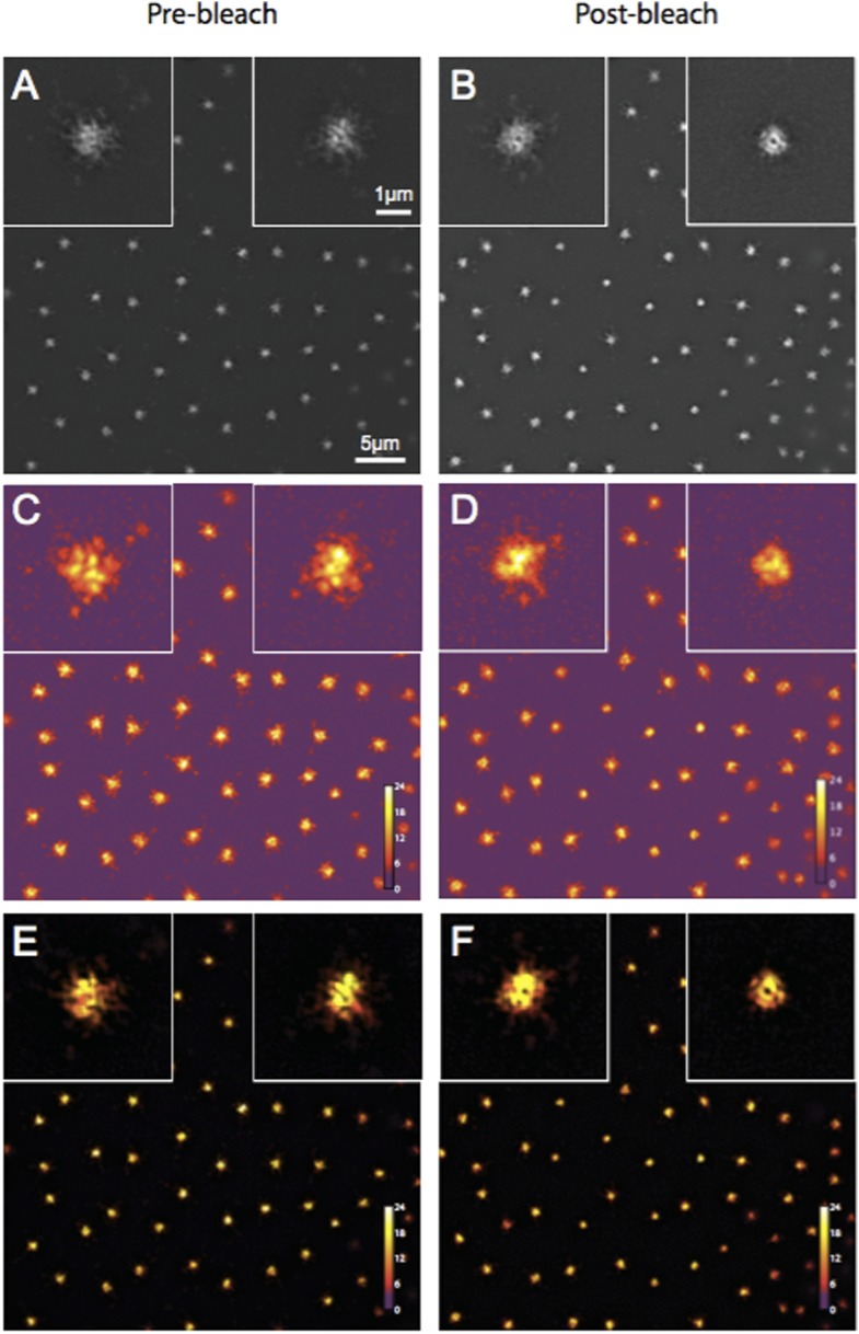
The reviewer thought that our 3D-SIM images of Spd-2-GFP looked strange. We now describe our 3D-SIM acquisition and reconstruction methods in more detail in the Experimental Procedures (subsection “3D-structured illumination (Sub-diffraction resolution) microscopy”). We also provide Author response image 1 showing a SIM-Check (a tool developed to specifically analyse the quality of SIM images) analysis of the Spd-2-GFP OMX data. This shows that the modulation contrast-to-noise ratios at centrosomes have scores of ∼12 and ∼11 for pre and postbleach images, respectively—well above the acceptable value of 3, and indicative of high local contrast between the stripes (generated as part of the SI imaging) and the signal. This confirms that the centrosomal Spd-2-GFP signal in the final reconstructed images is likely to represent true localization rather than reconstruction artifacts. We also note that the appearance of Spd-2-GFP in this paper is similar to that in our previous eLife paper (Conduit et al., 2014).

Author response image 1. Quality control of Spd-2-GFP 3D-SIM images using SIM-Check.

Author response image 1.

(A, B) Reconstructed images of centrosomes prior to photobleaching (A) and 5 min post photobleaching (B) in an embryo expressing Spd-2-GFP. Left and right insets in each image show a control (unbleached) and the experimental (photobleached) centrosome, respectively. (C, D) Modulation contrast-to-noise ratio (MCNR) images of the raw data used to produce the images and insets in (A) and (B). The Color LUT indicates varying MCNR values, with anything above 3 (purple) being acceptable. The Average feature (centrosomal) MCNR in the prebleached (C) and postbleached (D) images is ∼12 and ∼11, respectively. (E, F) Images of the reconstructed data shown in (A) and (B) colour coded according to the underlying MCNR of the raw data shown in (C) and (D).

DOI: http://dx.doi.org/10.7554/eLife.08483.015

2) Concerning the isolation of S-CAP complexes. My understanding is that the protocol used here is different. How does this differ from the previous work from Gopalakrishnan and colleagues? Could it be that other large cytosolic complexes (e.g. S-CAP complexes) are removed during this process? Did the authors ever perform sucrose gradients on extracts depleted or not of centrosomes? It would seem important to clarify what the differences are between the two studies and perhaps include a side-by-side comparison. In the Methods section the authors mention: “however, we cannot rule out the possibility that S-CAP complexes are less stable and possibly fall apart under these conditions, perhaps explaining the difference between our findings and those of Gopalakrishnan et al., 2011.” This disclaimer seems out of place...do S-CAP complexes exist or not?

The reviewer asks for a comparison between our IP protocol and that of Gopalakrishnan et al., and asks for clarification as to whether we think S-CAP complexes exist or not. As discussed above, we include a comparison of the methodologies as an Clearly we cannot detect significant levels of these complexes under the conditions we use here, but we cannot directly compare our results to those of Gopalakrishnan et al., who provided no information about the percentage of each cytoplasmic protein that co-immunoprecipitated with Sas-4.

Reviewer 2:

[…] I find it difficult to assess the merit of this manuscript. On the one hand it is well executed and corrects a mistaken notion of Sas-4 as a direct participant in PCM assembly. On the other, this notion never found widespread acceptance in the field because it contradicted what we have known about Sas-4 function for many years from work in Drosophila and other experimental models. Further, as discussed below Sas-4 may still interact with PCM components in the context of the centriolar wall, something the manuscript does not address. It is therefore not clear to me that this work makes a significant enough contribution to our understanding of either Sas-4 function or PCM assembly to merit publication in eLife.

1) The key observation reported in the current manuscript is that Sas-4 is not coordinately recruited with the PCM components Cnn and Spd2. This indeed runs counter to the idea of cytoplasmic Sas-4-containing ‘S-CAP’ complexes functioning in PCM assembly. However, the best evidence against such a role for Sas-4 is the complete lack of a PCM phenotype in Sas-4-depletions, as reported already in the very first studies on this protein in C. elegans (Kirkham et al., 2003; Leidel et al., 2003) and in numerous other studies since, as also mentioned by the authors in the concluding paragraph.

The reviewer questions the significance of our work, arguing that the idea that S-CAP complexes directly participate in PCM assembly never found widespread acceptance in the field, and pointing out that early studies in worm embryo effectively ruled out the idea that cytoplasmic Sas-4 was required for PCM assembly. We agree that these early worm experiments and our own early experiments in fly embryos (Stevens et al., Curr. Biol. 2007) made an S-CAP model unlikely. Nevertheless, we disagree with the suggestion that the S-CAP model was not taken seriously in the field for the following reasons:

A) Both S-CAP papers were published in high impact journals (Nature Communications and Nature Cell Biology), well after the original papers indicating that this model was unlikely to be correct.

B) We have had trouble publishing several papers in the past few years as reviewers questioned our data because it appeared to contradict the S-CAP model.

C) S-CAP complexes have been discussed as a serious model in virtually every recent review of centrosome/centriole assembly (e.g. Brito et al., Curr. Op. Cell Biol., 2012, Gonczy, Nat. Rev. Mol. Cell Biol., 2012; Mahen et al., Curr. Op. Cell Biol., 2012; Mennella et al., TICB, 2013; Woodruff et al., Philos. Trans. R Soc. Lond. B Biol. Sci. 2014) and also in all four of the first super-resolution papers describing how the PCM is organised around the centrioles (Mennella et al., NCB; Lawo et al., Curr. Biol.; Fu et al., Open Biol.; Sonnen et al., Biol. Open - all 2012).

D) In the paper that fails to detect cytoplasmic S-CAP complexes in worm embryos (Wueseke et al., Mol. Biol. Cell, 2014), the authors conclude that more work is required to assess whether this is due to species-specific differences between worms and flies.

2) The manner in which the spatial pattern of Sas-4/Cnn/Spd-2 recruitment was analyzed, by FRAP at standard resolution and centroid analysis similar to the way kinetochore protein localization was examined by Ted Salmon and colleagues (Wan et al., 2009) and by 3D-SIM FRAP, is indeed elegant and novel for centrosomes. However, the result comes as no surprise. We know at the very least from the original super-resolution studies in Drosophila (Mennella et al., 2012; Fu and Glover, 2012) that PCM proteins including Cnn and Spd-2, but also Asl, localize exclusively around the mother centriole. We also know that Sas-4 on mother centrioles is stably incorporated and does not exchange with the cytoplasmic pool, while daughter centrioles incorporate Sas-4 during S phase (Leidel et al., 2003; Novak et al., 2014). Given those steady state distributions and incorporation patterns, how could the FRAP results be any different?

The reviewer points out that several studies had previously concluded that Sas-4 is irreversibly incorporated into daughter centrioles during their formation, while other studies had concluded that Asl, Cnn and Spd-2 localise around mother centrioles. Thus, one could already infer that Sas-4 does not shuttle these proteins to the mother centriole. The Reviewers’ description of Sas-4 dynamics in fly embryos is, however, not quite correct. In the paper to which the reviewer refers (Novak et al. 2014) we showed (using a microscope with standard resolution) that an excess of Sas-4 builds up at centrioles during S-phase (when centrosomes are growing in size) and this excess is then gradually lost during mitosis (when centrosomes have reached their full size). Thus, although a significant fraction of Sas-4 molecules are clearly incorporated irreversibly into daughter centrioles, it was entirely possible that the excess Sas-4 molecules (that are not irreversibly incorporated into daughters) could have been shuttling S-CAP complexes to mother centrioles. Our super-resolution studies now prove that this is not the case. We clarify this point in the revised manuscript (subsection “The centrosomal recruitment of Sas-4, Cnn and Spd-2 differs in space and time”).

3) There is little to be gained from the immunoprecipitation data presented in Figure 4, showing weak or no interactions between Sas-4 and Asl, Cnn and γ-tubulin. This is essentially a repeat of previous work by the authors (Conduit et al., 2010), although arguably conducted in a more careful manner. A negative result is always difficult to interpret, even if the authors were to present a positive control (i.e. a protein that does interact with Sas-4 under their conditions). Furthermore, ‘S-CAP’ complexes could represent only a small fraction of the total Cnn or γ-tubulin population, without invalidating the original model of Gopalakrishnan and co-workers.

As we discuss above, we agree with the reviewer that our biochemical analysis adds little definitive information, as these studies can neither prove nor disprove the S-CAP model. Nevertheless, we thought it important to include these studies to show that we do not detect S-CAP complexes under the conditions we normally use when looking for protein-protein interactions. The results, although negative, support our imaging data and we suspect that not including the data would lead readers to wonder what proportion of Asl, Cnn and Spd-2 molecules could be part of S-CAP complexes (something that Gopalakrishnan et al. did not address).

4) Finally, it should be noted that the most recent work by Gopalakrishnan (Zheng et al., 2014) proposes that the interactions between Sas-4, γ-tubulin and other PCM components occur in the context of the well-described role of Sas-4 as a centriole duplication protein and centriolar microtubule wall component. It is unclear how such interactions could be detected in cytoplasmic extracts, but there is thus a plausible explanation for at least some of the original findings without invoking a direct, coordinated recruitment of Sas-4 with PCM components.

The reviewer suggests that the recent paper from the Gopalakrishnan lab (Zheng et al., 2014) shows how Sas-4 could play a role in “tethering” PCM components to the centriole wall. We entirely agree that Sas-4 in the centriole is likely to play an important part in tethering the PCM, and our own experiments strongly support this idea, as we find that the Sas-4 already at the centriole helps recruit Asl to centrioles (Novak et al., 2014), which then helps recruit the PCM (Conduit et al., 2014). However, this is a completely different concept from the S-CAP model proposed by Gopalakrishnan and colleagues – a model which is not disputed by Zheng et al.

Other points:

1) It should be clarified that the FRAP experiments in this manuscript do not distinguish between recruitment and cytoplasmic turnover.

The reviewer points out that we need to clarify that our FRAP experiments do not distinguish between recruitment and turnover. Given how the Reviewer uses the term “recruitment” in point 2 below, we suspect that he/she uses “recruitment” to mean the net accumulation of molecules at the centrosome and “turnover” to mean the exchange of centrosome molecules for cytoplasmic molecules. We agree that our FRAP data does not distinguish between these two phenomena. However, we use the term recruitment to include both of these phenomena (i.e. we count a molecule as being “recruited” to centrosomes every time a molecule is added to centrosomes from the cytoplasm, irrespective of whether this adds a new molecule to the centrosome or if it replaces an existing molecule). We now explicitly define how we use the term recruitment (Results, first paragraph).

2) Potentially explained by point 1 above, it is striking that the pattern of Cnn recovery reported here (constant across the cell cycle, Figure 2) does not match the pattern of recruitment reported previously (continuous recruitment in S phase, stable levels in mitosis, Figure 2, Conduit et al., 2010).

The reviewer believes that the pattern of GFP-Cnn recruitment in the current study does not match that reported in our 2010 paper. This is not the case, and we believe this confusion arises from our different definitions of recruitment (see above). In our earlier paper we showed that the total number of GFP-Cnn molecules at centrosomes increases during S-phase (for the reviewer this is “recruitment”) and then stays constant during M-phase (so the reviewer concludes that no recruitment occurs during M-phase). We now show that GFP-Cnn fluorescence recovers after bleaching during both S- and M-phase. Using the reviewers’ definitions, this is due to both recruitment and turnover in S-phase, but only due to turnover in M-phase. However, by our definition, GFP-Cnn molecules are still being recruited to centrosomes during M-phase. Thus, the pattern of GFP-Cnn dynamics in the current study is fully compatible with our earlier analysis. We hope that defining our use of the term “recruitment” helps to clarify this point (Results, first paragraph).

3) The authors should show that there is no residual shift between red and green channels by imaging the same protein (ideally Sas-4) in both channels.

The reviewer thought that we should show that there is no shift between the red and green channels on our confocal microscope by imaging the same protein (ideally Sas-4) in both channels. We apologise for not making this clear but we have performed this control using TetraSpeck beads that fluoresce in both red and green channels; this is the “gold standard” for channel alignment. We have now clarified this point in both the main text (Results, second paragraph) and the Experimental Procedures (subsection “FRAP experiments at standard resolution”).

[Editors' note: further revisions were requested prior to acceptance, as described below.]

Reviewer 1:

In the revised version of their manuscript the authors have tried to address some of the issues raised in my initial review. They have also addressed some of the issues raised by the other reviewer. Although they have not added much in terms of data they have clarified sufficiently well, I believe, some of the major criticisms. I would still recommend they further discuss the technical differences between their work and that of others, but I would leave this to their discretion and to better contrast the difference observed, and how they relate to models of PCM assembly. I therefore support publication of this manuscript in its current form.

Although Reviewer 1 thought that we should further discuss the differences between our work and that of others, he/she kindly left this to our discretion and supported publication of the manuscript in its current form. We agree that an extensive discussion would be of interest to the community, but it is always difficult to accurately assess why two different groups have performed similar experiments but reached different conclusions. We highlight that there are important differences between the two studies, but we prefer to let readers reach their own opinion as to why this might be the case. We have therefore left the paper unchanged in this regard.

Reviewer 2:

Since my criticisms of the original manuscript by Conduit and Raff centered largely on the significance of their results and thus the suitability of their manuscript for publication in eLife, I do not have much to add to my original comments. I agree with Reviewer 1 that there is merit in a paper that clearly refutes the existence of cytoplasmic pre-complexes involving Sas-4 and PCM proteins. However, I fear that S-CAP complexes will persist in the centrosome literature no matter how definitively these complexes are shown not to exist.

With regard to the experimental data and clarifications in the revised manuscript, I'm generally satisfied with those revisions. While a further biochemical characterization of Sas-4 in the cytoplasm along the lines of Wueseke et al., 2014 would be informative, this is clearly beyond the scope of this paper and I agree that sucrose gradients alone would not add significantly to what is primarily a paper built around high-resolution live imaging.

I do, however, think that the authors need to experimentally address the potential for shifts between red and green channels in their SIM/SIM-FRAP experiments. Correcting for chromatic shifts using TetraSpeck beads and then using those same beads to demonstate an absence of a shift is circular and does not exclude the possibility of small but significant shifts in the context of the live embryo. Imaging and quantitating the (preferably diffraction limited) signal for the same protein in both channels would eliminate this possibility and increase confidence in their measurements of Sas-4/PCM protein localization/recruitment, which are at the heart of their paper.

Reviewer 2 thought that we needed to experimentally address the potential for shifts between red and green channels in our SIM/SIM-FRAP experiments, pointing out that the use of TetraSpeck beads does not exclude the possibility of small but significant shifts in the context of the living embryo. To eliminate this possibility, the reviewer suggested that we image the signal from the same protein in both channels to confirm that they align. From these comments we realize that we originally misunderstood the reviewers concern: we thought we needed to address whether the red and green channels were systemically misaligned (which is addressed by the TetraSpeck bead experiment), but the reviewer was actually asking us to address whether the two channels were misaligned because the centrioles have moved slightly during acquisition. The reviewer is correct that this is an important issue, and we apologise that we originally misunderstood this point.

The centrioles can indeed move slightly between the acquisition of the green and red channels (which, for technical reasons, have to be acquired sequentially). This slight movement does not, however, affect the interpretation of our data. To prove this point, we now measure the distance between the center of the Asl toroid (marking the center of the mother centriole acquired in the red channel) and the center of each Sas-4 dot (marking the center of both the mother and daughter centrioles, acquired in the green channel). Prior to photobleaching, one Sas-4 centriole (the presumptive mother) is on average ∼80nm from the center of the Asl toroid, while the other (the presumptive daughter) is on average ∼210nm from the center of the Asl toroid. Thus, the mother centriole shifts by an average of ∼80nm during the time between acquisition of the two channels. After photobleaching, the Sas-4 signal recovers at an average distance of ∼260nm from the center of the Asl toroid: statistically, this distance is significantly different from the average distance the mother centriole shifts during acquisition (∼80nm) but is not significantly different from the average distance between the Asl toroid and the daughter centriole (∼210nm). This new analysis (shown in new Figure 3D and 3E) strongly supports our conclusion that Sas-4 is recruited to daughter centrioles rather than mother centrioles.

Associated Data

    This section collects any data citations, data availability statements, or supplementary materials included in this article.

    Supplementary Materials

    Figure 1—source data 1. Measuring the spatial offset between recovering GFP-Cnn and Sas-4-mCherry, Spd-2-GFP and Sas-4-mCherry, and Spd-2-GFP and RFP-Cnn during S-phase.

    (S1) This sheet includes the raw data used to calculate the offset between the centroids of the green and red fluorescent signals of sub-resolution beads. (S2) This sheet includes the raw data used to calculate the offset between the centroids of the GFP-Cnn and Sas-4-mCherry fluorescent signals shown in Figure 1H, and the raw data used to plot and offset the centrosomal profiles of GFP-Cnn and Sas-4-mCherry shown in Figure 1C. (S3) This sheet includes the raw data used to calculate the offset between the centroids of the Spd-2-GFP and Sas-4-mCherry fluorescent signals shown in Figure 1H, and the raw data used to plot and offset the centrosomal profiles of Spd-2-GFP and Sas-4-mCherry shown in Figure 1E. (S4) This sheet includes the raw data used to calculate the offset between the centroids of the Spd-2-GFP and RFP-Cnn fluorescent signals shown in Figure 1H, and the raw data used to plot and offset the centrosomal profiles of Spd-2-GFP and RFP-Cnn shown in Figure 1G. (S5) This sheet collates the offset data calculated in S2, S3 and S4 in order to plot the graph seen in Figure 1H.

    DOI: http://dx.doi.org/10.7554/eLife.08483.003

    elife08483s001.xlsx (594.6KB, xlsx)
    DOI: 10.7554/eLife.08483.003
    Figure 2—source data 1. A comparison of the centrosomal fluorescence recovery after photobleaching of GFP-Cnn, Spd-2-GFP and Sas-4-mCherry during M-phase and the following S-phase.

    (S1) This sheet includes the raw data used to calculate the fluorescence recovery of GFP-Cnn and Sas-4-mCherry during M-phase and the following S-phase. (S2) This sheet includes the raw data used to calculate the fluorescence recovery of Spd-2-GFP and Sas-4-mCherry during M-phase and the following S-phase. (S3) This sheet combines the Sas-4-mCherry recovery data from S1 and S2. (S4) This sheet collates the data from S1, S2 and S3 in order to plot the final recovery graph seen in Figure 2D.

    DOI: http://dx.doi.org/10.7554/eLife.08483.007

    elife08483s002.xlsx (249.6KB, xlsx)
    DOI: 10.7554/eLife.08483.007
    Figure 3—source data 1. Measuring the spatial offset between recovering Sas-4-GFP and Asl-mCherry at super resolution.

    This flie includes the raw data used to calculate the offset between the centroids of the two Sas-4-GFP foci and the single Asl-mCherry foci prior to photobleaching and the offset between the centroids of the single Sas-4-GFP foci and the single Asl-mCherry foci 5 min post-photobleaching. The data is used to plot the graph shown in Figure 3E.

    DOI: http://dx.doi.org/10.7554/eLife.08483.011

    elife08483s003.xlsx (53.9KB, xlsx)
    DOI: 10.7554/eLife.08483.011

    Articles from eLife are provided here courtesy of eLife Sciences Publications, Ltd

    RESOURCES