Abstract
Formation of black triangles between teeth due to loss of interdental palpilla is one of the common problems encountered in routine clinical practice, as extreme importance is given to esthetics. This paper discusses two different surgical approaches in treating three cases with papillary loss in the first case the reconstruction of papilla was achieved by using a semilunar coronally repositioned papilla technique and in second and the third case reconstruction of the papilla was achieved by modification of Nodland's microsurgical technique. In all the three cases a free connective tissue graft was used to reconstruct the lost volume of interdental papilla. Complete reconstruction of the lost papilla was achieved in all the three cases 6 months postoperatively.
Keywords: Black triangle, connective tissue graft, interdental papilla
Introduction
Interdental papilla (IDP) occupies the space created between two adjacent teeth. IDP acts as a biological barrier which protects the underlying periodontal structures, apart from playing an important role in esthetics.
The loss of IDP as a result of periodontal disease or of periodontal therapy leads to esthetic, phonetic, and food impaction problems. Among the periodontal surgical procedures, one of the most complex procedures to be performed are the papillary augmentation procedures, which aims to fill the black spaces created by a papillary loss in the interproximal area.
Several techniques were attempted previously to augment the IDP, however, a predictable technique for the reconstruction of lost IDP remains elusive. Beagle described a pedicle graft procedure utilizing the soft tissues palatal to the IDP to reconstruct new papilla. In 1996, Han and Takei described the use of a facial approach with a semilunar incision to gain access to the papillary area for augmentation of the papilla. Cortellini et al. proposed a simplified papilla preservation flap that requires a releasing incision in the papillary area and placement of a barrier membrane under the surgical site.[1] Azzi et al. have described techniques to gain access to augment the connective tissue and bone under the deficient papilla.[2] In all these techniques the blood supply to the recipient site might get jeopardized, Miller emphasized the significance of blood supply to the donor tissue in his classic publications.[3]
All these techniques mentioned above hindered the blood supply as they used releasing incisions. Placing releasing incisions close to the connective tissue graft, bone graft are barrier membrane can cause tenting of the papilla, which can result in exposure of the connective tissue graft, bone graft, and membrane. Apart from resulting in scarring releasing incisions given in patients with thin gingival biotype increases the risk of failure.
Root coverage procedures using predictable techniques aim at enhancing the blood supply at the recipient site, which supplements the donor tissue survival.[4] Previous studies have shown that procedures like tunneling surgical approach which eliminate the need for releasing incision have improved vascularity to the surgical area and better postsurgical outcomes, compared to techniques using releasing incisions.[5]
This paper describes a report of three cases, in which the first case was based on the technique described by Han and Takei[6] were releasing incisions are not used and the other two cases were based on the modification of microsurgical technique described by Nordland et al.[7] where the surgery was done without the use of releasing incisions, which resulted in better donor tissue survival and tissue trauma, bleeding, scarring, and pain.
Case Report
Preparation of the preoperative site
The Nordland and Tarnow classification system was used for the classification of the initial preoperative IDP,[8] which helped in determining the required tissue volume.
The IDP was anesthetized along the facial and palatal gingiva, with xylocaine with adrenaline (1:80,000). Root conditioning was done with tetracycline hydrochloride paste application for 60 s to sterilize the root surface and to achieve root demineralization. The patients were instructed to do a preprocedural oral rinse with 0.2% chlorhexidine digluconate solution for 30 s.
Surgical procedure
Case 1
A 24-year-old female patient was referred from the Department of Orthodontics to the Department of Periodontics, Faculty of Dental Sciences, Sri Ramachandra University for the reconstruction of lost IDP between the maxillary left central and lateral incisors. She was undergoing orthodontic treatment to correct deep overbite and spacing between her teeth. Clinical examination revealed papillary loss due to spacing between the above-mentioned teeth [Figure 1]. The space between 21 and 22 was closed orthodontically, and a contact point was created between the two teeth at the incisal third so that the distance between contact point to bone crest was 5 mm. Radiographic assessment was done to check the bone levels; radiograph revealed no significant bone loss, a complete reconstruction of the papilla was expected. The surgical procedure was explained to the patient, and informed consent was obtained.
Figure 1.
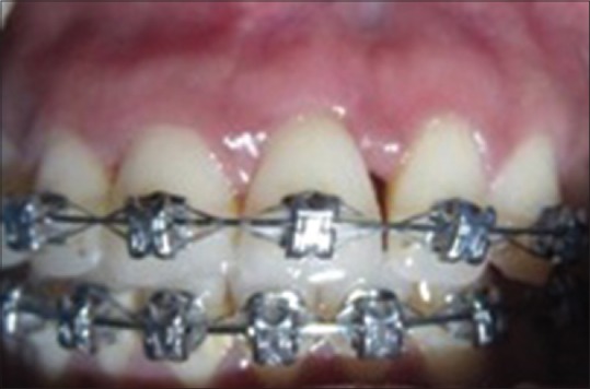
Preoperative image showing papillary loss
After administration of local anesthesia, a split thickness semilunar incision was performed from the distal buccal line angle of tooth 22 to mesial buccal line angle of tooth 21. Approximately 6–10 mm apical from the gingival margin extending into the alveolar mucosa using ophthalmic blades no. 001572, 082622, and fine stainless surgical blade SM 63 (Swann-Morton Ltd., Sheffield S6 ZBJ, England).
The intrasulcular incisions were made around the mesial and distal half of the two adjacent teeth to free the connective tissue from the root surface. Following this, the donor connective tissue graft was harvested from the palate and the donor tissue was sutured with 3-0 black silk suture. The harvested graft was trimmed according to the requirements of the recipient site. It was then tucked in [Figure 2] and pushed coronally to support and provide bulk to the coronally positioned interdental tissue. Then the flap was stabilized with 5-0 vicryl sutures, and a suspensory suture [Figure 3] was given as described by Nordland et al.[7]
Figure 2.
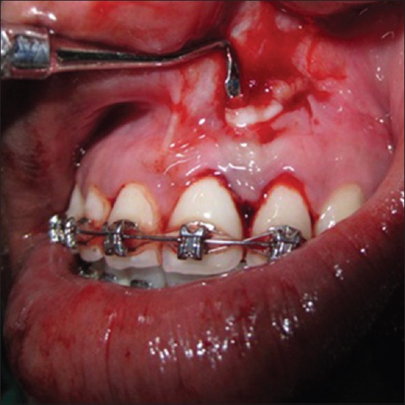
Harvested connective tissue graft being tucked into the recipient site
Figure 3.
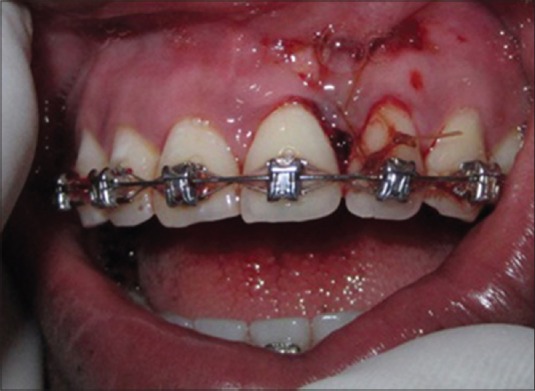
Suspensory suture
The patient was placed on analgesics and 0.2% chlorhexidine digluconate mouthwash twice daily for 2 weeks with no mechanical cleaning of the surgically treated interproximal area.
Postoperative follow-up
The sutures were removed 2 weeks after the procedure. The surgical site was examined for uneventful healing. Healing was found to be satisfactory. The patient was reviewed at regular intervals, normal anatomy and shape with the complete reconstruction of the IDP was achieved after 1 year [Figure 4].
Figure 4.
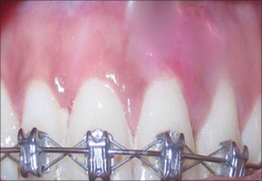
One-year postoperative image showing complete Papillary reconstruction (case 1)
Cases 2 and 3
A circumferential sulcular incision was made to the crest of the bone using ophthalmic blades no. 001572 and 082622 and fine stainless surgical blade SM 63(Swann-Morton Ltd., Sheffield S6 ZBJ, England). A sulcular incision was made to the crestal bone, severing the marginal gingiva. This incision extends circumferentially around adjacent teeth.
Following the minimal circumferential sulcular incision, a split thickness flap is prepared, extending facially up to and past the mucogingival junction, the mobility of the undermined tissue can be achieved. Mobility is usually to achieve space under the papilla to receive a dense fibrous connective tissue graft.
An adequate volume of donor connective tissue was obtained from the palate, and a “lasso” suture was used to pull and position the donor tissue. A 5-0 resorbable vicryl suture was used to assist in positioning of the graft under the deficient papilla [Figures 5–10].
Figure 5.
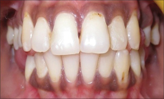
Case 2 preoperative image showing papillary loss
Figure 10.
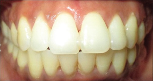
One-year postoperative image showing complete papillary reconstruction (case 3)
Figure 6.
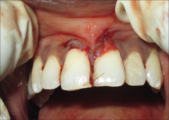
Suspensory suturing done
Figure 8.
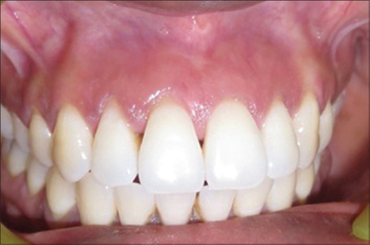
Case 3 preoperative image showing papillary loss
Figure 9.
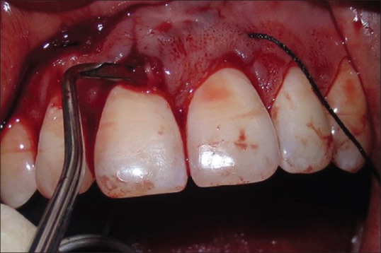
“Lasso” suture used to pull and position the donor tissue
The suspensory suture
A suspensory suture Figures 3 and 7 was given as described by Nordland et al.[7]
Figure 7.
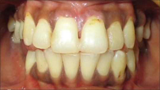
One-year postoperative image showing complete papillary reconstruction (case 2)
Wound tension can apply forces to the graft and surrounding tissues as they heal and also pressure created by lip muscles and tissue memory can create pressure and tension on the overlying tissue and create a tendency to pull the papillary tissue back to its original position. To preserve this new papillary tissue position, a “suspensory suture” is used. A suspensory suture that begins at the base of the papilla and is anchored around the interproximal contact and is essential to maintain the new position and height of the papilla.
The suspensory suture starts by piercing the facial papillary base, proceeds through the connective tissue graft, and exits through the base of the palatal papilla. Composite bonding material was added at the interproximal contacts of the adjacent teeth, which helped in preventing the suspensory suture from slipping through the interproximal contact. The suspensory suture is anchored by being looped around the bonded contact point. If this suture is not used, then tissue retraction usually occurs. Coe pack was given, and the patient was asked to use chlorhexidine mouth rinse during tissue healing and maturation.
Discussion
The presence or absence of the IDP is influenced by various factors. Some of the detrimental factors include crestal alveolar bone height, interradicular distance, contact area and dimensions of the interproximal space.[9]
When the distance between the bone crest and contact point is <5 mm, surgical interpretations to increase the volume of IDP is justified.[10] In situations where the distance from the bone crest to contact point is >5 mm a combination of nonsurgical (restorative, orthodontic) and surgical interventions would be appropriate.
Several techniques have been proposed in the literature for reconstruction of the lost IDP, most of which are based on the use of a connective tissue graft. Each technique was found to have its advantages and disadvantages.[11] One of the important criteria to be considered for the success of papillary reconstruction is the adequacy of blood supply to the recipient site.
The IDP is a small area of tissue with blood supply arising from various sources, originating from one direction toward the base.[11] This is an important factor to be considered in papillary augmentation techniques. Most techniques proposed horizontal incisions which interrupt blood supply that comes from the gingival connective tissue to the papilla.[12] This paper describes a report of three cases in which releasing incisions were eliminated to improve vascularity to the surgical area and improve the postoperative outcome.
In the Case 1, which is based on the technique described Han and Takei[6] a better blood supply from the flap to the graft was achieved, which helped in maintaining the papillary integrity and helped in avoiding flap necrosis. The procedure was done as, postorthodontic treatment the patient was concerned about the black triangle and as the problem was a class-I defect complete surgical reconstruction of IDP was achieved. Similar findings were reported by Deepalakshmi et al.[13]
In Cases 2 and 3 modification to the microsurgical technique described by Nodland were done, wherein we used ophthalmic blades no. 001572 and 082622 and fine stainless surgical blade SM 63 (Swann-Morton Ltd., Sheffield S6 ZBJ, England) to give the sulcular and interdental incisions and for extension of the incision beyond the mucogingival junction to create a pouch and tunnel, hence unnecessary releasing incisions which compromise the blood supply were eliminated.
In the first case, orthodontic tooth movement was used to close space and create a contact between the two teeth. In all the three cases a suspensory suture described by Nodland which starts by piercing the facial papillary base, proceeds through the connective tissue graft and exits through the base of palatal papilla was used and composite bonding material was used at the interproximal contacts of adjacent teeth helped in the prevention of this suspensory suture from slipping through the interproximal contact. The suspensory suture was anchored by looping around the bonded contact point, which helped in preventing the tissues from retracting back to its original position.[5]
The suspensory suture also helped in maintaining the donor tissue in a coronal position until the maturation of overlying flap at its desired postsurgical position, thereby preventing apical migration or displacement of the graft.
The present report showed three successfully treated cases due to proper planning and evaluation of soft and hard tissues that are required for optimal outcomes in papilla reconstruction techniques. However, the predictability and long term stability of these treatment approaches need to be evaluated by further clinical studies.
Financial support and sponsorship
Nil.
Conflicts of interest
There are no conflicts of interest.
References
- 1.Cortellini P, Pini Prato GP, Tonetti MS. The modified papilla preservation technique. A new surgical approach for interproximal regenerative procedures. J Periodontol. 1995;66:261–6. doi: 10.1902/jop.1995.66.4.261. [DOI] [PubMed] [Google Scholar]
- 2.Azzi R, Takei HH, Etienne D, Carranza FA. Root coverage and papilla reconstruction using autogenous osseous and connective tissue grafts. Int J Periodontics Restorative Dent. 2001;21:141–7. [PubMed] [Google Scholar]
- 3.Miller PD., Jr Root coverage using a free soft tissue autograft following citric acid application. Part 1: Technique. Int J Periodontics Restorative Dent. 1982;2:65–70. [PubMed] [Google Scholar]
- 4.Langer B, Calagna LJ. The subepithelial connective tissue graft. A new approach to the enhancement of anterior cosmetics. Int J Periodontics Restorative Dent. 1982;2:22–33. [PubMed] [Google Scholar]
- 5.Zabalegui I, Sicilia A, Cambra J, Gil J, Sanz M. Treatment of multiple adjacent gingival recessions with the tunnel subepithelial connective tissue graft: A clinical report. Int J Periodontics Restorative Dent. 1999;19:199–206. [PubMed] [Google Scholar]
- 6.Han TJ, Takei HH. Progress in gingival papilla reconstruction. Periodontol 2000. 1996;11:65–8. doi: 10.1111/j.1600-0757.1996.tb00184.x. [DOI] [PubMed] [Google Scholar]
- 7.Nordland WP, Sandhu HS, Perio C. Microsurgical technique for augmentation of the interdental papilla: Three case reports. Int J Periodontics Restorative Dent. 2008;28:543–9. [PubMed] [Google Scholar]
- 8.Nordland WP, Tarnow DP. A classification system for loss of papillary height. J Periodontol. 1998;69:1124–6. doi: 10.1902/jop.1998.69.10.1124. [DOI] [PubMed] [Google Scholar]
- 9.De Oliveira JD, Storrer CM, Sousa AM, Lopes TR, de Sousa Vieira J, Deliberador TM. Papillary regeneration: Anatomical aspects and treatment approaches. RSBO. 2012;9:448–56. [Google Scholar]
- 10.Tarnow DP, Magner AW, Fletcher P. The effect of the distance from the contact point to the crest of bone on the presence or absence of the interproximal dental papilla. J Periodontol. 1992;63:995–6. doi: 10.1902/jop.1992.63.12.995. [DOI] [PubMed] [Google Scholar]
- 11.Carranza N, Zogbi C. Reconstruction of the interdental papilla with an underlying subepithelial connective tissue graft: Technical considerations and case reports. Int J Periodontics Restorative Dent. 2011;31:e45–50. [PubMed] [Google Scholar]
- 12.Nobuto T, Suwa F, Kono T, Hatakeyama Y, Honjou N, Shirai T, et al. Microvascular response in the periosteum following mucoperiosteal flap surgery in dogs: 3-dimensional observation of an angiogenic process. J Periodontol. 2005;76:1339–45. doi: 10.1902/jop.2005.76.8.1339. [DOI] [PubMed] [Google Scholar]
- 13.Deepalakshmi D, Ahathya RS, Raja S, Kumar A. Surgical reconstruction of lost interdental papilla a case report. PERIO. 2007;4:229–34. [Google Scholar]


