Abstract
Objective: To determine the diagnostic accuracy of optical coherence tomography for basal cell carcinoma and the proportion of biopsies that could be avoided if optical coherence tomography is used to rule-in surgery. Design: Multicenter, prospective, observational study. Setting: Dermatology clinics. Participants: Consecutive patients with clinically challenging pink lesions suspicious for basal cell carcinoma. Measurements: Clinical, dermoscopic, and optical coherence tomography images were obtained for all subjects. At each stage, the clinician made a diagnosis (pathology + subtype if applicable), and assessed his/her own confidence in the diagnosis. Results: Optical coherence tomography significantly (p<0.01) improved sensitivity and specificity over clinical or dermoscopic evaluation. The percentage of correct diagnoses was 57.4 percent (clinical), 69.6 percent (dermoscopy), and 87.8 percent (optical coherence tomography). Optical coherence tomography significantly increased the certainty of diagnosis; clinicians indicated they were certain (>95% confident) in 17 percent of lesions examined clinically, in 38.6 percent examined with dermoscopy, and in 70 percent examined with optical coherence tomography. With the use of optical coherence tomography in the diagnosis of basal cell carcinoma, more than 1 in 3 patients could avoid a diagnostic biopsy. Conclusion: In a population of clinically challenging lesions, optical coherence tomography improved diagnostic certainty by a factor of four over clinical examination alone and improved diagnostic accuracy by 50 percent (57-88%). The addition of optical coherence tomography to other standard assessments can improve the false-positive rate and give a high degree of certainty for ruling in a positive diagnosis for basal cell carcinoma. A reduction of 36 percent in overall biopsies could be achieved by sending high certainty basal cell carcinoma positive optical coherence tomography diagnoses straight to surgery.
Nonmelanoma skin cancers (NMSC) comprising basal cell carcinomas (BCC) and squamous cell carcinomas (SCC) constitute 80 percent of all skin cancers,1 with BCC forming the vast majority of those.2,3 There were an estimated total number of NMSCs in the United States population in 2012 of 5,434,193, and the total number of persons in the United States treated for NMSC in 2012 was 3,315,554.4 In the United States, the prevalence of NMSC exceeds that of all other cancers combined.4,5
The incidence of BCC has been increasing annually by between two and eight percent since 1960.6 A recently published study, based on US Medicare fee-for-service population data, indicated an increase in procedures of 14 percent between 2006 and 2012, a 2.2 percent per year increase in this population.4 As the incidence of BCC also rises with age,3,7 it is becoming a serious problem in the growing elderly population of the United States.
Clinical evaluation of BCC lesions is subject to error, and as such, biopsies are the gold standard for diagnosis. However, biopsies are invasive and can lead to poor cosmetic outcomes particularly for lesions on the face or neck. Furthermore, biopsies are costly: The national average Medicare cost in 2013 for a skin biopsy was $104.40, with each additional biopsy costing $32.89, along with an additional $72.94 for the pathologist.8 Due to the high volume of NMSCs in the United States, an increasing focus on novel diagnostic modalities is appropriate.
Optical coherence tomography (OCT) is a noninvasive imaging technique that provides a cross-sectional image of the tissue microarchitecture down to a depth of approximately 1.5mm.9-11 Recent studies have established that the images are sufficiently detailed to enable identification of morphological criteria for different NMSC, such as actinic keratosis (AK), SCC, and BCC.9,11-13
OCT has the potential to be a noninvasive alternative to biopsy. Noninvasive technology, such as OCT, could provide value by helping to “rule-in” lesions that are suspicious of skin cancer and require treatment. This could be particularly valuable where the patient has more lesions than could be clinically or ethically biopsied providing the opportunity to scan many suspicious lesions in a single session to determine those which require further attention. Further, by improving sensitivity in difficult lesion populations, lesions could be identified earlier and treated more successfully.
Diagnosis and surgery could potentially take place on the same visit, improving the experience for the patient, and there may also be cost savings. As increasing portions of the Medicare payment pool will be used to pay for alternative payment models, an integrated approach to diagnosis and treatment during the same encounter may be attractive to many stakeholders. The key criterion for success in this application is for the technique to have low false-positive rates in order to minimize the number of benign lesions being unnecessarily treated.
This multicenter, prospective, observational study aimed to determine the sensitivity, specificity, positive predictive value (PPV), and negative predictive value (NPV) for OCT in the diagnosis of BCC. The proportion of biopsies that could be avoided using OCT as a noninvasive alternative to biopsies taken to rule-in surgery was assessed. In addition, the relationship between diagnostic certainty and diagnostic accuracy for clinical examination, dermoscopic-assisted diagnosis, and OCT-assisted diagnosis was also investigated.
METHODS
Subjects. Subjects were recruited at three sites to allow the inclusion of differing patient populations and differing clinician experience of OCT.
Subjects, male or female ≥18 years of age, with a maximum of three lesions, were included if they had at least one clinically challenging lesion, on the head or neck, that was suspicious for BCC as defined below, and was therefore to be biopsied to rule BCC in or out, and if they were eligible for Mohs surgery.
Subjects were excluded if they had a history or evidence of metastatic lesions, if topical actinic field therapies had been used within the eight weeks prior to the visit, or if the lesion area also contained other skin conditions, ulceration, or inflammation. Local Ethics Committees approved the research protocol and all research was conducted according to the principles of the Declaration of Helsinki.
Examinations. Recruitment and examination followed the path outlined in Figure 1. Once enrolled in the study, clinical, dermoscopic, and OCT images were obtained for all subjects following which a judgement was made as to whether biopsy was also required for confirmation of diagnosis.
Figure 1.
Study design
During the clinical examination, lesions were photographed with a standard digital camera (Canon X70, Sony Cyber-shot 14.4 or Nikon J1). Digital dermoscopy was carried out using a Handyscope DermLite and DermLite II HR (3Gen Inc., San Juan Capistrano, California), focusing on the central area of the lesion. OCT was carried out with a VivoSight OCT Scanner (Michelson Diagnostics, Maidstone, Kent, United Kingdom). Images were obtained with a field view of 6mm and a depth of 2mm, with a spatial resolution of better than 7.5μm in all axes.10 The function “multi-1” setting automatically provided 60 lateral scans of 6mm length every 100μm.
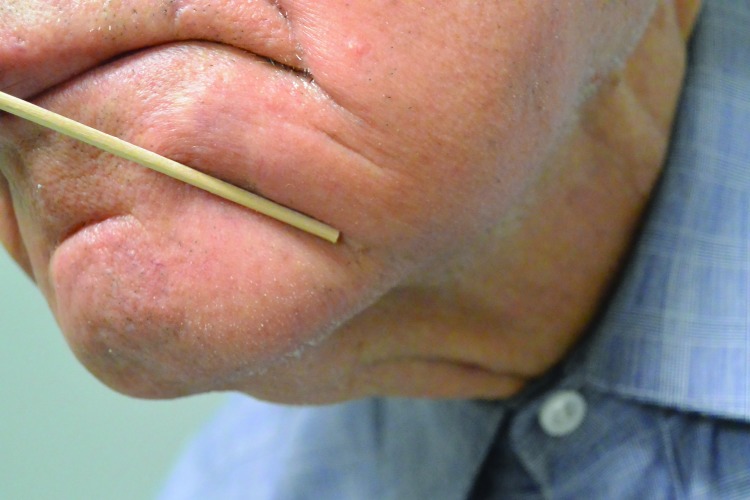
Clinical. Clinically challenging lesions were defined as lesions that did not have the usual characteristics of BCC, such as ulceration, bleeding, crusting, isolated pink scaly patches, or pearly papules. The lesions were diagnosed based on the patient’s clinical history of a nonhealing area of concern or the clinician’s inability to rule out BCC (Figures 2A and 3A).
Figure 2.
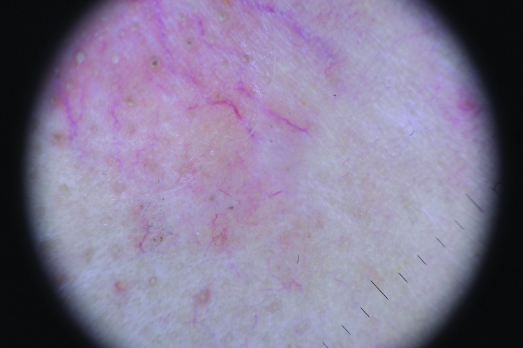
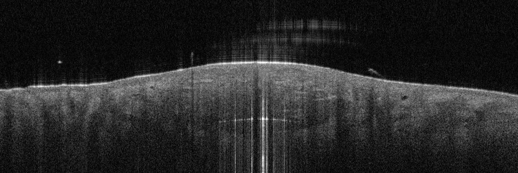
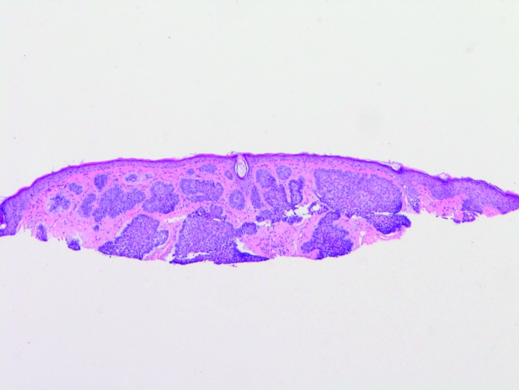
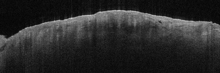
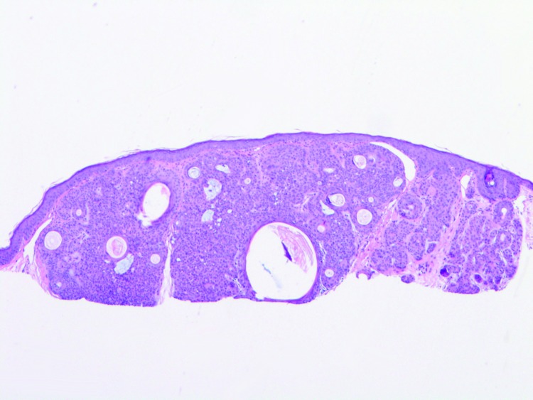
Clinically challenging basal cell skin cancer lesion, Patient 1. (A) Clinical image; (B) Dermoscopic image; (C) Optical coherence tomography (OCT) still image—note the hypo-echogenic tumor islands; (D) Histology (H & E)
Figure 3.
Clinically challenging basal cell skin cancer lesion, Patient 2. (A) Clinical image; (B) Dermoscopic image; (C) Optical coherence tomography (OCT) still image—note the hypo-echogenic tumor islands; (D) Histology (H & E)
Dermoscopy.14 Lesions were inspected for dermoscopic features consistent with BCC similar to the features seen in the two-step algorithm,15,16 including arborized vessels, pink-white shiny background, blue/grey ovoid nests, ash leaf pattern, dot-globular-like pattern, spoke wheel, and crystalline-like structures (Figures 2B and 3B).
Optical coherence tomography. OCT scans were inspected for features consistent with BCC. Specifically, the epidermis was analyzed for protrusions into the dermis with shadowing; the epidermal-dermal junction for lack of definition or rupturing; and the dermis for signal-poor ovoid structures, dark rims, ovoid structures with bright centers, dilated vessels, black areas or cysts, bright stroma, and/or small ovoid signal-poor structures (“fish shoal”) (Figures 2C and 3C). These features are consistent with those reported in previous studies.9,11,13
Assessments. At each stage, the following two main assessments were made:
Is the lesion BCC?
What is the diagnosis (pathology + subtype if applicable), how clear was the diagnosis, how much confidence does the clinician have in the diagnosis (Figures 2D and 3D)?
A recommendation for further treatment was made before the patient was returned for standard-of-care treatment. A biopsy was taken and the final diagnosis and lesion depth based on histopathology were recorded for comparison with the OCT diagnosis.
Outcomes. The primary outcome was to determine the proportion of biopsies that could have been avoided by using OCT as a noninvasive alternative to a biopsy taken for rule-in of surgical therapy.
The secondary outcomes were to determine the sensitivity, specificity, PPV, and NPV of OCT for diagnosis of clinically challenging BCC.
Estimation of sample size. Using data from a similarly challenging population of lesions,9 the best estimate of the proportion that will be “not indicated for Mohs” is 43.4 percent, calculated as: [proportion of patients with BCC x (1 - sensitivity)] + [proportion without BCC x specificity]. For a desired mean confidence interval (CI) of 20%, 101 lesions would be needed to detect a CI of ±10%. Confidence intervals were calculated using the Clopper-Pearson method.
RESULTS
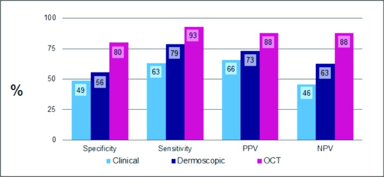
The diagnostic sensitivity, specificity, PPV, and NPV for the three techniques (clinical, dermoscopy, and OCT) are shown in Table 1 and Figure 4. OCT significantly (p<0.01) improved both sensitivity and specificity over clinical or dermoscopic evaluation (Tables 2 and 3).
TABLE 1.
Sensitivity, specificity, positive predictive value, and negative predictive value for all three examination techniques when all lesions are included
| SENSITIVITY (95% CI) | SPECIFICITY (95% CI) | PPV (95% CI) | NPV (95% CI) | |
|---|---|---|---|---|
| Clinical | 62.9% | 48.9% | 65.7% | 45.8% |
| Dermoscopy | 78.6% | 55.6% | 74.3% | 62.5% |
| OCT | 92.9% | 80.0% | 87.8% | 87.8% |
Sensitivity: the proportion of disease-positive lesions detected by a diagnostic test = TP/(TP + FN)
Specificity: the proportion of disease-negative lesions detected by a diagnostic test = TN/(TN + FP)
Positive predictive value (PPV) is the likelihood of a diagnostic test being correct when it indicates a disease-positive result = TP/(TP + FP)
Negative predictive value (NPV) is the likelihood of a diagnostic test being correct when it indicates a disease-negative result = TN/(TN + FN)
TP=true positive, FP=false positive, TN=true negative, FN=false negative
Figure 4.
Sensitivity, specificity, positive predictive value, and negative predictive value for all three examination techniques.
TABLE 2.
Statistical comparison of sensitivity
| OCT SENSITIVITY | OTHER SENSITIVITY | p | |
|---|---|---|---|
| OCT vs. clinical | 92.9% | 62.9% | 3.0x10⁻⁵ |
| OCT vs. dermoscopy | 92.9% | 78.6% | 0.0094 |
Both comparisons were statistically significant
TABLE 3.
Statistical comparison of specificity
| OCT SPECIFICITY | OTHER SPECIFICITY | p | |
|---|---|---|---|
| OCT vs. clinical | 80.0% | 48.9% | 0.0037 |
| OCT vs. dermoscopy | 80.0% | 55.6% | 0.0056 |
Both comparisons are statistically significant
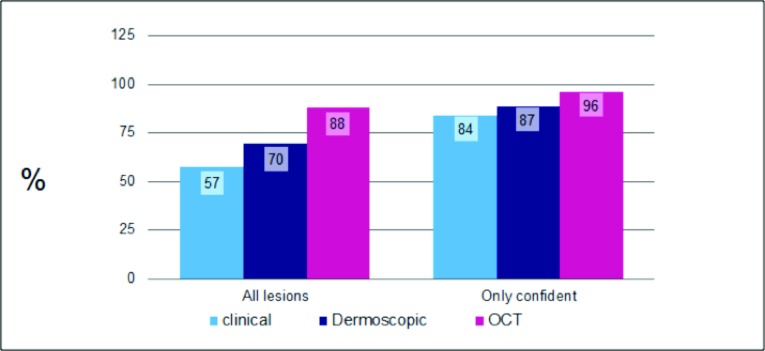
Of the 115 lesions examined, 70 were histologically confirmed as BCC. The most common subtype was nodular. The percentage of correct diagnoses was 57.4 (clinical), 69.6 percent (dermoscopy), and 87.8 percent (OCT) (Figure 5).
Figure 5.
The effect of diagnostic confidence on accuracy. OCT significantly increases not only the absolute % of correct diagnoses, but also the % correct when dermatologists are >95% confident in their diagnosis.
The use of OCT significantly increased the proportion of lesions where clinicians were certain about the diagnosis, a significant challenge in this difficult subset of lesions. Clinicians indicated that they were certain (>95% confident) in 17 percent of clinically examined lesions, increasing to 38.6 percent of lesions with the addition of dermoscopy and then to 70 percent with the further addition of OCT.
The increase in diagnostic certainty attributable to OCT also had a positive effect on accuracy, with an increase in correct diagnoses when the clinician was confident of their diagnosis (Figure 5). The accuracy of diagnosis with OCT increased from 88 to 96 percent when diagnostic certainty was accounted for.
DISCUSSION
This study aimed to determine whether OCT imaging could have a role in reducing the number of biopsies taken in daily dermatological practice. In US clinical practice, this would involve allowing lesions to be ruled-in for treatment without a biopsy being taken first.
Diagnostic accuracy, and particularly the false-positive rate, is important in this respect as this determines the risk of patients being sent for surgery where they would otherwise have only been subject to a surgical biopsy.
Diagnostic accuracy. OCT greatly improved diagnostic accuracy relative to clinical and dermoscopic examination in this difficult group of lesions that were suspicious for BCC. Overall accuracy was 57 percent with clinical examination alone, rising to 70 percent with dermoscopy and 88 percent with OCT.
Doctors were asked to rate their certainty in their diagnosis at each stage of the examination process. The intention was to evaluate whether using imaging increased their confidence and whether increased confidence in the diagnosis would correlate with better diagnostic results.
When only lesions where the doctor was >95 percent certain of their diagnosis were included, 84 percent of clinical diagnoses were correct, rising to 89 and 96 percent with dermoscopy and OCT, respectively.
The proportion of lesions where the clinician reached 95 percent plus diagnostic certainty increased fourfold, from 17 percent when only clinical diagnosis was used to 39 percent with dermoscopy and 68 percent once OCT was used.
These findings are similar to those of a study by Ulrich et al9 of similar design where OCT significantly improved both specificity and PPV and NPV over clinical and dermoscopic examination alone, in a difficult population consisting of lesions that were clinically suspicious for BCC, but not obviously benign nor obviously BCC (n=256).
In the Ulrich study, the PPV (the likelihood of a positive diagnosis being correct when applied to that population) was improved from 66 to 85 percent by the addition of OCT and dermoscopy, suggesting that OCT could safely be used to rule-in BCC for immediate surgical therapy, rather than having to wait for biopsy results.
Other studies have reported a good differentiation of BCC from healthy skin with high sensitivity (79-94%) and specificity (85-96%).11,17
Implications for biopsy avoidance. Critical to the rule-in decision is the false-positive rate, which determines how many lesions are sent incorrectly for surgery. In this challenging population, the false-positive rate was reduced from 7.8 to 1.3 percent when a diagnostic certainty threshold of 95 percent was applied. To calculate how this would affect biopsy avoidance figures, the authors examined the biopsy rate for two weeks at Mount Sinai Medical Center, New York, New York.
Over a 10-day period in February 2014, 120 lesions were biopsied on a rule-out BCC. Sixty-eight were confirmed histologically as BCC, and our results indicated that 44 of these biopsies could have been confidently avoided using OCT, an overall reduction of 36 percent. This is likely to be an underestimate as the difficult lesions recruited in this study are the minority of BCCs seen in the clinic, and specificity would be expected to be higher in the general lesion population.
Practical clinical uses of OCT. The benefits of using OCT to rule-in BCC, and to avoid biopsy for diagnosed lesions, include avoiding wounds and scars from an additional surgical procedure (biopsy) along with the anxiety generated by waiting for the biopsy results. As a high proportion of BCC lesions are found on the head or neck, the cosmetic impact of diagnosis is a very important consideration.
OCT has three primary benefits in the clinic. First, to increase the number of BCC lesions that can be identified early. This is achieved by scanning lesions that would not otherwise reach the threshold of suspicion to justify biopsy under standard clinical care and by improving sensitivity in difficult lesions by 50 percent, ensuring fewer BCCs are missed during the examination process.
Second, to reduce the number of biopsies that are taken to rule-in therapy—the authors found that more than 1 in 3 patients will avoid a diagnostic biopsy. This study has shown that the false-positive rate is less than 1 in 70 where clinicians are more than 95 percent certain in their OCT diagnosis.
Last, by identifying lesions earlier, clinical and cosmetic outcomes will be improved and morbidity will be reduced. Lesions that would not currently be treated can be identified at an early stage where the therapy is still straightforward. The patient will also benefit from receiving immediate treatment instead of having to return for treatment at a later date.
Limitations of the study. This was an observational study involving only clinically difficult lesions. This means that the proportion of biopsies avoided may be underestimated as, in general clinical practice, the proportion of “obvious” lesions is likely to be much higher. It is also likely that the specificity for “obvious” lesions would be higher than the 94.7 percent calculated in this study’s more challenging population.
The number of false positives at the 95 percent diagnostic certainty threshold was so low (one lesion) that the results did not reach significance over clinical or dermoscopic examination. This is reflected in the fact that the 95 percent upper confidence limit is 6.9 percent. This reduces quite slowly with sample size, assuming that the true rate of false positives remains the same, from 4.6 percent for 230 samples through 3.2 percent for 460 samples to 2.5 percent for 920.
CONCLUSION
In this study, the diagnostic value of OCT for BCC has been conclusively demonstrated: In a population of clinically challenging lesions, OCT improved diagnostic certainty by a factor of four over clinical examination alone, and improved diagnostic accuracy by 50 percent (57-88%). With OCT, 48 percent more BCCs could be detected than by clinical examination alone, and sensitivity in this population increased from 62.9 to 92.9 percent.
The addition of this noninvasive technique to other standard assessment techniques can improve the false-positive rate to such an extent that the clinician can have a high degree of certainty in ruling in a positive diagnosis for BCC. A reduction of 36 percent in overall biopsies could be achieved by sending high certainty BCC positive OCT diagnoses straight to surgery.
The benefits to the patient and the clinician of such an approach are significant, including earlier lesion detection, fewer invasive procedures, reduced morbidity, reduced waiting time for a diagnosis, and more rapid treatment. There may also be a reduced risk of performing surgery at the wrong site. All of this has the potential to improve patient satisfaction and their acceptance of the technology in the practice.
ACKNOWLEDGMENT
Statistical analysis carried out by Quantics, Edinburgh, United Kingdom. Editorial support provided by Jude Douglass, Healthcom Partners Ltd, Oxford, United Kingdom. Planning and analysis support provided by Dr. Gordon McKenzie of Michelson Diagnostics. Sinai biopsy data kindly provided by Jonathan L. Yao, MD, and Mary Chen, MD.
Footnotes
DISCLOSURE:This study was sponsored by Michelson Diagnostics, the manufacturers of the VivoSight OCT imaging device. Dr. Markowitz is a researcher for Michelson Diagnostics and Caliber ID and receives honorarium from 3-gen. Ms. Schwartz, Ms. Feldman, Ms. Bienenfeld, and Ms. Bieber, and Drs. Ellis, Alapati, Lebwohl, and Siegel report no relevant conflicts of interest.
REFERENCES
- 1.Debski T, Lembas L, Jethon J. Basal cell carcinoma. In: Agullo F, editor. Current Concepts in Plastic Surgery. Rijeka, Croatia: InTech; 2012. [Google Scholar]
- 2.Purdue MP, Freeman LB, Anderson WF, et al. Recent trends in incidence of cutaneous melanoma among US Caucasian young adults. J Invest Dermatol. 2008;128:2905. doi: 10.1038/jid.2008.159. [DOI] [PMC free article] [PubMed] [Google Scholar]
- 3.Lomas A, Leonardi-Bee J, Bath-Hextall F. A systematic review of worldwide incidence of nonmelanoma skin cancer. Br J Dermatol. 2012;166:1069–1080. doi: 10.1111/j.1365-2133.2012.10830.x. [DOI] [PubMed] [Google Scholar]
- 4.Rogers HW, Weinstock MA, Feldman SR, et al. Incidence estimate of nonmelanoma skin cancer (keratinocyte carcinomas) in the US population, 2012. J Am Acad Dermatol. doi: 10.1001/jamadermatol.2015.1187. 2015 Apr 30. Epub. [DOI] [PubMed] [Google Scholar]
- 5.Rogers HW, Weinstock MA, Harris AR, et al. Incidence estimate of nonmelanoma skin cancer in the United States, 2006. Arch Dermatol. 2010;146:283–287. doi: 10.1001/archdermatol.2010.19. [DOI] [PubMed] [Google Scholar]
- 6.Banzhaf CA, TTremstrup L, Ring HC, et al. Optical coherence tomography imaging of non-melanoma skin cancer undergoing iirdquimod therapy. Skin Res Technol. 2013;0:1–7. doi: 10.1111/srt.12102. [DOI] [PubMed] [Google Scholar]
- 7.Flohil SC, Seubring I, van Rossum MM, et al. Trends in basal cell carcinoma incidence rates: a 37-year Dutch observational study. J Invest Dermatol. 2013;133:913–918. doi: 10.1038/jid.2012.431. [DOI] [PubMed] [Google Scholar]
- 8. American Medical Association. RBRVS DataManager Online. In: American Medical Association; 2015.
- 9.Ulrich M, Maier T, Kurzen H, et al. The sensitivity and specificity of optical coherence tomography for the assisted diagnosis of non-pigmented basal cell carcinoma—an observational study. Br J Dermatol. doi: 10.1111/bjd.13853. 2015; in press. [DOI] [PubMed] [Google Scholar]
- 10.Tomlins PH, Woolliams P, Tedaldi M, et al. Measurement of the three-dimensional point-spread function in an optical coherence tomography imaging system. Proc SPIE. 2008 6847:68472Q. [Google Scholar]
- 11.Wahrlich C, Alawi SA, Batz S, et al. Assessment of a scoring system for basal cell carcinoma with multi-beam optical coherence tomography. J Eur Acad Dermatol Venereol. 2015;29(8):1562–1569. doi: 10.1111/jdv.12935. [DOI] [PubMed] [Google Scholar]
- 12.Boone MA, Norrenberg S, Jemec GB, et al. Imaging of basal cell carcinoma by high-definition optical coherence tomography: histomorphological correlation. A pilot study. Br J Dermatol. 2012;167:856–864. doi: 10.1111/j.1365-2133.2012.11194.x. [DOI] [PubMed] [Google Scholar]
- 13.Maier T, Braun-Faloc M, Hinz T, et al. Morphology of basal cell carcinoma in high definition optical coherence tomography: en-face and slice imaging mode, and comparison with histology. J Eur Acad Dermatol Venereol. 2013;(27):e97–el04. doi: 10.1111/j.1468-3083.2012.04551.x. [DOI] [PubMed] [Google Scholar]
- 14.Marghoob AA, Malvehy J, Braun R, editors. Atlas of Dermoscopy. 2nd ed. New York: Informa Healthcare; 2012. [Google Scholar]
- 15.Marghoob AA, Braun R. Proposal for a revised 2-step algorithm for the classification of lesions of the skin using dermoscopy. Arch Dermatol. 2010;146:426–428. doi: 10.1001/archdermatol.2010.41. [DOI] [PubMed] [Google Scholar]
- 16.Malvehy J, Puig S. Follow-up of melanocytic skin lesions wtih digital total-body photography and digital dermoscopy: a two-step method. Clin Dermatol. 2002;20:297–304. doi: 10.1016/s0738-081x(02)00220-1. [DOI] [PubMed] [Google Scholar]
- 17.Mogensen M, Joergensen TM, Nuernberg BM, et al. Assessment of optical coherence tomography imaging in the diagnosis of non-melanoma skin cancer and benign lesions versus normal skin: observer-blinded evaluation by dermatologists and pathologists. Dermatol Surg. 2009;35:965–972. doi: 10.1111/j.1524-4725.2009.01164.x. [DOI] [PubMed] [Google Scholar]


