Figure 5.
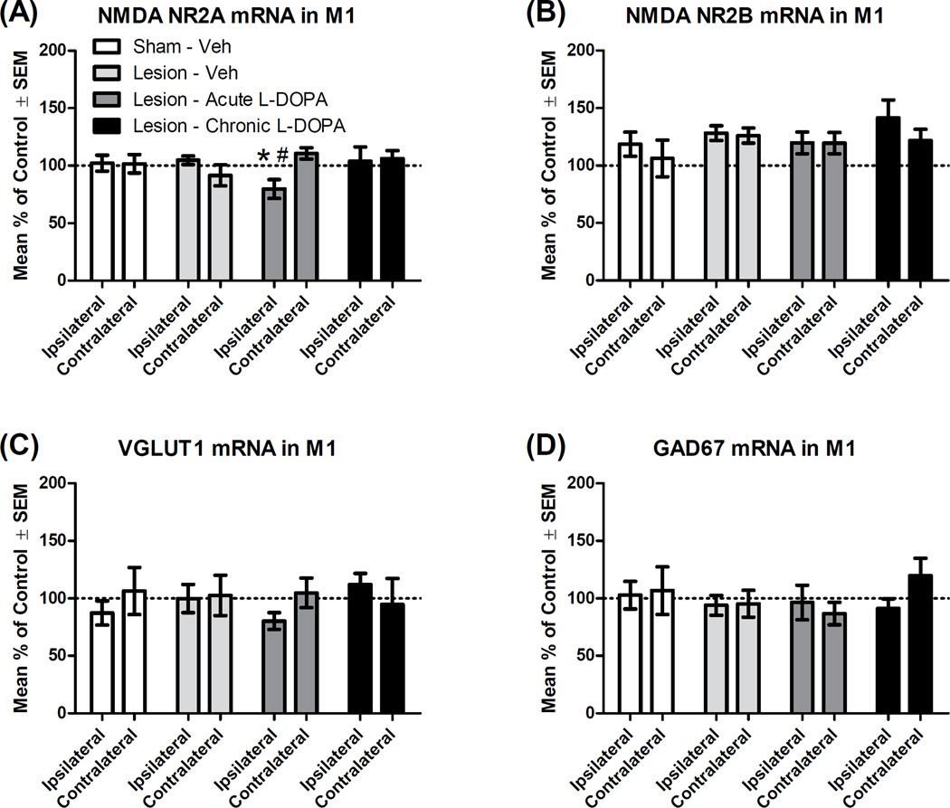
Effect of L-DOPA on mRNA transcription of the genes associated with glutamate and GABA signaling in the primary motor cortex (M1; n = 6–7 per group). Rats received a unilateral lesion with 6-hydroxydopamine (or sham). After recovery, rats were treated for 14 d with Vehicle (Veh), except rats in the “Chronic” condition, which received daily L-DOPA (6 mg/kg). The next day, rats in the “Acute” and “Chronic” groups received L-DOPA (6 mg/kg), while others received Veh. All rats were decapitated 65 min later and M1 tissue was analyzed via real-time polymerase chain reaction. Hemispheres are denoted as either ipsilateral or contralateral (to lesion or sham). Percent change in mRNA was normalized to control (“Sham - Veh [contralateral]”). (A) NMDA subunit NR2A mRNA. (B) NMDA subunit NR2B mRNA. (C) Vesicular glutamate transporter type I (VGLUT1) mRNA. (D) Glutamic acid decarboxylase 67 kDa (GAD67) mRNA. * p < .05 vs. Lesion - Veh (ipsilateral); # p < .05 vs. own contralateral hemisphere.

