Abstract
In recent years, Mongolia has experienced recurrent epizootics of equine influenza virus (EIV) among its 2·1 million horses and multiple incursions of highly pathogenic avian influenza (HPAI) virus via migrating birds. No human EIV or HPAI infections have been reported. In 2009, 439 adults in Mongolia were enrolled in a population‐based study of zoonotic influenza transmission. Enrollment sera were examined for serological evidence of infection with nine avian, three human, and one equine influenza virus strains. Seroreactivity was sparse among participants suggesting little human risk of zoonotic influenza infection.
Keywords: Agriculture, communicable diseases, emerging, influenza A virus, occupational exposure, seroepidemiologic studies, zoonoses
With vast and diverse terrains, harsh climates, and large populations of wild and domestic animals, Mongolia is home to numerous zoonotic disease problems. Influenza A viruses have caused considerable morbidity and deaths to horses and migrating birds. Approximately every 10 years, Mongolia experiences large epizootics of equine influenza virus (EIV) among its 2·1 million horses.1 Anecdotal reports suggest that Mongolian children become sick when horses are sick with EIV. Since first detections were noted in 2005, Mongolia's large migrating bird populations harbor both highly pathogenic and low pathogenic avian influenza viruses (AIV).2, 3, 4, 5 As both EIV6 and AIV are known to infect man, we sought to study Mongolians for evidence of EIV and AIV infection.
Methods
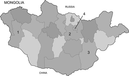
Four institutional review boards approved the study. Eligible participants (≥18 years old and self‐reporting no immunocompromising conditions) were recruited from seven soums (counties) within three aimags (provinces; Figure 1). In general, participants worked in livestock, agriculture, and mining industries. Consenting participants were interviewed at their home by study staff who completed enrollment forms and collected blood samples via venipuncture. Demographic information and medical history, including prior receipt of influenza vaccines and recent respiratory illness history, were assessed. Participants reported community, household, and occupational animal exposures. Reports of disease outbreaks in the participants' flocks/herds were also recorded. Data from the enrollment questionnaire were used to dichotomously classify domestic and wild animal exposures based upon a cut point of ≥5 cumulative hour/week during one's lifetime. Non‐animal‐exposed controls with no self‐reported household and occupational animal exposure were recruited from the capital city of Ulaanbaatar.
Figure 1.

Country map of Mongolia showing aimags (provinces) where animal‐exposed participants were enrolled (1‐Khovd, 2‐Tuv, and 3‐Dornogovi). Most non‐exposed participants were enrolled in the capital city (4‐Ulaanbaatar).
Laboratory methods
Whole blood specimens were transported using cold chain within 24 hours after collection to local field laboratories in Khovd and Dornogovi provinces and to the National Influenza Center of the National Center of Communicable Diseases, in Ulaanbaatar. Upon arrival, blood specimens were accessioned, and serum was separated, aliquoted, and frozen at −80°C. Frozen sera were transported on dry ice to the University of Florida for testing.
Influenza virus strains were selected based upon the hemagglutinin (HA) type for their best geographic and temporal proximity to the study population (Table 1). A microneutralization (MN) assay adapted from previous reports by Rowe et al.7, 8, 9, 10, 11 was used to detect antibodies against a panel of avian and avian‐like influenza A viruses, as well as a Mongolian H3N8 EIV.
Table 1.
Viruses used in serological studies. Unless otherwise indicated, serologic study was performed using the microneutralization assay
| Avian viruses | Human viruses |
|---|---|
| A/Migratory duck/Hong Kong MPS180/2003(H4N6) | A/Brisbane/59/2007(H1N1)a |
| A/Cygnus/Mongolia/3/2009(H5N1) | A/Mexico/4108/2009(H1N1)a |
| A/Nopi/Minnesota/2007/462960‐2(H5N2) | A/Brisbane/10/2007(H3N2)a |
| A/Teal/Hong Kong/w312/97(H6N1) | |
| A/Water fowl/Hong Kong/Mpb127/2005(H7N7) | Equine virus |
| A/Migratory duck/Hong Kong/MP2553/2004(H8N4) | A/Equine/Mongolia/01/2008(H3N8) |
| A/Hong Kong/1073/1999(H9N2)b | |
| A/Migratory duck/Hong Kong/MPD268/2007(H10N4) |
Virus studied with hemagglutination inhibition assay.
Virus of avian origin but isolated from a human.
Due to a low prevalence of elevated antibodies against the various avian and equine influenza viruses, rapidly waning titers,12 and the inability to determine when an infection might have occurred, a low threshold of antibody titer (≥1:10) was chosen as evidence of previous infection with a strain of EIV or AIV. Additionally, cross‐reactions from previous infection with human influenza viruses might confound the serology; therefore, potential confounding was controlled by also testing sera for antibodies against three human influenza viruses, using the hemagglutination inhibition (HI) assay as previously described.13, 14 A HI titer ≥1:40 was used as a cut point in including elevated antibody against human virus for multivariate modeling. MN and HI assay methods are reported as supplemental information.
Statistical methods
Analyses were performed using sas, version 9.2 (SAS Institute, Inc., Cary, NC, USA). Comparisons of participant demographics between the exposure groups were made using binary logistic regression. An exact conditional method was used for sparse data.
Results
Between January and June, 2009, 439 participants were enrolled: 358 (81·5%) reported household and/or occupational exposure to animals, and 81 (18·5%) were non‐animal‐exposed subjects. The cohort's median age was 39, and 52·2% were male (Table 2). Seventeen participants (4·0%) reported having previously received a seasonal influenza vaccine, with five receiving vaccines within a year of study enrollment.
Table 2.
Characteristics of study subjects upon enrollment, Mongolia, 2009. Unadjusted odds ratio for animal‐exposed participants compared with non‐animal‐exposed against control participants with binary logistic regression
| Variables | Total N | Exposed N (%) | Controls N (%) | Unadjusted OR (95% CI) |
|---|---|---|---|---|
| Age (years) | ||||
| 18–32 | 148 | 130 (36·3) | 22 (27·2) | 1·2 (0·6–2·2) |
| 33–45 | 139 | 105 (29·3) | 34 (42·0) | 0·6 (0·4–1·1) |
| 46–75 | 152 | 123 (34·4) | 25 (30·9) | Ref |
| Gender | ||||
| Female | 210 | 179 (50·0) | 31 (38·3) | 1·6 (1·0–2·6) |
| Male | 229 | 179 (50·0) | 50 (61·7) | Ref |
| Indoor water missinga | ||||
| Yes | 412 | 353 (99·2) | 59 (72·8) | 43·3 (12·4–232·8)b , c |
| No | 25 | 3 (0·8) | 22 (27·2) | Ref |
| Ever received vaccination for human influenzaa | ||||
| No | 411 | 346 (96·7) | 65 (92·9) | 2·2 (0·6–7·0)b |
| Yes | 17 | 12 (3·4) | 5 (7·1) | Ref |
| Heart disease, hypertension, or strokea | ||||
| Yes | 72 | 68 (19·0) | 4 (6·1) | 3·6 (1·3–10·3)c |
| No | 352 | 290 (81·0) | 62 (93·4) | Ref |
| Chronic breathing problemsa | ||||
| Yes | 43 | 38 (10·6) | 5 (7·3) | 1·5 (0·6–4·0) |
| No | 384 | 320 (89·4) | 64 (92·8) | Ref |
| Other chronic medical problemsa | ||||
| Yes | 65 | 58 (16·3) | 7 (10·0) | 1·8 (0·8–4·0) |
| No | 361 | 298 (83·7) | 63 (90·0) | Ref |
| Ever used tobacco productsa | ||||
| Yes | 115 | 100 (27·9) | 15 (22·1) | 1·4 (0·7–2·5) |
| No | 311 | 258 (72·1) | 53 (77·9) | Ref |
| Developed a respiratory illness in the last 12 monthsa | ||||
| Yes | 190 | 159 (44·4) | 31 (44·9) | 1·0 (0·6–1·6) |
| No | 237 | 199 (55·6) | 38 (55·1) | Ref |
Covariate has some missing data.
Fisher's exact method used.
Statistically significant data (P <0·05).
For the 358 animal‐exposed participants, lifetime animal exposure included the following: horses (76·0%), camels (39·4%), goats (55·0%), sheep (49·4%), cattle (48·3%), pigs (15·4%), and domestic poultry (9·5%). The majority (91%) of the exposed participants reported their animal exposures to have occurred recently (since 2003). Seventy‐five participants (17·1%) reported recent disease outbreaks in their horses or camels.
Elevated MN titers against EIV were sparse. Enrollment sera from four participants had elevated antibody titers against A/Equine/Mongolia/01/2008(H3N8; Table 3); of which 3 (75%) had a titer of 1:10 and reported recent occupational exposure to horses. One participant was a 27‐year‐old male who had been a horse and camel herder for the last 10 years. The other two participants were members of the same family household (47‐year‐old male and 46‐year‐old female), and both had been horse herders for the last 18 years. All three participants reported recent disease outbreaks in their equine herds and also farmed cattle, sheep, and goats. While none reported ever receiving a seasonal influenza vaccine, all three had elevated antibody titers (≥1:40) against the A/Brisbane/10/2007(H3N2) human influenza virus.
Table 3.
Distribution of elevated MN titers against equine and avian influenza viruses and HI titers against human influenza among seropositive study participants in Mongolia
| Participant | A/Equine/Mongolia /01/2008(H3N8) | A/Cygnus/Mongolia/3/2009(H5N1) | A/Teal/Hong Kong/ w312/97(H6N1) | A/Hong Kong/1073/ 1999(H9N2) | A/Brisbane/10/ 2007(H3N2) |
|---|---|---|---|---|---|
| 27‐year‐old male | 10 | <10 | <10 | <10 | 320 |
| 47‐year‐old male | 10 | <10 | <10 | <10 | 40 |
| 46‐year‐old female | 10 | <10 | <10 | <10 | 40 |
| 32‐year‐old female | 20 | <10 | 10 | <10 | 160 |
| 69‐year‐old male | <10 | 10 | <10 | <10 | 80 |
| 52‐year‐old male | <10 | <10 | <10 | 10 | 80 |
| 63‐year‐old male | <10 | <10 | <10 | 10 | <10 |
| 23‐year‐old male | <10 | <10 | <10 | 10 | <10 |
MN, Microneutralization assay; HI, Hemagglutination inhibition assay.
The fourth EqH3N8 seropositive (1:20) participant was a 32‐year‐old female who worked as a national health official. While she reported working with cows for the last 17 years and pigs for the last 2 years, she reported no exposure to horses or camels. Furthermore, this participant also had an elevated titer (1:10) against the A/Teal/Hong Kong/w312/97(H6N1) AIV; she was the only seropositive subject. She reported no exposure to poultry or wild birds. She did report receiving a seasonal influenza vaccine in 2006 and was seropositive (1:160) against A/Brisbane/10/2007(H3N2). She reported experiencing 1–2 episodes of a respiratory illness (fever and cough or sore throat) in the 30 days prior to her enrollment and reported 3–5 episodes of respiratory illness among household family members (six adults and two children) within the same time period.
Seroreactivity against the remaining AIV strains was also very sparse. Three participants had elevated titers (1:10) against A/Hong Kong/1073/1999(H9N2). One participant had a prior 4‐year history of household exposure to chickens, but none cited recent poultry or wild bird exposure. Only one AvH9N2 seropositive participant was also seropositive against A/Brisbane/10/2007(H3N2), and all three reported never receiving an influenza vaccination.
One participant, a 69‐year‐old male, had an elevated antibody titer (1:10) against A/Cygnus/Mongolia/3/2009(H5N1) HPAI virus. This participant was a herder of horses, camels, sheep, goats, and cattle, but he reported no poultry or wild bird exposure. He had never received an influenza vaccine, but he did have an elevated antibody titer (1:80) against A/Brisbane/10/2007(H3N2).
In examining serological reactivity against human influenza virus strains, 287 participants (65·8%) had elevated titers (≥1:40) against A/Brisbane/10/2007(H3N2). Forty‐two participants (9·6%) had elevated titers against A/Brisbane/59/2007(H1N1). Seven participants (1·6%) had elevated titers against the 2009 pandemic influenza virus, A/Mexico/4108/2009(H1N1). Six of the seven were enrolled in May–June 2009, and one participant was enrolled in January 2009.
Discussion
A 1966 report by US National Institutes of Health researchers clearly documented experimental EIV infections in humans.6 While 4 (12%) of 33 inoculated subjects experienced clinical illness, 20 subjects (61%) had evidence of infection by an observed antibody titer increase. As Mongolia has large populations of horses that roam its vast plains, comingling with aquatic birds that occasionally introduce HPAI, it seems prudent to study Mongolia's rural people for evidence of zoonotic influenza virus infection.
Our study data, and human influenza surveillance data in Mongolia, suggest that if AIV or EIV infections occur in Mongolians, they likely occur infrequently. It seems possible that the low MN titers detected might reflect EIV infections during the 2007–2008 H3N8 equine epizootic that had waned12 by human blood draw in 2009. However, it seems equally plausible that the low tiers may be explained by cross‐reactivity from previous human influenza A virus infection, especially that due to human H3N2 virus.
The sparse serological evidence for human EIV and AIV infections in this study population suggests that neither EIV nor AIV are currently posing a significant human health risk to rural Mongolians. Nevertheless, Mongolia with its large populations of horses and migrating birds seems an important place to continue to conduct surveillance for the emergence of novel influenza strains.
Authorship addendum
Nyamdavaa Khurelbaatar involved in the study design, Mongolia principal investigator, Mongolian study coordination. Whitney S. Krueger involved in IRB approval, study coordination, data management, statistical analysis, and manuscript preparation. Gary L. Heil involved in the laboratory assay design and laboratory analyses. Badarchiin Darmaa managed enrollment and data collection at one site in Mongolia. Daramragchaa Ulziimaa involved in the IRB approval, managed enrollment, and data collection at one site in Mongolia. Damdindorj Tserennorov managed enrollment and data collection at one site in Mongolia. Ariungerel Baterdene involved in the data management and entry in Mongolia. Benjamin D. Anderson prepared data summaries and manuscript. Gregory C. Gray involved in the study design, overall principal investigator, data interpretation, and manuscript preparation.
Supporting information
Data S1.Influenza serological assay methods.
Acknowledgements
This research was supported by multiple grants from the US Department of Defense Armed Forces Health Surveillance Center's Global Emerging Infections Surveillance and Response Program (Dr. Gray, principal investigator) and an ARRA supplement grant from the National Institute of Allergy and Infectious Diseases (R01 AI068803‐Dr. Gray). The study was made possible through collaborations between the Global Pathogens Laboratory, University of Florida Emerging Pathogens Institute, USA; Center for Emerging Infectious Diseases, University of Iowa, College of Public Health, USA; and the authors' various organizations in Mongolia.
We thank Sharon F. Setterquist, Mark G. Lebeck, Troy McCarthy, and Dwight Ferguson for their technical contributions to this research while working at the University of Iowa's Center for Emerging Infectious Diseases and Danielle Peters, John Friary, Clint McDaniel, Robin Derby, and John Burks of the Global Pathogens Laboratory for their technical contributions. We especially want to thank the US Centers for Disease Control and Prevention, Dr. Malik Peiris (Chair, Department of Microbiology, University of Hong Kong, Hong Kong China), Dr. Richard Webby (St Jude Children's Research Hospital, Memphis, Tennessee), and Dr. Dennis Senne (National Veterinary Services Laboratories, Ames, Iowa) for sharing their viruses and antisera.
Khurelbaatar et al. (2013) Sparse evidence for equine or avian influenza virus infections among Mongolian adults with animal exposures. Influenza and Other Respiratory Viruses 7(6), 1246–1250.
References
- 1. Yondon M, Heil GL, Burks JP et al Isolation and characterization of H3N8 equine influenza A virus associated with the 2011 epizootic in Mongolia. Influenza Other Respir Viruses 2013. [DOI] [PMC free article] [PubMed] [Google Scholar]
- 2. Sakoda Y, Sugar S, Batchluun D et al Characterization of H5N1 highly pathogenic avian influenza virus strains isolated from migratory waterfowl in Mongolia on the way back from the southern Asia to their northern territory. Virology 2010; 406:88–94. [DOI] [PubMed] [Google Scholar]
- 3. Kang HM, Kim MC, Choi JG et al Genetic analyses of avian influenza viruses in Mongolia, 2007 to 2009, and their relationships with Korean isolates from domestic poultry and wild birds. Poult Sci 2011; 90:2229–2242. [DOI] [PubMed] [Google Scholar]
- 4. Ishtiaq F, Gilbert M, Brown J et al Antibodies to influenza A virus in wild birds across Mongolia, 2006‐2009. J Wildl Dis 2012; 48:768–775. [DOI] [PMC free article] [PubMed] [Google Scholar]
- 5. Gilbert M, Jambal L, Karesh WB et al Highly pathogenic avian influenza virus among wild birds in Mongolia. PLoS ONE 2012; 7:e44097. [DOI] [PMC free article] [PubMed] [Google Scholar]
- 6. Alford RH, Kasel JA, Lehrich JR, Knight V. Human responses to experimental infection with influenza A/Equi 2 virus. Am J Epidemiol 1967; 86:185–192. [DOI] [PubMed] [Google Scholar]
- 7. Rowe T, Abernathy RA, Hu‐Primmer J et al Detection of antibody to avian influenza A (H5N1) virus in human serum by using a combination of serologic assays. J Clin Microbiol 1999; 37:937–943. [DOI] [PMC free article] [PubMed] [Google Scholar]
- 8. Gill JS, Webby R, Gilchrist MJ, Gray GC. Avian influenza among waterfowl hunters and wildlife professionals. Emerg Infect Dis 2006; 12:1284–1286. [DOI] [PMC free article] [PubMed] [Google Scholar]
- 9. Myers KP, Setterquist SF, Capuano AW, Gray GC. Infection due to 3 avian influenza subtypes in United States veterinarians. Clin Infect Dis 2007; 45:4–9. [DOI] [PMC free article] [PubMed] [Google Scholar]
- 10. Gray GC, McCarthy T, Capuano AW, Setterquist SF, Alavanja MC, Lynch CF. Evidence for avian influenza A infections among Iowa's agricultural workers. Influenza Other Respi Viruses 2008; 2:61–69. [DOI] [PMC free article] [PubMed] [Google Scholar]
- 11. Khuntirat BP, Yoon IK, Blair PJ et al Evidence for subclinical avian influenza virus infections among rural Thai villagers. Clin Infect Dis 2011; 53:e107–e116. [DOI] [PMC free article] [PubMed] [Google Scholar]
- 12. Buchy P, Vong S, Chu S et al Kinetics of neutralizing antibodies in patients naturally infected by H5N1 virus. PLoS ONE 2010; 5:e10864. [DOI] [PMC free article] [PubMed] [Google Scholar]
- 13. Myers KP, Olsen CW, Setterquist SF et al Are swine workers in the United States at increased risk of infection with zoonotic influenza virus? Clin Infect Dis 2006; 42:14–20. [DOI] [PMC free article] [PubMed] [Google Scholar]
- 14. Kayali G, Setterquist SF, Capuano AW, Myers KP, Gill JS, Gray GC. Testing human sera for antibodies against avian influenza viruses: horse RBC hemagglutination inhibition vs. microneutralization assays. J Clin Virol 2008; 43:73–78. [DOI] [PMC free article] [PubMed] [Google Scholar]
Associated Data
This section collects any data citations, data availability statements, or supplementary materials included in this article.
Supplementary Materials
Data S1.Influenza serological assay methods.


