Abstract
Please cite this paper as: Khandaker et al. (2012) Comparing the use of, and considering the need for, lumbar puncture in children with influenza or other respiratory virus infections. Influenza and Other Respiratory Viruses DOI:10.1111/irv.12039.
Background The clinical presentation of influenza in infancy may be similar to serious bacterial infection and be investigated with invasive procedures like lumbar puncture (LP), despite very limited evidence that influenza occurs concomitantly with bacterial meningitis, perhaps because the diagnosis of influenza is very often not established when the decision to perform LP is being considered.
Methods A retrospective medical record review was undertaken in all children presenting to the Children’s Hospital at Westmead, Sydney, Australia, in one winter season with laboratory‐confirmed influenza or other respiratory virus infections (ORVIs) but excluding respiratory syncytial virus, to compare the use of, and reflect on the need for, the performance of invasive diagnostic procedures, principally LP, but also blood culture, in influenza and non‐influenza cases. We also determined the rate of concomitant bacterial meningitis or bacteraemia.
Findings Of 294 children, 51% had laboratory‐confirmed influenza and 49% had ORVIs such as parainfluenza viruses (34%) and adenoviruses (15%). Of those with influenza, 18% had a LP and 71% had a blood culture performed compared with 6·3% and 55·5% in the ORVI group (for both P < 0·01). In multivariate analysis, diagnosis of influenza was a strong independent predictor of both LP (P = 0·02) and blood culture (P = 0·05) being performed, and, in comparison with ORVIs, influenza cases were almost three times more likely to have a LP performed on presentation to hospital. One child with influenza (0·9%) had bacteraemia and none had meningitis.
Interpretation Children with influenza were more likely to undergo LP on presentation to hospital compared with those presenting with ORVIs. If influenza is confirmed on admission by near‐patient testing, clinicians may be reassured and less inclined to perform LP, although if meningitis is clinically suspected, the clinician should act accordingly. We found that the risk of bacterial meningitis and bacteraemia was very low in hospitalised children with influenza and ORVIs. A systematic review should be performed to investigate this across a large number of settings.
Keywords: Bacterial meningitis, children, influenza, lumbar puncture, respiratory viral infection
Introduction
Influenza in children poses a significant burden to healthcare services and often requires escalated medical care. 1 , 2 A large proportion of the children with influenza admitted to hospital receive antibiotics for suspected bacterial infection and undergo multiple invasive procedures. For instance, during the winter of 2006, among children aged <5 years admitted to the Children’s Hospital at Westmead (CHW) with laboratory‐confirmed influenza, 68% received intravenous antibiotics and 23% underwent lumbar puncture (LP). 3 The following year, 2007, similar proportions of hospitalised children with confirmed influenza were given antibiotics (67%) and underwent LP (23%). 1
Pneumonia and secondary bacterial infections are recognised to follow influenza infection in children. 4 However, studies have shown that concomitant serious bacterial infections (SBI), especially meningitis, occur rarely in infants during the course of infection with respiratory viruses, including influenza and respiratory syncytial virus (RSV). 5 , 6 , 7 , 8 Lumbar puncture is now not specifically recommended in infants presenting with RSV bronchiolitis, the policy having evolved in recent years. 9 , 10 Although comparisons have been made between the clinical characteristics of influenza and RSV, 11 there are few studies that compare the use of invasive diagnostic procedures, especially LP, in children with influenza with respiratory viral infections other than RSV (ORVIs); the rates of concomitant bacterial infection are also uncommonly reported. To this end, we have compared the clinical characteristics, the rates of invasive diagnostic procedures, particularly LP, and the incidence of bacterial meningitis and bacteraemia in children presenting with influenza or with ORVIs at the CHW, a tertiary paediatric hospital in Greater Sydney, Australia.
Methods
Study population and setting
The study was approved by the CHW Human Research Ethics Committee. Subjects for this study were identified by searching the database of the department of virology at the CHW for positive test results with influenza A, influenza B, parainfluenza viruses 1, 2, 3 and adenoviruses from nasopharyngeal aspirates and/or nose/throat swabs collected between 1 January and 31 December 2007. This year was chosen as it was a peak influenza year. 12 Respiratory syncytial virus infections were excluded from the study as it is already established that the rate of RSV and concomitant bacteraemia and meningitis is very low in children. 8 , 11
Medical records for all children thus identified were reviewed to confirm that they had respiratory illness; additional data on clinical presentation, investigations performed and the course of illness were collected for analysis.
Baseline data on the influenza burden at our hospital for the same year have been published elsewhere. 1 Our primary outcome measures for this study were the ‘need for’ (performance of) invasive diagnostic procedures, primarily LP but also blood culture, in children with influenza and ORVIs and whether bacterial meningitis or bacteraemia were concomitantly present.
Bacterial meningitis and bacteraemia were defined as the growth of a single known bacterial pathogen in the respective specimen cultures from normally sterile sites, cerebrospinal fluid or blood. Urinary tract infection (UTI) was defined as the growth of a single pathogenic organism with 108 colony‐forming units/l of urine collected either by supra‐pubic aspiration, in‐and‐out catheter or a clean catch. UTI was considered as coincidental infection (bacterial infection with a definite non‐pulmonary focus). A culture was considered contaminated if there was a growth of bacteria that are usually considered contaminants (e.g. coagulase‐negative staphylococcus) or if >1 bacterial organism was isolated.
Laboratory methods
Children presenting to CHW with respiratory symptoms underwent the collection of a nasopharyngeal aspirate or a nose/throat swab according to the clinical judgement of the treating paediatrician; it was transported to the virology laboratory in viral transport medium at 4°C. Specimens were then batched and tested for respiratory viruses using Simulfluor Respiratory Screen (Chemicon International, Temecula, CA, USA). Positive samples had influenza A confirmed with antibodies (Imagen; DakoCytomation, Ely, UK) using direct fluorescent microscopy. Samples that were negative for influenza A by direct immunofluorescence were cultured on R‐Mix cells (Diagnostic Hybrids, Athens, OH, USA) at 37°C for 3 days and then labelled with antibodies to influenza A and influenza B (Imagen; DakoCytomation) using direct fluorescent microscopy. A laboratory diagnosis was defined by identification of a virus either by direct fluorescent antibody (DFA) or by culture fluorescence.
Statistical analysis
Data analysis was carried out using the Statistical Package for the Social Sciences (IBM SPSS Statistics 19 Inc, Somers, NY, USA). Comparisons were made between groups on several variables (e.g. type of respiratory viral infection, clinical presentation and presence of any SBI) using the chi‐square test (with Yeats correction) or Fisher’s exact test and multinominal logistic regression analysis where appropriate. Tests for normality were performed (Kolmogorov–Smirnov test), and when data were normally distributed, the differences in means between groups were compared by the independent samples t‐test. The relative risk and corresponding 95% confidence interval (CI) were also calculated. A P‐value of ≤0·05 was considered as significant.
Results
Demography and clinical presentation
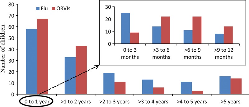
In 2007, influenza and ORVIs were laboratory‐confirmed in 294 children who are presented to the CHW. Their median age was 14·5 months (range 13 days–19 years); 150 (51%) children had influenza (144 influenza A and six influenza B) and 144 (49%) had ORVIs (89 parainfluenza 3, 44 adenovirus, six parainfluenza 1 and five parainfluenza 2). The demographics and the presenting symptoms of these children are presented in Table 1. Of the influenza cases, 25 (16·6%) were aged <3 months compared with nine of 144 (6·2%) in the ORVI group (P < 0·01). The distribution of respiratory viruses by age group is shown in Figure 1. Mean body temperature on presentation was significantly higher in the influenza group compared with that in the ORVI group (38·6 versus 38·0°C, P < 0·01) as was the mean heart rate (160 versus 152, P < 0·01). There were no significant differences in terms of gender, oxygen saturation and respiratory rate on presentation between the groups.
Table 1.
Comparison of demographics and presenting symptoms in both groups
| Variables | ORVIs | Influenza | Risk ratio (95% CI) | P‐value |
|---|---|---|---|---|
| Median age in months (range) | 13 (0·5–18) | 18 (0·5–19) | – | 0·13 |
| Age ≤3 months, n (%) | 9 (6·2) | 25 (16·6) | 0·8 (0·8–0·9) | <0·01 |
| Age >3–≤12 months, n (%) | 58 (40·3) | 33 (22·0) | 1·3 (1·1–1·5) | <0·01 |
| Age >12–≤60 months, n (%) | 63 (43·7) | 75 (50·0) | 0·8 (0·7–1·1) | 0·29 |
| Age >60 months, n (%) | 14 (9·7) | 17 (11·3) | 0·9 (0·9–1·0) | 0·71 |
| Male:female | 79:65 | 81:69 | 1·0 (0·7–1·3) | 0·91 |
| Mean temperature (°C) | 38·0 | 38·6 | – | <0·01 |
| Seizure on presentation, n (%) | 8 (5·5) | 12 (8) | 0·97 (0·9–1) | 0·54 |
| Lowest SpO2 in emergency presentation (mean) | 96·2 | 96·3 | – | 0·59 |
| Supplemental O2 requirements, n (%) | 33 (22·9) | 20 (13·3) | 1·1 (1·0–1·2) | 0·03 |
| Highest HR in emergency presentation (mean) | 152·4 | 159·7 | – | 0·02 |
| Highest RR in presentation (mean) | 39·5 | 37·7 | – | 0·24 |
SpO2, pulse oximeter oxygen saturation; HR, heart rate; ORVIs, other respiratory virus infections; RR, respiratory rate.
Figure 1.

Distribution of respiratory viruses by age groups.
Investigations on presentation and SBI
Of the 150 influenza cases, 27 (18%) had a LP as part of assessment compared with nine of 144 (6·3%) in the other group (P < 0·01); 71% (107/150) of those with influenza had a blood culture performed compared with 56% (80/144) in the ORVI group (P < 0·01; Table 2).
Table 2.
Comparison of investigations, antibiotic use and hospitalisation rates in both groups
| Variables | ORVIs (n = 144) | Influenza (n = 150) | Risk ratio (95% CI) | P‐value |
|---|---|---|---|---|
| LP, n (%) | 9 (6·2) | 27 (18·0) | 0·8 (0·8–0·9) | <0·01 |
| Blood culture, n (%) | 80 (55·5) | 107 (71·3) | 0·6 (0·4–0·8) | <0·01 |
| Urine culture, n (%) | 47 (32·6) | 66 (44·0) | 0·8 (0·6–0·9) | 0·05 |
| Received IV antibiotics, n (%) | 65 (45·1) | 70 (46·6) | 0·9 (0·7–1·2) | 0·81 |
| Hospitalised, n (%) | 118 (81·9) | 122 (81·3) | 1·0 (0·6–1·6) | 0·89 |
| ICU admission, n (%) | 6 (4·1) | 12 (8·0) | 0·9 (0·9–1·0) | 0·22 |
| Mean LOS hospital days (SD) | 4·9 (±8·4) | 3·7 (±5·1) | – | 0·04 |
LP, lumbar puncture; IV, intravenous; ICU, intensive care unit; LOS, length of stay; ORVIs, other respiratory virus infections; SD, standard deviation.
There were no cases of bacterial meningitis in either group. Only one child (0·9%) with laboratory‐confirmed influenza had bacteraemia (Enterobacter cloacae), and none in the ORVIs had bacteraemia (P = 0·9). Among those tested, 7 (10·6%) children with influenza had UTI diagnosed and another 7 (14·9%) children with ORVIs had UTI (P = 0·57; Table 3).
Table 3.
Serious bacterial infections and coincidental bacteraemia in cases of influenza or other respiratory viruses
| Variable | ORVIs (n = 144) | Influenza (n = 150) | ||
|---|---|---|---|---|
| n/N | % (95% CI) | n/N | % (95% CI) | |
| Bacteraemia* | 0/80 | 0 | 1/107 | 0·9 (0·1–5) |
| Meningitis | 0/9 | 0 | 0/27 | 0 |
| UTI | 7/47 | 14·9 (7·4–27·6) | 7/66 | 10·6 (5–20) |
N, total number of children needing the procedure; n, number of culture‐positive children; ORVIs, other respiratory virus infections.
*Organism isolated: Enterobacter cloacae with influenza A.
Predictors of invasive procedures on presentation
In the multivariate analysis, we explored the likelihood of an invasive procedure based on several predictors including the presenting symptoms, age and respiratory viral diagnosis. A diagnosis of influenza (P = 0·02) and age ≤3 months (P < 0·01) were the only significant independent predictors of a LP, whereas a high temperature (≥39·5°C) or having a febrile convulsion was not predictive.
Independent predictors of having a blood culture performed on presentation were as follows: a diagnosis of influenza (P = 0·05) and temperature of ≥39·5°C (P < 0·01), whereas age ≤3 months did not reach the level of significance (P = 0·06). In comparing influenza with ORVIs, influenza‐positive cases were almost three times more likely (OR 2·8, 95% CI 1·4–5·9) to have had a LP performed and 1·3 times (95% CI 1·0–1·5) more likely to have a blood culture on presentation to hospital.
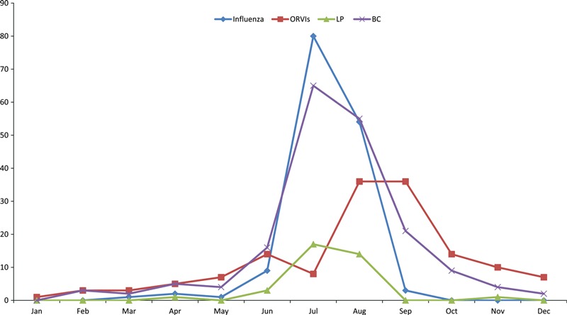
Most influenza cases were presented during July and August, whereas the ORVI cases were predominant in an overlapping period between August and October. Figure 2 shows the seasonal distribution of influenza and ORVIs and the rate of invasive procedures according to the date of presentation. A distinct seasonal pattern of the rate of performance of invasive procedures corresponding to the peak of influenza is apparent.
Figure 2.

Number of influenza and other respiratory viral infections and invasive procedures according to the month of presentation.
Discussion
Our study suggests that, whereas the risk of SBI (bacterial meningitis or bacteraemia) was very low in children with either influenza or ORVIs, the rate of performing invasive procedures was significantly higher in children with influenza compared with those with ORVIs. Furthermore, whereas clinicians are more inclined to perform LP if, for example, a child is younger, has had a febrile convulsion or has a high fever, multivariate analysis showed that the only two independent predictors of LP were a diagnosis of influenza and age <3 months. Other studies have shown that the risk of SBI is very low in children with respiratory viral infection, but have not addressed whether influenza is a strong predictor of LP being performed. 6 , 7 , 13 For example, a prospective multicentre cross‐sectional study, conducted in the USA over 3 years from 1998 to 2001, showed that the risk of SBI in young infants was significantly lower in children with influenza compared with those without influenza. 6 Another retrospective study, conducted over four consecutive influenza seasons in the USA, demonstrated that febrile children with influenza had a lower prevalence of bacteraemia, UTI, consolidative pneumonia or any SBI compared with those without influenza. 7 The rate of SBI in young infants with RSV was also lower compared with those without RSV. 8
Despite a very low prevalence of SBI, this study demonstrates that children with influenza had significantly higher utilisation of invasive diagnostic procedures, particularly LP. This can partly be explained by the fact that children with influenza presented appearing more unwell as manifested by their higher mean temperature (38·6 versus 38°C, P < 0·01) and mean heart rates (160 versus 152, P < 0·01). This suggests that the children with influenza look clinically more unwell, and their presentation mimics SBI, including meningitis, and so full septic workups, including LP, are performed on presentation. This is consistent with other studies conducted in our hospital in a previous winter and one conducted elsewhere, both showing that about one‐fourth of the children with influenza underwent a LP. 3 , 7
A seasonal pattern for performing blood cultures and LP was observed in this study, the peak of which corresponded with that of influenza infections (Figure 2). There were, however, no cases of meningitis associated with influenza, although admittedly the numbers in our study are not large, and in our view, a systematic review of influenza and serious concomitant infection is indicated to further address this question. Our study is important in finding that the risk of SBI in children with influenza or ORVIs was low during a busy winter season, and yet, children with influenza undergo significantly higher number of invasive diagnostic procedures on presentation that might be reduced by improved ascertainment of influenza directly on admission. There are high rates of immunisation against pneumococcal, Hib and meningococcal C disease in Western Sydney, which explains why bacterial meningitis is now rare. 14
Our study shows that about 17% of the children in the influenza group were younger than 3 months of age, while only 6% in the ORVI group were <3 months (P < 0·01). However, overall, about half of the children in either group were aged <1 year (Figure 1). This is consistent with findings from other studies. 15 , 16 , 17 Many of the influenza cases in our study were too young to be immunised against influenza. Considering the high burden of influenza in young children and the frequent use of invasive diagnostic procedures, it is important to consider how to rapidly screen febrile children with respiratory symptoms during the influenza season, for example, with a rapid diagnostic test. Indeed, a rapid diagnostic test (QuickVue Influenza A + B; Quidel Corporation, San Diego, CA, USA) performed on nasopharyngeal aspirates evaluated at our hospital during the 2009 pandemic season was found to be both sensitive and specific (84% and 98·4%, respectively) compared with nucleic acid testing. 18 Studies conducted elsewhere also have shown good sensitivity and specificity, 19 but some rapid diagnostic tests perform better than others and their sensitivities, particularly, may vary according to the swab type, influenza virus strain and patients’ age. 20 As children with confirmed influenza were very unlikely to suffer from meningitis, rapid screening for influenza during the influenza season may greatly reduce the need for LP; this may have economic benefits too. A study from Spain has shown that patients positive for influenza, using a rapid diagnostic test in the emergency department, had a significantly reduced rate of hospitalisation compared with those who were negative. 21 A US study found that using influenza status alone, as a screening test for infants with SBI (with a positive influenza test being indicative of low risk for SBI), resulted in a negative predictive value of 97·5% (95% CI 93·0–99·2%). 6 From a clinical perspective, if the admitting consultant paediatrician were to review in person all cases being considered for LP, as occurs, for example, in Finland (T. Heikkinen, personal communication), the proportion undergoing the procedure may well be reduced. Notwithstanding, if meningitis is clinically suspected, the clinician should act accordingly, irrespective of the outcome of influenza diagnostic tests.
Other respiratory virus infections were more likely to present between 3 and 12 months of age, to require more oxygen on admission and a longer stay in hospital: this may relate to a greater proportion having bronchiolitis or viral pneumonitis than the influenza cases did. Most of the influenza cases (89·3%) in our study presented during July and August, the southern hemisphere winter influenza season. Clinicians should consider a diagnosis of influenza in febrile unwell children presenting during peak months of influenza activity.
There are some limitations in our study. Firstly, DFA and virus culture were employed for diagnosing respiratory viruses as that was the practice in 2007 at our hospital. Owing to lower sensitivity of these tests compared with nucleic acid tests, 22 some respiratory viral infections may have been missed; however, that being the case, it may help to better explain the apparent close association between the peaks of influenza diagnosis and performance of LP. Direct fluorescent antibody and other rapid antigen tests may provide more rapid turnaround times than nucleic acid testing, thus allowing LP to be avoided. Another limitation is that other respiratory viruses like rhinovirus, coronavirus, enterovirus, human metapneumovirus (hMPV) and human bocavirus were not looked for.
A systematic review addressing the risk of concomitant influenza and bacterial meningitis should be performed. Rapid screening of febrile infants with respiratory symptoms during periods of known influenza activity may help to avert the use of LP, particularly if these cases are clinically reviewed by the senior admitting consultant, who, if still suspicious of concomitant bacterial meningitis, can ensure the LP is performed.
Conflict of interests
RB received financial support from pharmaceutical companies CSL, Sanofi, GSK, Novartis, Roche and Wyeth to conduct research and attend and present at scientific meetings. Any funding received is directed to an NCIRS research account at The Children’s Hospital at Westmead and is not personally accepted by Professor Booy.
Author contributions
Dr. Gulam Khandaker retrieved and analysed the data and wrote the manuscript; Dr. Leon Heron supervised data analysis and manuscript writing; Dr. Harunor Rashid analysed data and wrote the discussion section; Dr. Jean Li‐Kim‐Moy contributed to all sections of the manuscript; Dr. David Lester‐Smith and Dr. Mary McCaskill provided data and contributed to the manuscript; Professor Alison Kesson performed laboratory testing and wrote the methods; Professor Cheryl Jones and Professor Elizabeth J Elliott provided data and revised the manuscript; Dr. Yvonne Zurynski retrieved data and contributed to the manuscript; Professor Dominic E Dwyer performed laboratory testing and revised the manuscript; Professor Robert Booy as the lead investigator was responsible for conception and overseeing the study and revising the report.
Institution where the work has been carried out: The Children’s Hospital at Westmead, Sydney, NSW 2145, Australia.
References
- 1. Lester‐Smith D, Zurynski YA, Booy R, Festa MS, Kesson AM, Elliott EJ. The burden of childhood influenza in a tertiary paediatric setting. Commun Dis Intell 2009; 33:209–215. [PubMed] [Google Scholar]
- 2. Milne BG, Williams S, May ML, Kesson AM, Gillis J, Burgess MA. Influenza A associated morbidity and mortality in a paediatric intensive care unit. Commun Dis Intell 2004; 28:504–509. [PubMed] [Google Scholar]
- 3. Iskander M, Kesson A, Dwyer D et al. The burden of influenza in children under 5 years admitted to the Children’s Hospital at Westmead in the winter of 2006. J Paediatr Child Health 2009; 45:698–703. [DOI] [PubMed] [Google Scholar]
- 4. Zurynski YA, Lester‐Smith D, Festa MS, Kesson AM, Booy R, Elliott EJ. Enhanced surveillance for serious complications of influenza in children: role of the Australian paediatric surveillance unit. Commun Dis Intell 2008; 32:71–76. [PubMed] [Google Scholar]
- 5. Hsiao AL, Chen L, Baker MD. Incidence and predictors of serious bacterial infections among 57‐ to 180‐day‐old infants. Pediatrics 2006; 117:1695–1701. [DOI] [PubMed] [Google Scholar]
- 6. Krief WI, Levine DA, Platt SL et al. Influenza virus infection and the risk of serious bacterial infections in young febrile infants. Pediatrics 2009; 124:30–39. [DOI] [PubMed] [Google Scholar]
- 7. Smitherman HF, Caviness AC, Macias CG. Retrospective review of serious bacterial infections in infants who are 0 to 36 months of age and have influenza A infection. Pediatrics 2005; 115:710–718. [DOI] [PubMed] [Google Scholar]
- 8. Levine DA, Platt SL, Dayan PS et al. Risk of serious bacterial infection in young febrile infants with respiratory syncytial virus infections. Pediatrics 2004; 113:1728–1734. [DOI] [PubMed] [Google Scholar]
- 9. Zorc JJ, Hall CB. Bronchiolitis: recent evidence on diagnosis and management. Pediatrics 2010; 125:342–349. [DOI] [PubMed] [Google Scholar]
- 10. Liebelt EL, Qi K, Harvey K. Diagnostic testing for serious bacterial infections in infants aged 90 days or younger with bronchiolitis. Arch Pediatr Adolesc Med 1999; 153:525–530. [DOI] [PubMed] [Google Scholar]
- 11. Meury S, Zeller S, Heininger U. Comparison of clinical characteristics of influenza and respiratory syncytial virus infection in hospitalised children and adolescents. Eur J Pediatr 2004; 163:359–363. [DOI] [PubMed] [Google Scholar]
- 12. Khandaker G, Lester‐Smith D, Zurynski Y, Elliott EJ, Booy R. Pandemic (H1N1) 2009 and seasonal influenza A (H3N2) in children’s hospital, Australia. Emerg Infect Dis 2011; 17:1960–1962. [DOI] [PMC free article] [PubMed] [Google Scholar]
- 13. Byington CL, Enriquez FR, Hoff C et al. Serious bacterial infections in febrile infants 1 to 90 days old with and without viral infections. Pediatrics 2004; 113:1662–1666. [DOI] [PubMed] [Google Scholar]
- 14. Hull B, Dey A, Campbell‐Lloyd S, Menzies RI, McIntyre PB. NSW annual immunisation coverage report, 2010. N S W Public Health Bull 2011; 22:179–195. [DOI] [PubMed] [Google Scholar]
- 15. Moore DL, Vaudry W, Scheifele DW et al. Surveillance for influenza admissions among children hospitalized in Canadian immunization monitoring program active centers, 2003–2004. Pediatrics 2006; 118:e610–e619. [DOI] [PubMed] [Google Scholar]
- 16. Poehling KA, Edwards KM, Weinberg GA et al. The underrecognized burden of influenza in young children. N Engl J Med 2006; 355:31–40. [DOI] [PubMed] [Google Scholar]
- 17. Iskander M, Booy R, Lambert S. The burden of influenza in children. Curr Opin Infect Dis 2007; 20:259–263. [DOI] [PubMed] [Google Scholar]
- 18. Andresen DN, Kesson AM. High sensitivity of a rapid immunochromatographic test for detection of influenza A virus 2009 H1N1 in nasopharyngeal aspirates from young children. J Clin Microbiol 2010; 48:2658–2659. [DOI] [PMC free article] [PubMed] [Google Scholar]
- 19. Heinonen S, Silvennoinen H, Lehtinen P, Vainionpaa R, Heikkinen T. Feasibility of diagnosing influenza within 24 hours of symptom onset in children 1–3 years of age. Eur J Clin Microbiol Infect Dis 2011; 30:387–392. [DOI] [PubMed] [Google Scholar]
- 20. Chu H, Lofgren ET, Halloran ME, Kuan PF, Hudgens M, Cole SR. Performance of rapid influenza H1N1 diagnostic tests: a meta‐analysis. Influenza Other Respi Viruses 2012; 6:80–86. [DOI] [PMC free article] [PubMed] [Google Scholar]
- 21. Mintegi S, Garcia‐Garcia JJ, Benito J et al. Rapid influenza test in young febrile infants for the identification of low‐risk patients. Pediatr Infect Dis J 2009; 28:1026–1028. [DOI] [PubMed] [Google Scholar]
- 22. Playford EG, Dwyer DE. Laboratory diagnosis of influenza virus infection. Pathology 2002; 34:115–125. [DOI] [PubMed] [Google Scholar]


