Abstract
Aim:
Parkin has been shown to exert protective effects against 6-hydroxydopamine (6-OHDA)-induced neurotoxicity in different models of Parkinson disease. In the present study we investigated the molecular mechanisms underlying the neuroprotective action of parkin in vitro.
Methods:
HEK293, HeLa and PC12 cells were transfected with parkin, parkin mutants, p62 or si-p62. Protein expression and ubiquitination were assessed using immunoblot analysis. Immunoprecipitation assay was performed to identify the interaction between parkin and scaffold protein p62. PC12 and SH-SY5Y cells were treated with 6-OHDA (200 μmol/L), and cell apoptosis was detected using PI and Hoechst staining.
Results:
In HEK293 cells co-transfected with parkin and p62, parkin was co-immunoprecipitated with p62, and parkin overexpression increased p62 protein levels. In parkin-deficient HeLa cells, transfection with wild-type pakin, but not with ligase activity-deficient pakin mutants, significantly increased p62 levels, suggesting that parkin stabilized p62 through its E3 ligase activity. Transfection with parkin or p62 significantly repressed ERK1/2 phosphorylation in HeLa cells, but transfection with parkin did not repress ERK1/2 phosphorylation in p62-knockdown HeLa cells, suggesting that p62 was involved in parkin-induced inhibition on ERK1/2 phosphorylation. Overexpression of parkin or p62 significantly repressed 6-OHDA-induced ERK1/2 phosphorylation in PC12 cells, and parkin overexpression inhibited 6-OHDA-induced apoptosis in PC12 and SH-SY5Y cells.
Conclusion:
Parkin protects PC12 cells against 6-OHDA-induced apoptosis via ubiquitinating and stabilizing scaffold protein p62, and repressing ERK1/2 activation.
Keywords: Parkinson disease, parkin, p62, ERK1/2, 6-OHDA, PC12 cells
Introduction
Parkinson disease (PD) is the second most common age-associated neurodegenerative disease, affecting approximately 1% of 65-year-old people1. The pathological features of PD include the selective loss of dopaminergic (DA) neurons in the substantia nigra pars compacta (SNpc) and the presence of Lewy bodies (LBs)2. Both environmental and genetic factors are associated with PD. Exposure to pesticides and neurotoxins has been implicated in the etiology of PD3. In recent years, the role of genetic mutations in PD-related genes has provided crucial insight into the pathogenesis of PD. Several genes with loss-of-function mutations have been identified in familial PD4. Among these genes, PARK2, which encodes parkin, is most commonly associated with the recessive forms of PD5. Parkin functions as an E3 ubiquitin ligase, targeting ubiquitin to specific substrates. PARK2 mutations lead to the loss of its function and therefore to an accumulation of its substrates in the brain; such accumulation is related to the occurrence of PD6. It has been reported that parkin, but not its pathogenic mutants, can protect DA neurons against neurotoxicity7.
As an E3 ligase, parkin participates in mitophagy upon being transferred from the cytosol to the mitochondria to ubiquitinate mitochondrial substrates after carbonyl cyanide m-chlorophenylhydrazone (CCCP) treatment8. The ubiquitinated mitochondria are then recognized by p62, which links ubiquitinated substrates/mitochondria to phagophores and leads to the engulfment of the ubiquitinated substrates/mitochondria by phagophores to form autophagosomes9,10. As a signaling adaptor protein, p62 has multiple domains11 and is associated with many diseases. Mutations in the gene encoding p62 lead to Paget's disease of bone (PDB)12. Furthermore, the accumulation of p62 promotes tumorigenesis13.
Previous studies have suggested that the PB1 domain of p62 interacts with extracellular signal-regulated kinase (ERK) and maintains low ERK activity under normal conditions11,14,15. Consistent with these findings, ERK1/2 activity is enhanced in p62-deficient mice15. ERK1/2 is a member of the family of mitogen-activated protein kinases (MAPKs) and mediates different cellular events through signaling pathways16,17. Usually, the activation of ERK1/2 has a positive effect on neuronal cell survival18,19. However, persistent ERK1/2 activation is toxic to DA neurons20. ERK1/2 activation is observed in both cellular and animal models after the administration of neurotoxins such as 6-hydroxydopamine (6-OHDA) and levodopa, which elicit oxidative injury21,22,23. The inhibition of ERK1/2 activity ameliorates DA neuron loss and PD-associated phenotypes24. These studies suggest the involvement of ERK1/2 activation in PD pathogenesis. Interestingly, parkin protects DA neurons against rotenone-induced cell death by repressing ERK activation25. However, the mechanism mediating the effects of parkin on ERK1/2 is completely unknown.
Here, we demonstrate that parkin ubiquitinates and stabilizes p62, leading to the inhibition of ERK1/2 activity, thereby protecting DA neurons against 6-OHDA-induced cell death.
Materials and methods
Plasmids
The EGFP, EGFP-parkin, EGFP-parkin K161N, EGFP-parkin T240R, Flag, Flag-parkin, and EGFP-p62 constructs have been previously described26,27.
Cell culture, transfection and treatment
HeLa, HEK293 or PC12 cells were cultured in Dulbecco's modified Eagle's medium (DMEM; Gibco, Los Angeles, CA, USA) supplemented with 10% fetal bovine serum (Gibco), 100 μg/mL penicillin and 100 μg/mL streptomycin. SH-SY5Y cells were grown in DMEM/F12 (Gibco). All of these cells were maintained at 37 °C with 5% CO2. To achieve transient overexpression, the cultured cells were transfected with plasmids using Lipofectamine 2000 (Invitrogen, Carlsbad, CA, USA) according to the manufacturer's protocol. Twenty-four hours after transfection, the cells were harvested for immunoblot analyses or immunoprecipitation assays. PC12 or SH-SY5Y cells transfected with p62 or parkin were treated with 6-OHDA (Sigma, St Louis, MO, USA) at 200 μmol/L. Twenty-four hours later, the treated cells were collected for immunoblot analyses. For propidium iodide (PI; Sigma) staining, cells transfected with EGFP or EGFP-parkin were treated with 6-OHDA for 12 h. The cells were then stained with PI and observed using an inverted fluorescent microscope (IX71, Olympus, Tokyo, Japan).
Immunoblot analysis and antibodies
The cells were lysed in cell lysis buffer containing 50 mmol/L Tris-HCl (pH7.5), 150 mmol/L NaCl, 1% NP40, 0.5% sodium deoxycholate, and a complete protein inhibitor cocktail (Roche, Mannheim, Germany). The samples were separated by SDS-PAGE and then transferred onto polyvinylidene difluoride membrane (Millipore, Billerica, MA, USA). The following primary antibodies were used: monoclonal anti-α-tubulin antibody, anti-GAPDH antibody (Millipore), monoclonal anti-Flag antibody (Sigma), monoclonal anti-p-ERK1/2 antibody, anti-GFP antibody, anti-ubiquitin (Ub) antibody (Santa Cruz Biotechnology, Santa Cruz, CA, USA), polyclonal anti-ERK1/2 antibody, and anti-p62 antibody (Santa Cruz Biotechnology). Anti-mouse or anti-rabbit IgG-HRP was used as the secondary antibody (Jackson ImmunoResearch, West Grove, PA, USA). Immunoreactive bands were detected using an ECL detection kit (Thermo, Rockford, IL, USA).
Immunoprecipitation
HEK293 cells were co-transfected with EGFP or EGFP-p62 and Flag or Flag-parkin. Twenty-four hours after transfection, the cells were collected and sonicated in cell lysis buffer. Then, the cellular debris was removed by centrifugation at 12 000×g for 15 min at 4 °C. After discarding the precipitate, the supernatants were incubated with monoclonal anti-GFP (Roche) and Protein-G agarose overnight at 4 °C. After incubation, the immunoprecipitates were washed five times with cell lysis buffer. Then, the proteins were eluted with SDS sample buffer and analyzed by SDS-PAGE.
RNA interference
Double-stranded oligonucleotides were designed against the 5′-CATGTCCTACGTGAAGGATGATT-3′ region of p62. Non-specific oligonucleotides served as a negative control. The oligonucleotides were transfected with RNAiMAX (Invitrogen) into confluent cells. Briefly, a mixture of Opti-MEM, RNAiMAX and siRNA was incubated for 20 min at room temperature before transfection. Twelve hours after transfection, the medium was replaced with fresh complete medium. The cells were collected 72 h after transfection for further analysis.
Statistical analysis
Densitometric analyses of immunoblots from three independent experiments were performed using Photoshop 7.0 (Adobe, San Jose, CA, USA). The data were analyzed using Origin 6.0 (Originlab, Northampton, MA, USA). The quantitative data are presented as the mean±SEM. Statistical significance was assessed via one-way ANOVA and significance was set at P<0.05.
Results
Parkin interacts with and ubiquitinates p62
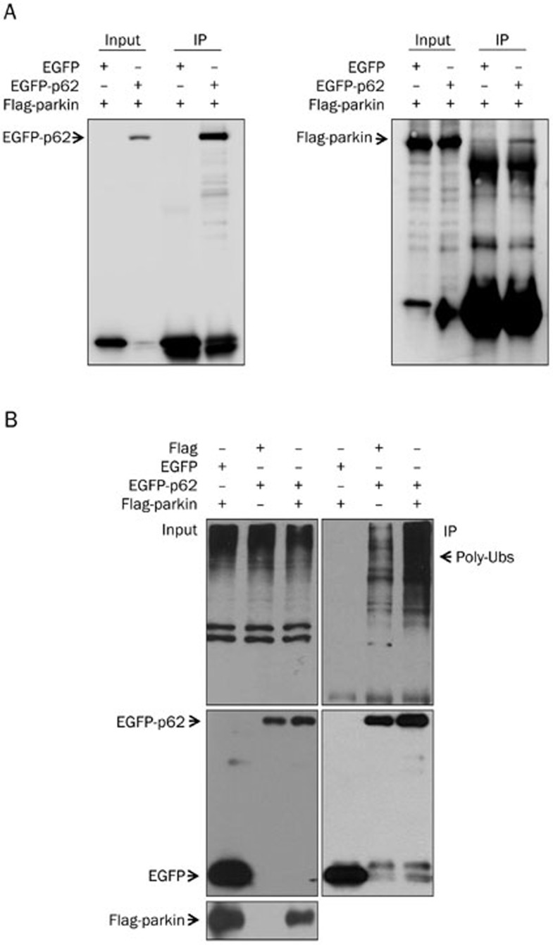
Parkin is an E3 ubiquitin ligase that ubiquitinates its target proteins28. It has been reported that both parkin and p62 function in mitophagy29. p62 recognizes the substrates that are ubiquitinated by parkin; then, with the help of LC3, p62 delivers these substrates to the autophagosomes for degradation. As parkin is an E3 ligase that binds to its substrates, we hypothesized that parkin and p62 directly interact with each other. Thus, we performed immunoprecipitation assays. In cells that were co-transfected with EGFP-p62 and Flag-parkin, Flag-parkin was co-immunoprecipitated when EGFP-p62 was precipitated with anti-EGFP antibody (Figure 1A). However, Flag-parkin was not co-immunoprecipitated in cells that were co-transfected with EGFP (Figure 1A). In addition, the ubiquitination level of p62 was significantly increased in cells transfected with EGFP-parkin (Figure 1B).
Figure 1.
Parkin interacts with and ubiquitinates p62. (A) HEK293 cells were co-transfected with Flag-parkin and EGFP or EGFP-p62, respectively. The cells were collected 24 h after transfection and immunoprecipitated with an anti-GFP antibody. The inputs and immunoprecipitates were subjected to immunoblot analysis using an anti-Flag or anti-GFP antibody. (B) HEK293 cells were co-transfected with EGFP or EGFP-p62 and Flag or Flag-parkin. Twenty-four hours after transfection, the cells were collected for immunoprecipitation analysis using an anti-GFP antibody. The inputs and immunoprecipitates were subjected to immunoblot analysis using anti-Flag, anti-EGFP and anti-ubiquitin (Ub) antibodies.
Parkin stabilizes p62 through its E3 ubiquitin ligase activity
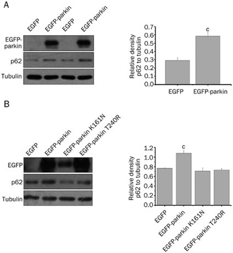
Ubiquitination usually acts as a degradation signal for some proteins. We hypothesized that the increased ubiquitination of p62 by parkin promotes its degradation. We overexpressed EGFP or EGFP-parkin in parkin-deficient HeLa cells, which harbor parkin exon deletions30, to identify the effects of parkin on p62 degradation. Interestingly, endogenous p62 was not decreased but was significantly increased following the overexpression of EGFP-parkin (Figure 2A). We next investigated whether the increase in p62 is associated with the E3 ligase activity of parkin. We transfected cells with wild-type EGFP-parkin or two ligase activity-deficient mutants (EGFP-parkin K161N and EGFP-parkin T240R). Our results showed that EGFP-parkin, but not EGFP-parkin K161N or EGFP-parkin T240R, increased p62 levels (Figure 2B). These results suggest that parkin stabilizes p62 through its E3 ligase activity.
Figure 2.
Parkin stabilizes p62 through its E3 ubiquitin ligase activity. (A) HeLa cells were transfected with EGFP or EGFP-parkin for 24 h. Cell lysates were subjected to immunoblot analysis with anti-GFP, anti-p62 and anti-tubulin antibodies. The quantitative analysis of the relative density of p62 compared with the loading control (tubulin) is shown in the right panel. The data are presented as the mean±SEM from three independent experiments. cP<0.01 vs EGFP group, one-way ANOVA. (B) HeLa cells expressing EGFP, EGFP-parkin, EGFP-parkin K161N or EGFP-parkin T240R. Twenty-four hours after transfection, the cells were subjected to immunoblot analysis with the indicated antibodies. The band density of p62 relative to tubulin is shown in the right panel. The data are presented as the mean±SEM from three independent experiments. cP<0.01 vs EGFP group, one-way ANOVA.
Parkin represses ERK1/2 activity by regulating p62
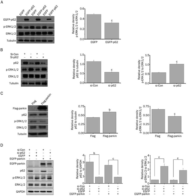
Previous studies have reported that p62 influences ERK1/2 activation14 and that parkin represses ERK activation25. We hypothesized that the effects of parkin on ERK1/2 are media ated by its regulation of p62. First, we examined the effects of p62 on ERK1/2 activation. In HeLa cells transfected with EGFP or EGFP-p62, pan-ERK1/2 levels were not changed, but ERK1/2 phosphorylation was decreased in cells transfected with EGFP-p62 (Figure 3A). By contrast, in p62-knockdowon cells, ERK1/2 phosphorylation was increased (Figure 3B), suggesting that p62 inhibits ERK1/2 activation. Next, we examined the relationship between parkin, p62 and ERK1/2. In parkin-overexpressing cells, p62 was increased, whereas p-ERK1/2 was decreased (Figure 3C). Furthermore, in p62-knockdown cells, parkin failed to repress ERK1/2 activation (Figure 3D), suggesting that p62 mediates the parkin-dependent effects on ERK1/2 activation.
Figure 3.
Parkin represses ERK1/2 activity by regulating p62. (A) HeLa cells were transfected with EGFP or EGFP-p62. Cell lysates were subjected to immunoblot analysis with anti-GFP, anti-pERK1/2, anti-ERK1/2 and anti-tubulin antibodies. The band density of p-ERK1/2 relative to pan-ERK1/2 is shown in the right panel. Mean±SEM. cP<0.01 vs EGFP group. (B) HeLa cells were transfected with si-con or si-p62 for 48 h followed by immunoblot analysis with the indicated antibodies. The band densities of p-ERK1/2 relative to pan-ERK1/2 and p62 relative to tubulin are shown in the middle and right panels. Mean±SEM. cP<0.01 vs si-con group. (C) HeLa cells were transfected with Flag or Flag-parkin for 24 h followed by immunoblot analysis. The band densities of p-ERK1/2 relative to pan-ERK1/2 and p62 relative to tubulin are shown. Mean±SEM. bP<0.05, cP<0.01 vs Flag group. (D) HeLa cells were first transfected with si-con or si-p62 for 24 h and then with EGFP or EGFP-parkin for another 24 h followed by immunoblot analysis. The band densities of p-ERK1/2 relative to pan-ERK1/2 and p62 relative to GAPDH are shown in the middle and right panels. Mean±SEM. bP<0.05, cP<0.01.
p62 represses 6-OHDA-induced ERK1/2 activation
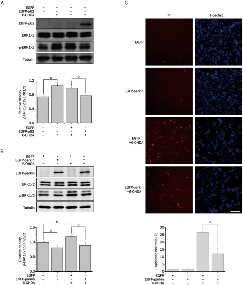
6-OHDA is a neurotoxin that induces massive oxidative stress, leading to the specific degeneration of DA neurons31,32,33. It has been reported that 6-OHDA-induced oxidative stress promotes sustained ERK1/2 activation34. We determined whether p62 or parkin could inhibit ERK1/2 activation in 6-OHDA-treated cells. In cells that were transfected with EGFP-p62 (Figure 4A) or EGFP-parkin (Figure 4B), the overexpression of EGFP-parkin or EGFP-p62 decreased 6-OHDA-induced ERK1/2 activation, suggesting that parkin and p62 have similar effects on ERK1/2 activation. To further identify the protective effects of parkin on 6-OHDA-induced cell death, we transfected SH-SY5Y cells with EGFP or EGFP-parkin and treated the cells with or without 6-OHDA (Figure 4C). Parkin overexpression significantly decreased 6-OHDA-induced cell death, as shown by the decreased number of dead cells stained with PI.
Figure 4.
p62 and parkin inhibit 6-OHDA-induced ERK1/2 activation. (A) PC12 cells were transfected with EGEP or EGFP-p62 and then treated with 200 μmol/L 6-OHDA for 24 h. The cell lysates were subjected to immunoblot analysis with the indicated antibodies. The band density of p-ERK1/2 relative to pan-ERK1/2 is shown in the lower panel. The values are the mean±SEM from three independent experiments. bP<0.05, cP<0.01, one-way ANOVA. (B) The cells were transfected with EGFP or EGFP-parkin for 24 h and treated with 6-OHDA for another 24 h. The total cell lysates were subjected to Western blotting analysis with the indicated antibodies. The band density of p-ERK1/2 relative to pan-ERK1/2 is shown in the lower panel. The values are the mean±SEM from three independent experiments. bP<0.05, one-way ANOVA. (C) SH-SY5Y cells were transfected with EGFP or EGFP-parkin for 24 h and treated with 6-OHDA for another 12 h. The cells were stained with PI (red) and Hoechst (blue). The scale bar represents 20 μm. The percentage of apoptotic cells was calculated and plotted in the lower panel. cP<0.01, one-way ANOVA.
Discussion
Genetic mutations and environmental toxins play important roles in DA neuronal degeneration. 6-OHDA is a neurotoxin that induces the specific degeneration of DA neurons and is usually used to prepare PD animal models35. It has been reported that 6-OHDA produces reactive oxygen species (ROS) and impairs mitochondrial function36. Although ERK activation usually promotes cell survival, oxidative stress induced by 6-OHDA activates MAPK family members such as ERK1/2 and promotes DA neuronal death34,37,38. A previous study showed that persistent glutamate-induced ERK1/2 activation promotes cell death in a hippocampal cell line and primary neurons39. Abnormal activation of ERK1/2, along with interactions with abnormal mitochondria, is present in the substantia nigra in the brains of PD patients40. Moreover, the inhibition of ERK1/2 activation could reduce 6-OHDA-induced cell death37. Thus, abnormal ERK1/2 activation plays a critical role in PD pathogenesis.
Parkin is documented to mediate neuroprotection against 6-OHDA-induced neurotoxicity7,21,41,42. As parkin is an E3 ubiquitin ligase, its neuroprotective effect may be executed via its substrates. In the present study, we found that p62 is a substrate of parkin. Parkin directly interacts with p62 and ubiquitinates p62 via its E3 ligase activity. However, the ubiquitination of p62 by parkin does not promote p62 degradation. By contrast, the stability of p62 is increased, leading to upregulated p62 protein levels in cells. However, parkin mutants (K161N and T240R) that have lost E3 ligase activity fail to stabilize p62, suggesting that the ubiquitination of p62 by parkin does not provide a degradation signal. Polyubiquitination is specifically linked to different lysine (K) sites in the substrate, primarily K48 or K6343. A K48-linked polyubiquitin chain is considered to be a proteasome degradation signal. However, K63-linked polyubiquitin chains do not mediate proteasomal degradation, but instead drive other cellular processes, such as endocytic trafficking, translation and DNA repair44. Recently, ESCRT0 (endosomal sorting complex required for transport) and other factors have been identified as molecules that selectively bind to K63-linked polyubiquitin chains and inhibit their binding to the proteasome45. Thus, K63-linked polyubiquitin chains may block their substrates from being degraded by the proteasome. In our study, the p62 that is polyubiquitinated by parkin is not degraded, but rather is stabilized, suggesting that the polyubiquitin chains may be K63-linked.
p62 is an important scaffold protein that has multiple domains. Those domains enable p62 to regulate kinase-activated and ubiquitin-mediated signaling pathways46. The PB1 domain of p62 interacts with some protein kinases, including ERK1/2, MEKK3 and MEK511. It has been reported that p62 is involved in regulating ERK1/2 activation. In p62-deficient mice, the basal level of ERK1/2 activity is enhanced14. However, p62 overexpression dramatically inhibits ERK1/2 activation15. We also observed that p62 overexpression represses and p62 knockdown activates the phosphorylation of ERK1/2. Furthermore, we found that parkin overexpression represses ERK1/2 activation, similar to p62 overexpression. Importantly, parkin overexpression fails to repress ERK1/2 activation in p62-knockdown cells, suggesting that p62 mediates the parkin-dependent effects on ERK1/2 activation. In addition, both parkin and p62 repress 6-OHDA-induced ERK1/2 activation in neuronal cells, further suggesting that parkin and p62 act on the same pathway. Thus, our study provides a mechanistic explanation of how parkin represses ERK1/2 activation.
In summary, this study provides evidence that p62 is a substrate of parkin. Parkin ubiquitinates and stabilizes p62 to repress ERK1/2 activation, thereby attenuating 6-OHDA-induced ERK1/2 activation.
Author contribution
Feng GAO and Guang-hui WANG designed the experiments; Xiao-ou HOU and Jian-min SI performed the main experiments; Hai-gang REN, Dong CHEN and Hong-feng WANG prepared the plasmids; Zheng YING and Qing-song HU contributed analytic tools, Feng GAO and Guang-hui WANG analyzed the data; Xiao-ou HOU drafted the manuscript; and Feng GAO and Guang-hui WANG revised the manuscript.
Acknowledgments
This work was supported in part by the National Natural Sciences Foundation of China (No 31330030 and 81371393), National Basic Research Program of China (973 Program) (2011CB504102), the Natural Science Foundation of Jiangsu Province (BK20140328), and a project funded by the Priority Academic Program Development of Jiangsu Higher Education Institutions.
References
- 1Farrer MJ. Genetics of Parkinson disease: paradigm shifts and future prospects. Nat Rev Genet 2006; 7: 306–18. [DOI] [PubMed] [Google Scholar]
- 2Thomas B, Beal MF. Parkinson's disease. Hum Mol Genet 2007; 16: R183–94. [DOI] [PubMed] [Google Scholar]
- 3Dekker MC, Bonifati V, van Duijn CM. Parkinson's disease: piecing together a genetic jigsaw. Brain 2003; 126: 1722–33. [DOI] [PubMed] [Google Scholar]
- 4Bossy-Wetzel E, Schwarzenbacher R, Lipton SA. Molecular pathways to neurodegeneration. Nat Med 2004; 10 Suppl: S2–9. [DOI] [PubMed] [Google Scholar]
- 5Tan EK, Skipper LM. Pathogenic mutations in Parkinson disease. Hum Mutat 2007; 28: 641–53. [DOI] [PubMed] [Google Scholar]
- 6Mata IF, Lockhart PJ, Farrer MJ. Parkin genetics: one model for Parkinson's disease. Hum Mol Genet 2004; 13: R127–33. [DOI] [PubMed] [Google Scholar]
- 7Jiang H, Ren Y, Zhao J, Feng J. Parkin protects human dopaminergic neuroblastoma cells against dopamine-induced apoptosis. Hum Mol Genet 2004; 13: 1745–54. [DOI] [PubMed] [Google Scholar]
- 8Youle RJ, Narendra DP. Mechanisms of mitophagy. Nat Rev Mol Cell Biol 2011; 12: 9–14. [DOI] [PMC free article] [PubMed] [Google Scholar]
- 9Ding WX, Ni HM, Li M, Liao Y, Chen X, Stolz DB, et al. Nix is critical to two distinct phases of mitophagy, reactive oxygen species-mediated autophagy induction and Parkin-ubiquitin-p62-mediated mitochondrial priming. J Biol Chem 2010; 285: 27879–90. [DOI] [PMC free article] [PubMed] [Google Scholar]
- 10Narendra D, Tanaka A, Suen DF, Youle RJ. Parkin-induced mitophagy in the pathogenesis of Parkinson disease. Autophagy 2009; 5: 706–8. [DOI] [PubMed] [Google Scholar]
- 11Manley S, Williams JA, Ding WX. Role of p62/SQSTM1 in liver physiology and pathogenesis. Exp Biol Med (Maywood) 2013; 238: 525–38. [DOI] [PMC free article] [PubMed] [Google Scholar]
- 12Cavey JR, Ralston SH, Hocking LJ, Sheppard PW, Ciani B, Searle MS, et al. Loss of ubiquitin-binding associated with Paget's disease of bone p62 (SQSTM1) mutations. J Bone Miner Res 2005; 20: 619–24. [DOI] [PubMed] [Google Scholar]
- 13Mathew R, Karp CM, Beaudoin B, Vuong N, Chen G, Chen HY, et al. Autophagy suppresses tumorigenesis through elimination of p62. Cell 2009; 137: 1062–75. [DOI] [PMC free article] [PubMed] [Google Scholar]
- 14Lee SJ, Pfluger PT, Kim JY, Nogueiras R, Duran A, Pages G, et al. A functional role for the p62-ERK1 axis in the control of energy homeostasis and adipogenesis. EMBO Rep 2010; 11: 226–32. [DOI] [PMC free article] [PubMed] [Google Scholar]
- 15Rodriguez A, Duran A, Selloum M, Champy MF, Diez-Guerra FJ, Flores JM, et al. Mature-onset obesity and insulin resistance in mice deficient in the signaling adapter p62. Cell Metab 2006; 3: 211–22. [DOI] [PubMed] [Google Scholar]
- 16Cagnol S, Chambard JC. ERK and cell death: mechanisms of ERK-induced cell death--apoptosis, autophagy and senescence. FEBS J 2010; 277: 2–21. [DOI] [PubMed] [Google Scholar]
- 17Bost F, Aouadi M, Caron L, Even P, Belmonte N, Prot M, et al. The extracellular signal-regulated kinase isoform ERK1 is specifically required for in vitro and in vivo adipogenesis. Diabetes 2005; 54: 402–11. [DOI] [PubMed] [Google Scholar]
- 18Lindgren N, Leak RK, Carlson KM, Smith AD, Zigmond MJ. Activation of the extracellular signal-regulated kinases 1 and 2 by glial cell line-derived neurotrophic factor and its relation to neuroprotection in a mouse model of Parkinson's disease. J Neurosci Res 2008; 86: 2039–49. [DOI] [PubMed] [Google Scholar]
- 19Wang XL, Xing GH, Hong B, Li XM, Zou Y, Zhang XJ, et al. Gastrodin prevents motor deficits and oxidative stress in the MPTP mouse model of Parkinson's disease: involvement of ERK1/2-Nrf2 signaling pathway. Life Sci 2014; 114: 77–85. [DOI] [PubMed] [Google Scholar]
- 20Lee E, Park HR, Ji ST, Lee Y, Lee J. Baicalein attenuates astroglial activation in the 1-methyl-4-phenyl-1,2,3,4-tetrahydropyridine-induced Parkinson's disease model by downregulating the activations of nuclear factor-kappaB, ERK, and JNK. J Neurosci Res 2014; 92: 130–9. [DOI] [PubMed] [Google Scholar]
- 21Benskey MJ, Manfredsson FP, Lookingland KJ, Goudreau JL. The role of parkin in the differential susceptibility of tuberoinfundibular and nigrostriatal dopamine neurons to acute toxicant exposure. Neurotoxicology 2015; 46: 1–11. [DOI] [PMC free article] [PubMed] [Google Scholar]
- 22Park HJ, Park KH, Shin KS, Lee MK. The roles of cyclic AMP-ERK-Bad signaling pathways on 6-hydroxydopamine-induced cell survival and death in PC12 cells. Toxicol In Vitro 2013; 27: 2233–41. [DOI] [PubMed] [Google Scholar]
- 23Santini E, Alcacer C, Cacciatore S, Heiman M, Herve D, Greengard P, et al. L-DOPA activates ERK signaling and phosphorylates histone H3 in the striatonigral medium spiny neurons of hemiparkinsonian mice. J Neurochem 2009; 108: 621–33. [DOI] [PubMed] [Google Scholar]
- 24Reinhardt P, Schmid B, Burbulla LF, Schondorf DC, Wagner L, Glatza M, et al. Genetic correction of a LRRK2 mutation in human iPSCs links parkinsonian neurodegeneration to ERK-dependent changes in gene expression. Cell Stem Cell 2013; 12: 354–67. [DOI] [PubMed] [Google Scholar]
- 25Ren Y, Jiang H, Yang F, Nakaso K, Feng J. Parkin protects dopaminergic neurons against microtubule-depolymerizing toxins by attenuating microtubule-associated protein kinase activation. J Biol Chem 2009; 284: 4009–17. [DOI] [PMC free article] [PubMed] [Google Scholar]
- 26Ren H, Fu K, Mu C, Li B, Wang D, Wang G. DJ-1, a cancer and Parkinson's disease associated protein, regulates autophagy through JNK pathway in cancer cells. Cancer Lett 2010; 297: 101–8. [DOI] [PubMed] [Google Scholar]
- 27Chen D, Gao F, Li B, Wang H, Xu Y, Zhu C, et al. Parkin mono-ubiquitinates Bcl-2 and regulates autophagy. J Biol Chem 2010; 285: 38214–23. [DOI] [PMC free article] [PubMed] [Google Scholar]
- 28Shimura H, Hattori N, Kubo S, Mizuno Y, Asakawa S, Minoshima S, et al. Familial Parkinson disease gene product, parkin, is a ubiquitin-protein ligase. Nat Genet 2000; 25: 302–5. [DOI] [PubMed] [Google Scholar]
- 29Geisler S, Holmstrom KM, Skujat D, Fiesel FC, Rothfuss OC, Kahle PJ, et al. PINK1/Parkin-mediated mitophagy is dependent on VDAC1 and p62/SQSTM1. Nat Cell Biol 2010; 12: 119–31. [DOI] [PubMed] [Google Scholar]
- 30Denison SR, Wang F, Becker NA, Schule B, Kock N, Phillips LA, et al. Alterations in the common fragile site gene Parkin in ovarian and other cancers. Oncogene 2003; 22: 8370–8. [DOI] [PubMed] [Google Scholar]
- 31Glinka Y, Gassen M, Youdim MB. Mechanism of 6-hydroxydopamine neurotoxicity. J Neural Transm Suppl 1997; 50: 55–66. [DOI] [PubMed] [Google Scholar]
- 32Blum D, Torch S, Lambeng N, Nissou M, Benabid AL, Sadoul R, et al. Molecular pathways involved in the neurotoxicity of 6-OHDA, dopamine and MPTP: contribution to the apoptotic theory in Parkinson's disease. Prog Neurobiol 2001; 65: 135–72. [DOI] [PubMed] [Google Scholar]
- 33Li LY, Zhao XL, Fei XF, Gu ZL, Qin ZH, Liang ZQ. Bilobalide inhibits 6-OHDA-induced activation of NF-kappaB and loss of dopaminergic neurons in rat substantia nigra. Acta Pharmacol Sin 2008; 29: 539–47. [DOI] [PubMed] [Google Scholar]
- 34Park SY, Kim do Y, Kang JK, Park G, Choi YW. Involvement of activation of the Nrf2/ARE pathway in protection against 6-OHDA-induced SH-SY5Y cell death by alpha-iso-cubebenol. Neurotoxicology 2014; 44: 160–8. [DOI] [PubMed] [Google Scholar]
- 35Wang T, Liu YY, Wang X, Yang N, Zhu HB, Zuo PP. Protective effects of octacosanol on 6-hydroxydopamine-induced Parkinsonism in rats via regulation of ProNGF and NGF signaling. Acta Pharmacol Sin 2010; 31: 765–74. [DOI] [PMC free article] [PubMed] [Google Scholar]
- 36Masoudi N, Ibanez-Cruceyra P, Offenburger SL, Holmes A, Gartner A. Tetraspanin (TSP-17) protects dopaminergic neurons against 6-OHDA-induced neurodegeneration in C. elegans. PLoS Genet 2014; 10: e1004767. [DOI] [PMC free article] [PubMed] [Google Scholar]
- 37Kulich SM, Chu CT. Sustained extracellular signal-regulated kinase activation by 6-hydroxydopamine: implications for Parkinson's disease. J Neurochem 2001; 77: 1058–66. [DOI] [PMC free article] [PubMed] [Google Scholar]
- 38Runden E, Seglen PO, Haug FM, Ottersen OP, Wieloch T, Shamloo M, et al. Regional selective neuronal degeneration after protein phosphatase inhibition in hippocampal slice cultures: evidence for a MAP kinase-dependent mechanism. J Neurosci 1998; 18: 7296–305. [DOI] [PMC free article] [PubMed] [Google Scholar]
- 39Stanciu M, Wang Y, Kentor R, Burke N, Watkins S, Kress G, et al. Persistent activation of ERK contributes to glutamate-induced oxidative toxicity in a neuronal cell line and primary cortical neuron cultures. J Biol Chem 2000; 275: 12200–6. [DOI] [PubMed] [Google Scholar]
- 40Zhu JH, Guo F, Shelburne J, Watkins S, Chu CT. Localization of phosphorylated ERK/MAP kinases to mitochondria and autophagosomes in Lewy body diseases. Brain Pathol 2003; 13: 473–81. [DOI] [PMC free article] [PubMed] [Google Scholar]
- 41Vercammen L, Van der Perren A, Vaudano E, Gijsbers R, Debyser Z, Van den Haute C, et al. Parkin protects against neurotoxicity in the 6-hydroxydopamine rat model for Parkinson's disease. Mol Ther 2006; 14: 716–23. [DOI] [PubMed] [Google Scholar]
- 42Hyun DH, Lee M, Halliwell B, Jenner P. Effect of overexpression of wild-type or mutant parkin on the cellular response induced by toxic insults. J Neurosci Res 2005; 82: 232–44. [DOI] [PubMed] [Google Scholar]
- 43Weissman AM. Themes and variations on ubiquitylation. Nat Rev Mol Cell Biol 2001; 2: 169–78. [DOI] [PubMed] [Google Scholar]
- 44Ikeda F, Dikic I. Atypical ubiquitin chains: new molecular signals. 'Protein Modifications: Beyond the Usual Suspects' review series. EMBO Rep 2008; 9: 536–42. [DOI] [PMC free article] [PubMed] [Google Scholar]
- 45Nathan JA, Kim HT, Ting L, Gygi SP, Goldberg AL. Why do cellular proteins linked to K63-polyubiquitin chains not associate with proteasomes? EMBO J 2013; 32: 552–65. [DOI] [PMC free article] [PubMed] [Google Scholar]
- 46Moscat J, Diaz-Meco MT, Wooten MW. Signal integration and diversification through the p62 scaffold protein. Trends Biochem Sci 2007; 32: 95–100. [DOI] [PubMed] [Google Scholar]


