

Abstract
Acute Hemorrhagic Edema of Infancy (AHEI) is a rare leukocytoclastic vasculitis, clinically characterized by the classical triad: palpable purpuric skin lesions, edema and fever, and is commonly misdiagnosed as Henoch-Schönlein purpura. In addition to its sudden onset, AHEI is also characterized by its self-limited course with complete and spontaneous recovery occurring between 1 and 3 weeks. Because of the scarcity of studies on therapy with corticosteroids, the conservative approach is usually recommended. The authors report an unusual case of an one-year-old boy who presented with typical cutaneous rash of AHEI and orchitis, the latter showing complete resolution after less than 24 hours of prednisolone therapy. The authors call attention to this entity mainly as a differential diagnosis of Henoch-Schönlein purpura and to the importance of new studies to establish the benefits of corticosteroid therapy for AHEI.
Keywords : Infant; Vasculitis, Leukocytoclastic, Cutaneous; Orchitis; Steroids; Purpura
INTRODUCTION
Acute Hemorrhagic Edema of Infancy (AHEI) is a rare leukocytoclastic vasculitis, which typically affects children between 4 and 24 months.1-3 It is commonly misdiagnosed as Henoch-Schönlein purpura (HSP) due to clinical similarities.4-6
AHEI is clinically characterized by the classical triad of palpable purpuric skin lesions, edema and fever.7 Visceral involvement is rare, and the scrotum is involved in less than 10 percent of cases.8 AHEI is usually considered non-responsive to corticosteroid therapy, hence a conservative approach is generally recommended.
CASE REPORT
An one-year-old boy was referred to the hospital with a four-day history of cough and rhinorrhea and a two-day history of low-grade fever and hemorrhagic cutaneous eruption. The skin rash started in both knees and spread to his upper extremities, feet, face and ears followed by swelling of knees, hands, feet and scrotum. There was no history of vomiting, urinary complaints or recent immunizations.
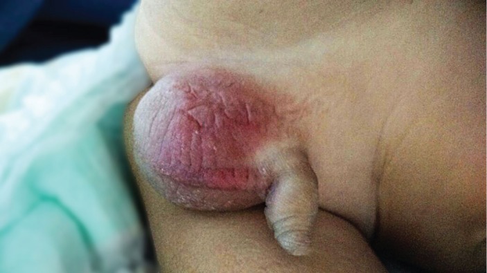
On clinical examination, he was in good overall clinical condition and afebrile. He had multiple rounded palpable purpuric patches over the upper and the lower limbs, face, oral cavity mucosa and both ears, sparing the trunk. (Figure 1) He also had edema over knees, hands, feet and hyperemia and edema in his scrotum (Figure 2). Neither passive restriction of the joints movements nor signs of joint effusion were present. There was no evidence of abdominal tenderness.
Figure 1. Multiple rounded palpable purpuric patches over upper (left) and lower (middle) limbs, ear and face (right).

Figure 2. Hyperemia and edema of the scrotum.
Scrotal ultrasonography showed marked bilateral scrotal skin thickening and small bilateral hydroceles, with normal testicular blood flow. Laboratory studies revealed normal blood count with elevated erythrocyte sedimentation rate and C-reactive protein. Urinalysis, coagulation tests and random protein-to-creatinine ratio were normal (as shown in Table 1).
Table 1. Laboratory work up.
| RV | RV | ||||
|---|---|---|---|---|---|
| Hemoglobin | 11.7 | 10.5-13.5 g/dL | ESR | 47 | < 10 mm 1st hour |
| Leukocytes | 11.87 | 6.0-17.5 × 10³/mm3 | CRP | 100 | < 5 mg/dL |
| Neutrophils | 3.917 | 1.5-8.5 × 10³/mm3 | Leukocytes (U) | 2000 | <10.000/mm3 |
| Lymphocytes | 6.184 | 4.0-10.5 ×10³/mm3 | Erythrocytes (U) | 2000 | <10.000/mm3 |
| Platelets | 433 | 150-450 ×103/mm3 | Random protein (U) | 23 | 1-14 mg/dL |
| aPTT | 1.13 | < 1.25 | Random creatinine (U) | 89.5 | 24-392 mg/dL |
| INR | 1.09 | 0.8-1.2 |
aPTT- activated partial thromboplastin time, CRP= c-Reactive protein, ESR= erythrocyte sedimentation rate, INR= international normalized ratio, U= urine.
Oral prednisolone was prescribed (2mg/kg/day) and the scrotal inflammatory signs subsided on the second day after treatment. No new skin lesions were noticed. The patient was discharged two days after admission for an outpatient follow-up.
DISCUSSION
Acute Hemorrhagic Edema of Infancy (AHEI) is a leukocytoclastic vasculitis commonly misdiagnosed or misinterpreted as Henoch-Schonlein Purpura (HSP). Although the current classification of childhood vasculitis9,10 does not include AHEI as a vasculitis identity, from the first publication by Irving M. Snow11 in 1913, to the year 2004, there were 100 cases of this disease reported worldwide.1 AHEI is typically characterized by the classical triad of palpable purpuric skin lesions, edema and fever7 and it is more common in males, between 4 and 24 months, without racial predominance.2,3
The clinical history usually begins with sudden appearance of well demarcated, annular, medallion-like, rosette shaped purpuric plaques, mostly observed on the face and the extremities, with relative sparing of the trunk.3,12 Visceral involvement is rare, but symptoms like arthritis, gastrointestinal bleeding and scrotal erythema and edema, as presented in our case, has been described in literature.13
Although the etiology remains unknown, it is believed that AHEI is an immune-complex mediated disease. This hypothesis is based on the observation that infections (mostly upper respiratory tract infections and urinary tract infections), drugs or immunizations preceded almost 75% of AHEI cases.14
Diagnosis of AHEI is majorly based on clinical grounds. In doubtful cases, the skin biopsy followed by histopathologic with immunofluorescence examination may be of great value for diagnosis.15 Laboratory tests are usually normal, although leukocytosis, thrombocytosis and eosinophilia may be observed.6 However, according to some authors6,13 when the diagnosis of AHEI is suspected, a complete blood count, urine analysis and clotting tests should be required to differentiate AHEI from other more severe diagnosis.
Histologic findings of the skin lesions are consistent with small-vessel vasculitis, showing typical leukocytoclastic vasculitis in the dermis with or without fibrinoid necrosis.3,5,13 Immunofluorescence testing can reveal C3, fibrinogen and IgM deposits and, less commonly, IgM, IgE or IgA, the latter a common finding in HSP.16,17
AHEI is a self-limited disease, running a benign course, with complete spontaneous recovery in 1 to 3 weeks.3 Although conservative management is the most frequently adopted approach, few publications have reported beneficial use of corticosteroids. These authors18,19 observed a clear improvement of symptoms after high-dose of corticosteroid therapy and recommend that, in severe cases of AHEI, corticotherapy should be considered to shorten the symptoms and give significant relief. In the case reported herein, the scrotal inflammation subsided in less than 24 hours after prednisolone therapy, and the skin lesions became undetectable after 2 weeks.
Among the differential diagnosis for AHEI, Henoch Schönlein Purpura (HSP) is probably the most important. Because of similarities involving both syndromes, there is a debate in literature if AHEI should be considered as a separate entity or as a variant of HSP.13 Currently, most authors5,6,13,20 consider them as different diseases based on clinical, pathological and epidemiological differences.
These differences involve the patient’s age; while AHEI afflicts more often younger children aging from 4 months up to 2 years, HSP occurs in older ones, from 3 to 6 years of age.20 Skin lesions in AHEI are rounded palpable purpuric plaques, frequently observed on the face and upper limbs, while in HSP lesions are uncharacteristic palpable purpura that distribute on a gravitational pattern, like the buttock and extensor surface of the legs, usually sparing the face.13,21 Visceral involvement is very rare in AHEI, in contrast to HSP, which frequently presents with gastrointestinal and renal complications.13,21 Perivascular IgA deposition is frequently seen in HSP and often absent in AHEI.3 Another important difference is that relapses are commonly observed in HSP and rarely reported in AHEI.5,7
It is likely that the AHEI’s low prevalence may be due to a number of misdiagnosis as Henoch-Schonlein Purpura (HSP). Nonetheless as the treatment of these two entities differs, it becomes important to accurately differentiate them. While corticosteroid therapy is indicated for HSP cases,22 most authors defend a conservative approach for AHEI. In the case presented herein, there was marked improvement of symptoms after corticosteroid therapy, noted by the rapid regression of scrotal erythema, edema and discontinuing the appearance of new lesions after treatment. However, larger series are required to accurately access the benefit of corticotherapy for AHEI with extracutaneous complications.
Footnotes
Cunha DFS, Darcie ALF, Benevides GN, et al. Acute Hemorrhagic Edema of Infancy: an unusual diagnosis for the general pediatrician. Autopsy Case Rep [Internet]. 2015;5(3):37-41. http://dx.doi.org/10.4322/acr.2015.020
REFERENCES
- 1.Roh MR, Chung HJ, Lee JH. A case of acute hemorrhagic edema of infancy. Yonsei Med J. 2004;45(3):523-6. http://dx.doi.org/10.3349/ymj.2004.45.3.523. PMid: [DOI] [PubMed] [Google Scholar]
- 2.Savino F, Lupica MM, Tarasco V, et al. Acute hemorrhagic edema of infancy: a troubling cutaneous presentation with a self-limiting course. Pediatr Dermatol. 2013;30(6):e149-52. http://dx.doi.org/10.1111/pde.12004. PMid: [DOI] [PubMed] [Google Scholar]
- 3.Jindal SR, Kura MM. Acute hemorrhagic edema of infancy-a rare entity. Indian Dermatol Online J. 2013;4(2):106-8. http://dx.doi.org/10.4103/2229-5178.110630. PMid: [DOI] [PMC free article] [PubMed] [Google Scholar]
- 4.Pelajo CF, Oliveira SKF. Edema hemorrágico agudo da infância: uma variante da púrpura de Henoch-Schönlein? Rev Bras Reumatol. 2007;47(1):69-71. http://dx.doi.org/10.1590/S0482-50042007000100014. [Google Scholar]
- 5.Saraclar Y, Tinaztepe K, Adaliolu G, Tuncer A. Acute hemorrhagic edema of infancy (AHEI)--a variant of Henoch-Schönlein purpura or a distinct clinical entity? J Allergy Clin Immunol. 1990;86(4 Pt 1):473-83. http://dx.doi.org/10.1016/S0091-6749(05)80202-7. PMid: [DOI] [PubMed] [Google Scholar]
- 6.Karremann M, Jordan AJ, Bell N, Witsch M, Dürken M. Acute hemorrhagic edema of infancy: report of 4 cases and review of the current literature. Clin Pediatr (Phila). 2009;48(3):323-6. http://dx.doi.org/10.1177/0009922808323113. PMid: [DOI] [PubMed] [Google Scholar]
- 7.McDougall CM, Ismail SK, Ormerod A. Acute haemorrhagic oedema of infancy. Arch Dis Child. 2005;90(3):316. http://dx.doi.org/10.1136/adc.2004.060632. PMid: [DOI] [PMC free article] [PubMed] [Google Scholar]
- 8.Fiore E, Rizzi M, Ragazzi M, et al. Acute hemorrhagic edema of young children (cockade purpura and edema): a case series and systematic review. J Am Acad Dermatol. 2008;59(4):684-95. http://dx.doi.org/10.1016/j.jaad.2008.06.005. PMid: [DOI] [PubMed] [Google Scholar]
- 9.Ozen S, Ruperto N, Dillon MJ, et al. EULAR/PReS endorsed consensus criteria for the classification of childhood vasculitides. Ann Rheum Dis. 2006;65(7):936-41. http://dx.doi.org/10.1136/ard.2005.046300. PMid: [DOI] [PMC free article] [PubMed] [Google Scholar]
- 10.Eleftheriou D, Dillon MJ, Brogan PA. Advances in childhood vasculitis. Curr Opin Rheumatol. 2009;21(4):411-8. http://dx.doi.org/10.1097/BOR.0b013e32832c49f2. PMid: [DOI] [PubMed] [Google Scholar]
- 11.Snow IM. Purpura, urticaria and angioneurotic edema of the hands and feet in a nursing baby. JAMA. 1913;61(1):18-9. http://dx.doi.org/10.1001/jama.1913.04350010020008. [Google Scholar]
- 12.Carder KR. Hypersensitivity reactions in neonates and infants. Dermatol Ther (Heidelb). 2005;18(2):160-75. http://dx.doi.org/10.1111/j.1529-8019.2005.05014.x. PMid: [DOI] [PubMed] [Google Scholar]
- 13.Moradinejad MH, Entezari P, Mahjoub F, Ziaee V. Acute hemorrhagic edema of infancy; a report of five Iranian infants and review of the literature. Iran J Pediatr. 2011;21(1):107-12. PMid: [PMC free article] [PubMed] [Google Scholar]
- 14.Ince E, Mumcu Y, Suskan E, Yalcinkaya F, Tümer N, Cin S. Infantile acute hemorrhagic edema: a variant of leukocytoclastic vasculitis. Pediatr Dermatol. 1995;12(3):224-7. http://dx.doi.org/10.1111/j.1525-1470.1995.tb00163.x. PMid: [DOI] [PubMed] [Google Scholar]
- 15.Alhammadi AH, Adel A, Hendaus MA. Acute hemorrhagic edema of infancy: a worrisome presentation, but benign course. Clin Cosmet Investig Dermatol. 2013;6:197-9. http://dx.doi.org/10.2147/CCID.S51525. PMid: [DOI] [PMC free article] [PubMed] [Google Scholar]
- 16.Krause I, Lazarov A, Rachmel A, et al. Acute haemorrhagic oedema of infancy, a benign variant of leucocytoclastic vasculitis. Acta Paediatr. 1996;85(1):114-7. http://dx.doi.org/10.1111/j.1651-2227.1996.tb13904.x. PMid: [DOI] [PubMed] [Google Scholar]
- 17.Emerich PS, Prebianchi PA, Motta LL, Lucas EA, Ferreira LM. Acute hemorrhagic edema of infancy: report of three cases. An Bras Dermatol. 2011;86(6):1181-4. http://dx.doi.org/10.1590/S0365-05962011000600019. PMid: [DOI] [PubMed] [Google Scholar]
- 18.Manzoni APDS, Viecili JB, Andrade CB, Kruse RL, Bakos L, Cestari TF. Acute hemorrhagic edema of infancy: a case report. Int J Dermatol. 2004;43(1):48-51. http://dx.doi.org/10.1111/j.1365-4632.2004.01820.x. PMid: [DOI] [PubMed] [Google Scholar]
- 19.Risikesan J, Koppelhus U, Steiniche T, Deleuran M, Herlin T. Methylprednisolone therapy in acute hemorrhagic edema of infancy. Case Rep Dermatol Med. 2014;2014:853038. PMid: [DOI] [PMC free article] [PubMed] [Google Scholar]
- 20.Garty BZ, Ofer I, Finkelstein Y. Acute hemorrhagic edema of infancy. Isr Med Assoc J. 2002;4(3):228-9. PMid: [PubMed] [Google Scholar]
- 21.Javidi Z, Maleki M, Mashayekhi V, Tayebi-Maybodi N, Nahidi Y. Acute hemorrhagic edema of infancy. Arch Iran Med. 2008;11(1):103-6. PMid: [PubMed] [Google Scholar]
- 22.Dillon MJ. Henoch-Schönlein purpura: recent advances. Clin Exp Rheumatol. 2007;25(1, Suppl 44):S66-8. PMid: [PubMed] [Google Scholar]