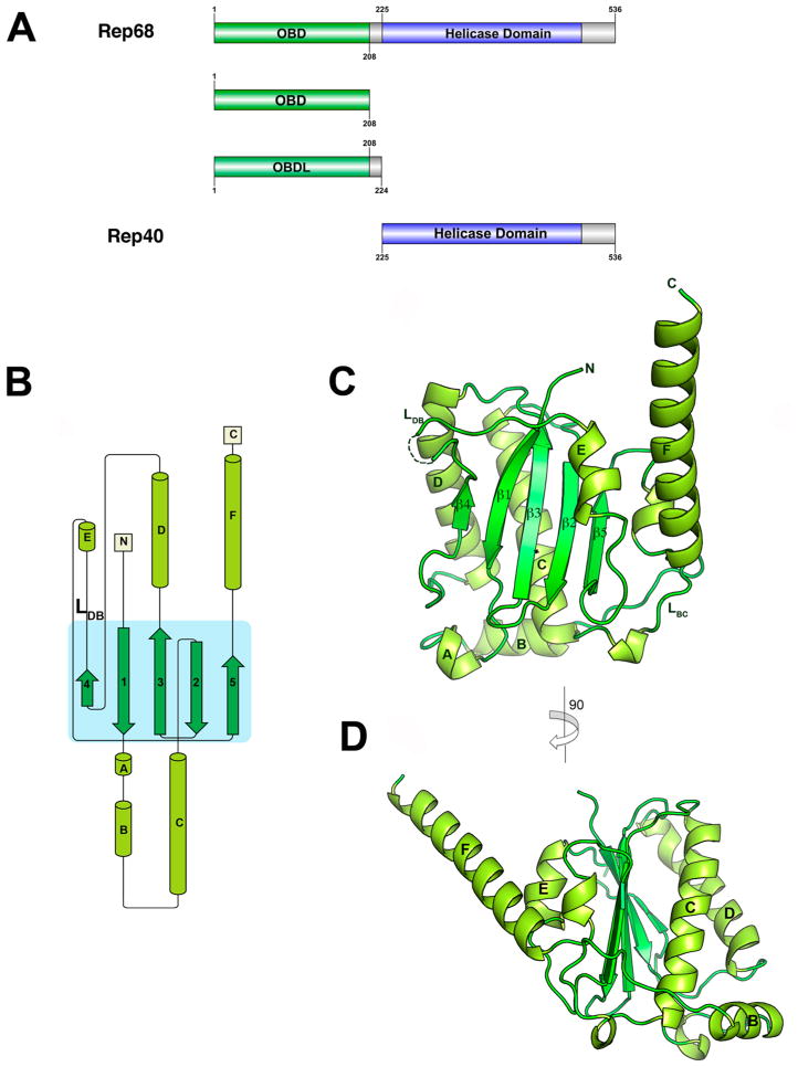
Figure 1.
Structure of AAV2 OBD. (A) Domain structure of AAV2 Rep68 protein: OBD is shown in green, Rep40 (SF3 helicase domain), in blue, and linker and C-terminal tail, in gray. The Rep40 protein spans residues 225–536 of Rep68. (B) Topology diagram of AAV2 OBD. (C) Ribbon diagram of the OBD structure. The α-helices are light green, and β-strands are dark green. Secondary structure elements are labeled with α-helices A–F, and β-sheets are numbered 1–5. The DNA binding loop LDB with missing residues in the structure is shown as a dotted line. (D) A view of the structure rotated by 90° clockwise. α-Helix F is shown protruding from the core of the structure at an angle of almost 45°.

