Abstract
This article describes the synthesis of silver nanoparticles using the aqueous extract of Alternanthera sessilis as a reducing agent by sonication, espousing green chemistry principles. Biologically synthesized nanoparticle-based drug delivery systems have significant potential in the field of biopharmaceutics due to its smaller size entailing high surface area and synergistic effects of embedded biomolecules. In the present work the cytotoxic effect of biosynthesized silver nanoparticles studied by MTT assay against breast cancer cells (MCF-7 cell line) showed significant cytotoxic activity with IC50 value 3.04 μg/mL compared to that of standard cisplatin. The superior activity of the silver nanoparticles may be due to the spherical shape and smaller particle size 10–30 nm as confirmed from transmission electron microscope (TEM) analysis. The data obtained in the study reveal the potent therapeutic value of biogenic silver nanoparticles and the scope for further development of anticancer drugs.
Keywords: Nanosilver, Cytotoxicity, Alternanthera sessilis, MCF-7 cell line
Introduction
Infection paves a pathway to non-communicable diseases such as cardiovascular disease and cancer. Cancer is a multifaceted genetic disease caused primarily by environmental factors and its treatment is usually a combination of numerous varied modalities. Different types of cancers can behave very differently. Among these lung cancer and breast cancer are very disparate diseases. Breast cancer is a malignant tumor that starts in the breast cells and occurs particularly in women (Alison 2001). The Times of India (12 Oct. 2012) estimates 1,00,000–1,25,000 new breast cancer cases in India every year. This statistical number is estimated to double by 2025. The mortality rate of patients and the must for cancer therapy coerces the need for technological breakthroughs in terms of easy availability, cost effectiveness and safety in terms of side effects. Paclitaxel, an FDA approved breast cancer drug is used widely in breast cancer treatments. It is alleged to have a string of side effects. These stipulate the need for herbal medicines in cancer therapy.
Nanomedicine is an upcoming field that could potentially make a major impact on human health (Teli et al. 2010). Nanoparticles possess unique chemical, physical and biological properties, and hence it finds use in various fields like business, therapeutics, electronics, cosmetics, catalysis and drug delivery (Sriram et al. 2012). It offers a new view for tumor detection, prevention and treatment. Nanoparticles eradicate cancer cells by flow and penetration to different regions of tumors through blood vessels and then to interstitial space to arrive at the target cells. The environmental and physiological characteristics vary from one tumor tissues to another. Hence nanoparticles should be designed in such manner, taking into account the target site and route of administration to generate optimal therapeutic effects (Wang et al. 2014).
Among nanoparticles, silver nanoparticles have an eye-catching role owing to their innumerable physical and chemical properties. Silver nanoparticles are more potent than the silver ions as revealed in avant-garde research. Silver nanoparticles are potential anticancer agents (Raghunandan et al. 2011). Cytotoxicity studies of silver nanoparticles using plant extracts: Melia dubia—human breast cancer cell line (Kathiravan et al. 2014), Malus domestica (apple) extract—MCF7 (Lokina et al. 2014), Inonotus obliquus (Chaga mushroom) extract—A549 human lung cancer (CCL185) and MCF7 human breast cancer (HTB22) cell lines (Nagajyothi et al. 2014), Erythrina indica—breast and lung cancer cell lines (Rathi Sre et al. 2015), Piper longum fruit—breast cancer cell lines (Reddy et al. 2014), Annona squamosa and Brassica Oleracea. var. botrytis—MCF-7 (Vivek et al. 2012; Ranjitham et al. 2013) are reported.
Silver nanoparticles synthesized using Acalypha indica Linn shows only 40 % cell inhibition against human breast cancer cells (MDA-MB-231) (Krishnaraj et al. 2014). The MCF-7 cells lose their 50 % viability with AgNPs (5 µg/mL) produced by Dendrophthoe falcata (Sathishkumar et al. 2014). Datura inoxia AgNPs inhibits 50 % proliferation of human breast cancer cell line MCF7 at 20 μg/mL after 24 h incubation by suppressing its growth, arresting the cell cycle phases, reducing DNA synthesis to induce apoptosis (Gajendran et al. 2014). Nuclear condensation, cell shrinkage and fragmentation are noticed for MCF-7 cells treated with Sesbania grandiflora mediated AgNPs (20 µg/mL) after 48 h in Hoechst staining.
Morinda citrifolia root extract-mediated AgNPs (100 µg) produced 100 % death of HeLa cell lines (Suman et al. 2013). Longer exposures to Eucalyptus chapmaniana AgNPs (0.02 mmol/mL) resulted in 85 % cell death after 24 h incubation (Sulaiman et al. 2013a, b). The viability of HL-60 cells decreased to 44 % after 6 h treatment with Rosmarinus officinalis AgNPs at 2 mM and cell death increased to 80 % after 24 h incubation (Sulaiman et al. 2013a, b). Cytotoxic activity was extremely sensitive to the size of the nanoparticles produced using Iresine herbstii leaf and the viability measurements decreased with increasing dosage (25–300 µg/mL) against the HeLa cell lines (Dipankar and Murugan 2012). Piper longum-mediated silver nanoparticles exhibit a significant cytotoxic effect (94.02 %) at 500 µg/mL on HEp-2 cell lines (Jacob et al. 2012). The therapeutic effect of silver nanoparticles may elicit through manipulation of their size, shape, elemental composition, charge and surface modification or functionalisation, leading target particles to specific organs (Thorley and Tetley 2013).
Owing to the significance of silver nanoparticles in cancer treatment and the necessity for newer breast cancer drugs, the present work spotlights the cytotoxic potential of the synthesized biogenic silver nanoparticles. Amid the cropping methods of nanosynthesis, biogenic synthesis finds healthier application in pharmacology due to the non-toxic nature of the source of capping material used.
Alternanthera sessilis is a weed growing on a variety of soil types. Its young shoots and leaves are ingested as vegetables. Phytochemical screening reveals the presence of reducing sugars, steroids, terpenoids, saponins, tannins and flavonoids in A. sessilis (Sahithi et al. 2011). The herb possesses antioxidant (Borah et al. 2011), anti-inflammatory (Sahithi et al. 2011), antipyretic (Nayak et al. 2010), haematinic (Arollado and Osi 2010), hepatoprotective (Lin et al. 1994), antiulcer (Purkayastha and Nath 2006), antimicrobial (Jalalpure et al. 2008), diuretic (Roy and Saraf 2008) and cytotoxic (Balasuriya and Dharmaratne 2007) activities. The herb is also reported as febrifuge, galactagogue, abortifacient, and used in the treatment of indigestion (Anandkumar and Sachidanand 2001). The plant is reported to contain lupeol, α and β-spinasterol, β-sitosterol, stigmasterol, campesterol, handianol, 24-methylenecycloartanol, cycloeucalenol and 5α-stigmasta-7-enol (Jou et al. 1979; Sinha et al. 1984).
High levels of ellagic acid and rutin are reported in the HPLC analysis of the ethanolic extract of A. sessilis (Mondal et al. 2015). Ellagic acid possesses a selective antiproliferative activity and induces apoptosis in Caco-2 colon, MCF-7, Hs 578T and DU145 cancer cells (Losso et al. 2004). Ellagic acid down-regulates the 17β-estradiol-induced hTERT α + β + mRNA expression and exerts chemopreventive effects in breast cancer (Strati et al. 2009).
With the aforesaid background necessitating research in newer breast cancer drugs, the present work is aimed at assessing the anticancer potential of plant-mediated silver nanoparticles in vitro, against human breast cancer cell lines MCF-7.
Materials and methods
Plant-mediated silver nanoparticles
Fresh aerial parts of A. sessilis were used to produce silver nanoparticles from silver nitrate. The aqueous extract of A. sessilis was treated with of silver nitrate (3 mM) solution (1:10) and sonicated using ultrasonic bath {Ultrasonics [1.5 L (H)]}. The optimized conditions for the formation of silver nanoparticles are reported in our earlier paper (Firdhouse and Lalitha 2013). The nanosilver formed was purified by repeated centrifugation and characterized.
Characterization of silver nanoparticles
The nanoparticle formation was ascertained by recording UV–visible spectra (double beam spectrophotometer 2202- Systronics). The morphology and the particle size of the A. sessilis extract-mediated silver nanoparticles were characterized by transmission electron microscopy (FEI’s TecnaiTM G2 transmission electron microscope (TEM)).
In vitro cytotoxicity study of silver nanoparticles
Cell culture
Human breast cancer cell line (MCF-7) was purchased from National Centre for Cell Science (NCCS), Pune. The cancer cells were grown in Eagle’s minimum essential medium (EMEM) containing 10 % fetal bovine serum (FBS) and maintained at 37 °C, 5 % CO2, 95 % air and 100 % relative humidity.
Cell treatment procedure
The monolayer cells were detached with trypsin–ethylene diamine tetra acetic acid (EDTA) to make single cell suspensions. The viable cells were counted using a hemocytometer. The cell solution was diluted with medium containing 5 % FBS to give final density of 1 × 10⁵ cells/mL. The cell suspensions (100 µL/well) were seeded onto 96-well plates, maintaining the plating density as 10,000 cells/well and incubated at 37 °C, 5 % CO2, 95 % air and 100 % relative humidity for cell attachment to the bottom of the wells. After 24 h, the cells were treated with serial concentrations of the nanosilver samples.
The nanosilver samples were passed through a 0.45-µm filter syringe. An aliquot (100 µL) of the sample solution was diluted to 1 mL with serum free medium. Twofold serial dilutions were made to provide a total of five sample concentrations. Varying concentrations (1.56, 3.12, 6.25, 12.5, 25 µL/mL) of silver nanoparticles were inoculated into grown cell containing 100 µL medium. Then the plates were incubated for 48 h at 37 °C, 5 % CO2, 95 % air and 100 % relative humidity. Varying concentrations (0.1, 1, 10, 50, 100 µM) of cisplatin were used as standard. The study was run in triplicate to ensure accuracy of the results.
MTT assay
The yellow solution of 3-[4,5-dimethylthiazol-2-yl] 2,5-diphenyltetrazolium bromide (MTT) (15 µL) was added to phosphate buffered saline (5 mg/mL) in each well. The plates were incubated at 37 °C for 4 h for the reduction of MTT. The resulting purple formazan crystals were solubilized in 100 µL of DMSO and the absorbance was measured at 570 nm using a micro plate reader (EMR500-Labomed). The cell inhibition (%) was calculated using the formula:
A non-linear regression graph was plotted between cell inhibition (%) and log10 concentration and the 50 % minimum inhibitory concentration (IC50) was determined using Graph Pad Prism software.
Results and discussion
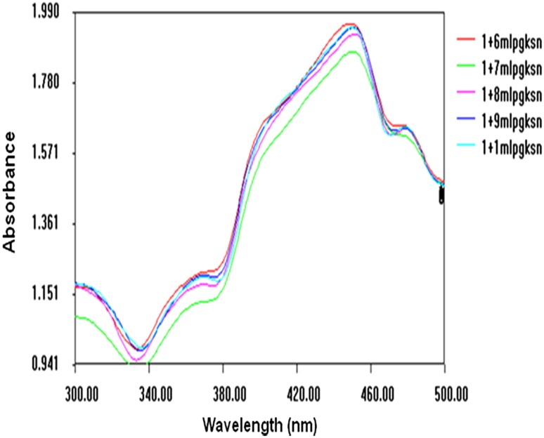
The fresh aqueous extract of A. sessilis and silver nitrate solution was mixed in 1:10 ratio and sonicated for 45 min. The yellow color solution changed to reddish-brown indicating the formation of silver nanoparticles. The completion of reduction of silver ions to nanosilver is evidenced from the broad surface plasmon resonance (SPR) band (420–450 nm) in the UV–visible spectra (Fig. 1). TEM analysis revealed the spherical morphology of the nanosilver with particle size in the range 10–30 nm (Fig. 2).
Fig. 1.
UV–visible spectrum of Alternanthera sessilis extract-mediated silver nanoparticles
Fig. 2.
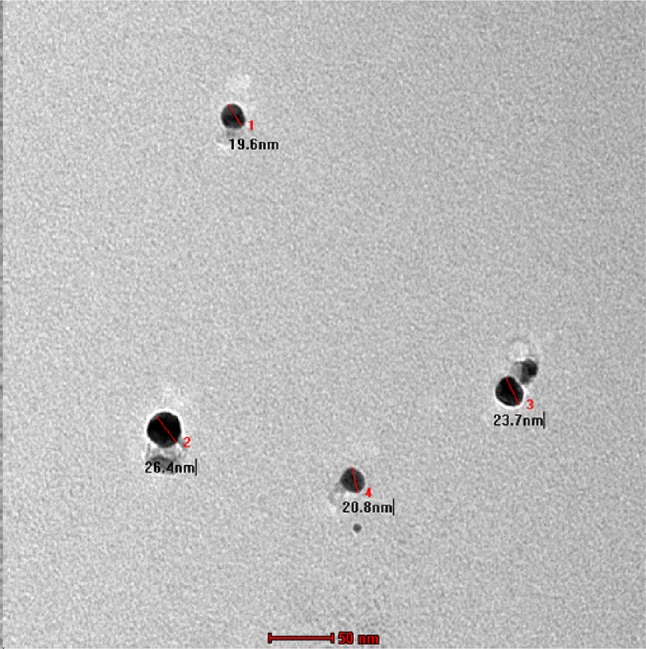
TEM micrograph of the biogenically synthesized silver nanoparticles
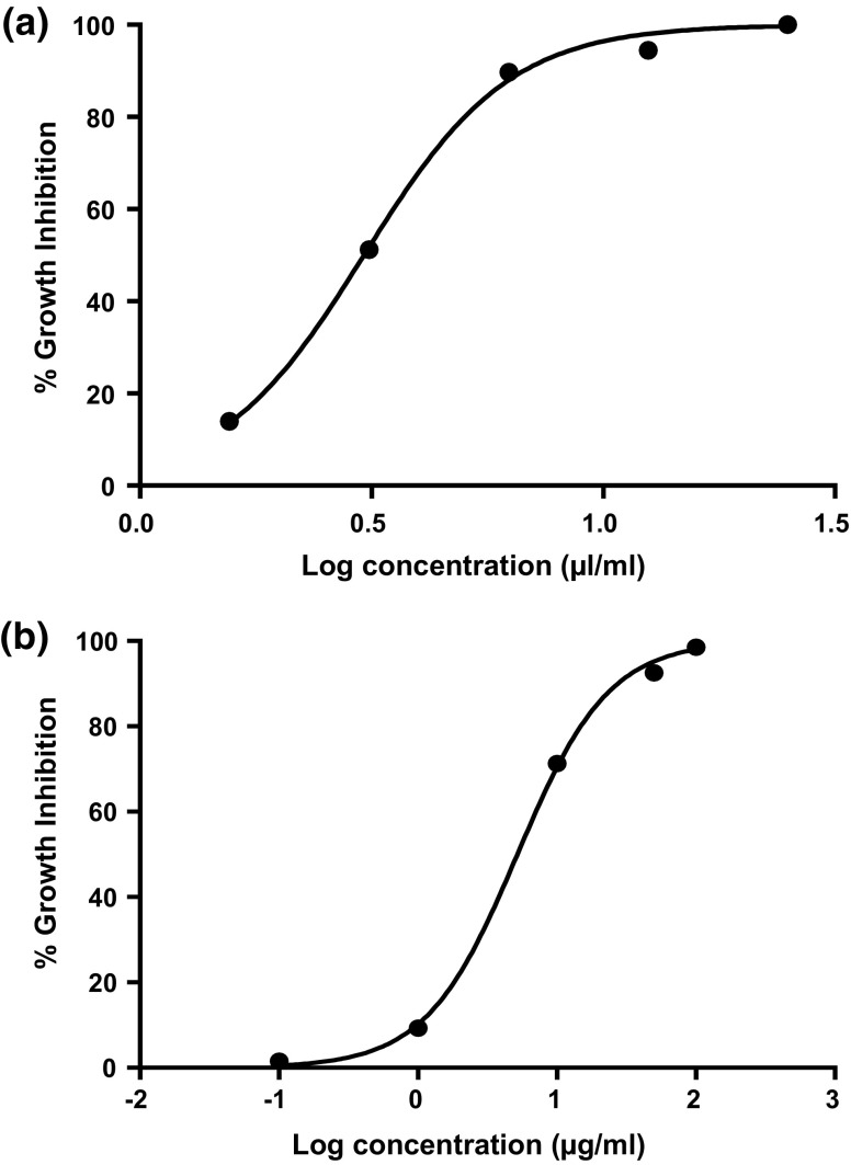
The in vitro cytotoxic effect of silver nanoparticles against breast cancer cell lines (MCF-7) and cell inhibition (%) was carried out by MTT assay and compared with the standard cisplatin. Cisplatin, the commercially available anticancer drug, was used as standard and its cytotoxicity is shown in Fig. 3a–c. Comparison of the cytotoxicity of synthesized AgNPs (1.56, 3.12, 6.25, 12.5, 25 µL/mL) and cisplatin (0.1, 1, 10, 50, 100 µM) disclosed similar mortality rate. The cytomorphological changes of AgNPs on MCF-7 cell lines at different concentrations (1.56, 6.25, 25 µL) involve intracellular suicide program possessing morphological changes like cell shrinkage, oxidative stress, coiling and biochemical response leading to apoptosis as shown in Fig. 4d–f. It is quite obvious from the results that the apoptosis rate of MCF-7 cell lines increases with increase in concentration of silver nanoparticles (Fig. 5a). A dose-dependent increase in cell inhibition is seen after 48 h exposure The IC50 of cell inhibition of silver nanoparticles was observed at 3.043 µL/mL. The complete cell inhibition (99 %) of breast cancer cell lines was obtained at a maximum concentration of 25 µg/mL. These results evidence the dose- and time-dependent increase in cytotoxicity. The IC50 value predicts that the plant-mediated nanosilver proves to be a promising drug for chemotherapeutic treatment. Complete apoptosis was observed with 25 µg/mL of AgNPs, whereas it is 30 µg/mL (or 100 µM) for cisplatin.
Fig. 3.
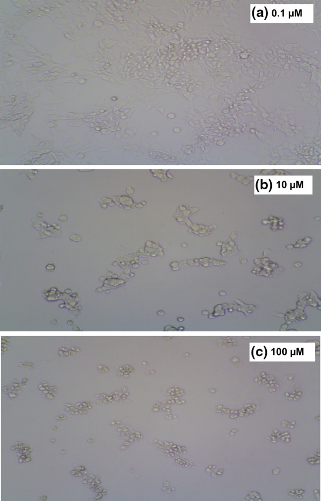
Cytomorphological changes and growth inhibition of cisplatin on MCF-7 cell line at a 0.1 µM, b 10 µM and c 100 µM concentrations
Fig. 4.
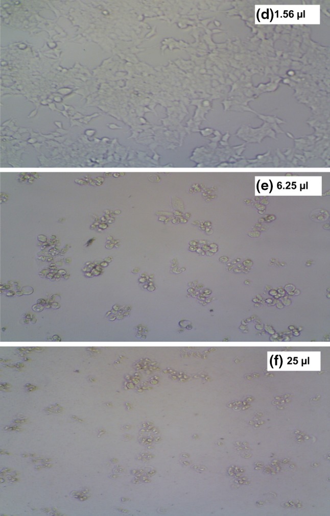
Cytomorphological changes and growth inhibition of silver nanoparticles on MCF-7 cell line at d 1.56 µL, e 6.25 µL and f 25 µL concentrations
Fig. 5.
Cytotoxicity of Alternanthera sessilis-embedded silver nanoparticles (a) and cisplatin (b) on human breast cancer MCF-7 cell lines
Alternanthera sessilis is enriched with flavonoids, glycosides, sugars, amino acids and steroids. Flavonoids are polyphenols which exhibit a wide variety of biological activities such as antioxidant, antibacterial, antiviral, anti-inflammatory and anticancer (Jogendra et al. 2012). There are reports on appreciable (86 %) cytotoxic activity of the chloroform fractionate of methanol extract of A. sessilis at maximum concentration (100 µg/mL) (Chan et al. 2008). Alternanthin B, a flavonoid isolated from Alternanthera philoxeroides species is known to possess antitumor activity (Zhou et al. 1988). Ragasa et al. (2002) reported that the chloroform extract of the air-dried leaves of A. sessilis afforded a mixture of diastereomers of new ionone derivatives which are anti-proliferative agents. The HPLC of ethanolic extract of A. sessilis evidences ellagic acid to be a prominent chemical constituent. Ellagic acid possesses antiproliferative activity.
Mode of action of drugs on cancer cells
A thorough review of literature embarks the different mechanisms of drug action on cancer cells. Interference of the electron transport mechanism of the cell by silver ions inhibits the respiratory mechanism as the silver cation readily oxidizes ATP resulting in the formation of silver (0). The replication and protein encoding of the bacteria get disrupted by the binding capability of silver to the DNA and RNA. The bacterial cell disruption aided by silver is similar to that of the action of platinum complex anticancer drug, cisplatin (Youngs et al. 2012).
Cisplatin, an anticancer drug has been commercially developed for selective killing of cancerous cells without any effect on non-cancerous or normal cells. Drugs used in cancer therapy enhance permeability and retention (EPR) effect, passively targeting the leaky vasculature of the tumor cells, since the cancer cells do not have a strong vasculature system compared to that of normal cells. Hence, the penetration of the drugs is easier and finally kills the cancer cells, but sometimes it enters into the normal cells and cause toxic effects (Jaracz et al. 2005; Blanco et al. 2009; Luo and Prestwich 2002; Leamon and Reddy 2004; Salazar and Ratnam 2007; Yoo and Park 2004).
To overcome these facets, the drugs can be loaded with nanoparticles and targeted moieties on the surface which will act against the particular receptor without affecting the normal cells. Many receptors have been discovered for cancer drug targeting, the commonly used one is folic acid (Kim 2006). The aqueous extract of Taxus baccata synthesized silver nanoparticles revealed potent anticancer effects on MCF-7 cells with an IC50 value of 0.25 μg/mL by MTT assay (Kajani et al. 2014). AntiABCG2 monoclonal antibody combined with AgNPs and Vincristine provide an efficient, targeted therapeutic method for inhibiting myeloma growth in mice (Dou et al. 2013). MDA-MB-231 breast cancer cells exposed to Ganoderma neojaponicum Imazeki-AgNPs after 24 h showed increased production, reactive oxygen species and hydroxyl radical. The apoptotic effects of AgNPs were further confirmed by the activation of caspase 3 and DNA nuclear fragmentation (Gurunathan et al. 2013a, b). The cytotoxicity of T. divaricata leaf extract synthesized AgNPs against human breast cancer cell indicates the presence of significant amounts of reducing entities (Devaraj et al. 2014).
Enhanced cytotoxic activity was observed for MCF-7 than A549 cells due to the increased cytotoxicity, decreased viability and proliferation which result in apoptosis through induced programmed cell death by irradiated AgNPs (MfouoTynga et al. 2014). The differential response of breast cancer cells to AgNPs induced hyperthermia, which implies AgNPs to be effective photothermal agents (Thompson et al. 2014). Apoptosis could be activated through Bax/BCl2 and caspase cascade mediated mitochondrial dysfunction, which potentially inhibits the proliferation of MCF-7 cells (Jeyaraj et al. 2015).
Proposed mode of action of nanosilver on MCF-7 cells
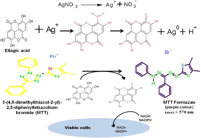
Figure 6 is the proposed mode of action of biogenically synthesized nanosilver embedded with ellagic acid on breast cancer cell lines. The nature of plant extract directly affects the physical, chemical and cytotoxic properties of the nanoparticles due to the interaction of nanoparticles with cells and intracellular macromolecules like proteins and DNA. Cellular uptake of nanoparticles leads to generation of reactive oxygen species which provoke oxidative stress. Cell damage by silver nanoparticles may be due to loss of cell membrane integrity, apoptosis and oxidative stress. A.sessilis is rich in beta ionones and flavonoid Alternanthin B the structures of which are given below. Molecules with ionone rings and Alternanthin B are known anti-tumor agents, though the mechanism of action is not well-defined in literature. 
Fig. 6.
Proposed mode of action of ellagic acid-embedded silver nanoparticles on MCF-7 cell line
β-Ionone is an end-ring analog of β-carotenoid which possesses potent antiproliferative activity. Mo and Elson (2004) demonstrated that β-ionone and a variety of isoprenoids can inhibit the growth of malignant cells in different experimental models. The cell cycle was arrested in the human gastric adenocarcinoma cancer cell lines treated with β-ionone by inhibition of cell growth and DNA synthesis in a dose-dependent manner and this may be regulated by mitogen-activated protein kinase pathways (Liu et al. 2004; Dong et al. 2013). The impaired activity of cyclin-dependent kinase (CDK) 2 and decreased expression of positive regulators of G1 to S phase progression aided by mevalonate depletion results in a G1 phase cell cycle arrest. The inhibition of HMG-CoA reductase activity also mediates the depletion of mevalonate which contributes to the cell cycle inhibitory and anti-proliferative effects of β-ionone on human breast cancer cells (MCF-7) (Duncan et al. 2004).
The flavonoid rings and ionone rings might have interfered with the gene expression, immune modulation and would have boosted the antioxidant effect resulting in apoptosis and death of cancer cells. Further studies are needed to optimize the mechanism of AgNPs on cancer cells. 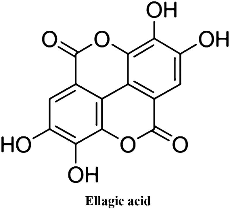
The mechanism of ellagic acid on MCF-7 cell lines is reported. The inhibition of proliferation of MCF-7 breast cancer cells is through the modulation of the TGF-β/Smad3 pathway associated with decreased phosphorylation of RB proteins (Zhang et al. 2014). Blueberry extracts, rich in ellagic acid, modulate the PI3K/AKT/NF kappa B pathway, and inhibits the growth and metastasis of MDA-MB-231 breast cancer cells (Adams et al. 2010). Treatment with ellagic acid arrests G0/G1 cell cycle and induces apoptosis in bladder cancer T24 cells (Li et al. 2005). It also induces apoptosis through G1/S cell cycle arrest in SW480 colon cancer cells (Narayanan and Re 2001). Han et al. (2006) reported ellagic acid to significantly reduce HOS cell proliferation and induce apoptosis as evidenced by chromosomal DNA degradation and through the upregulation of Bax and activation of caspase-3.
Ellagic acid metabolizes to urolithins-A in the gut, which exerts a remarkable antiproliferative activity in human colon cancer cells (Caco-2, SW-480 and HT-29) (González-Sarrías et al. 2005). The treatment of ellagic acid with PC3 cells results in a dose-dependent inhibition of cell growth/cell viability accompanied by induction of apoptosis and cleavage of poly(ADP-ribose) polymerase (PARP) and morphological changes. Ellagic acid induces apoptosis by upregulation of c-fos and pS2 protein in MCF-7; whereas, it follows intrinsic pathway in MDA-MB-231 cells. Hence, ellagic acid may have different pathways or antiproliferative activity on human breast cancer cell lines (Kim et al. 2009).
It has also been shown previously that NPs interfere with the MTT assay by adsorbing the tetrazolium salt (Wörle-Knirsch et al. 2006), or by releasing metal ions which modify the catalytic activity of the mitochondrial reductases (Kroll et al. 2009). Gurunathan et al. (2013a, b) revealed that the potential cytotoxic effect of biologically synthesized AgNPs in MDA-MB-231 cells accompanied by inhibiting the growth of cells, concentration-dependent activation of LDH, increased level of ROS generation and activation of caspase-3 are considered to be the most significant of the executioner caspases resulting in cellular apoptosis.
The mode of action of AgNPs and ellagic acid portrays AgNPs embedded with ellagic acid to inhibit the proliferation of MCF-7 cells through Bax/BCl2 and caspase cascade-mediated mitochondrial dysfunction accompanied by the TGF-β/Smad3 pathway. The combination of these pathways may be the reason for the increased rate of cell inhibition even at lower concentration. Thus, the AgNPs fabricated ellagic acid exhibits 99 % anti-proliferation at a concentration of 25 µL/mL. The present result was found to be valid on comparing with that of the cell inhibition acquired using individual components viz, ellagic acid and AgNPs.
Conclusion
Plant-mediated synthesis of silver nanoparticles using the extract of A. sessilis by sonication method advocates green nanotechnology. The results of the present cytotoxic study against human breast cancer MCF-7 cell lines by MTT assay revealed that silver nanoparticles serve as a potential anticancer drug compared to the standard cisplatin. Nanosilver showed excellent apoptosis rate due to their smaller size and spherical morphology. The present study contributes a novel and alternate approach in cancer therapy. The novelty of the present research is that complete cell inhibition (99 %) of breast cancer cell lines with plant-mediated nanosilver particles is obtained with 25 µg/mL, whereas it is 30 µg/mL for cisplatin, the standard drug used in breast cancer. Use of this edible plant extract eliminates the need to remove toxic byproducts during the formation of silver nanoparticles. Further studies are needed to optimize the mechanism of AgNPs on cancer cells.
Acknowledgments
The authors are sincerely thankful to the Avinashilingam Institute for Home Science and Higher Education for Women University, Coimbatore, Tamil Nadu, for providing research facilities and KMCH college of Pharmacy for the help rendered in cytotoxicity studies.
Compliance with ethical standards
Conflict of interest
The author(s) declare(s) that there is no conflict of interests.
References
- Adams LS, Phung S, Yee N, et al. Blueberry phytochemicals inhibit growth and metastatic potential of MDA-MB-231 breast cancer cells through modulation of the phosphatidylinositol 3-kinase pathway. Cancer Res. 2010;70:3594–3605. doi: 10.1158/0008-5472.CAN-09-3565. [DOI] [PMC free article] [PubMed] [Google Scholar]
- Alison MR. Cancer. Encyclopedia of life sciences. London: Nature Publishing Group; 2001. [Google Scholar]
- Anandkumar BH, Sachidanand YN. Treatment of acne vulgaris with new polyherbal formulation clarina cream. Indian J Dermatol. 2001;46:138–141. [Google Scholar]
- Arollado EC, Osi MO. Hematinic activity of Alternanthera sessilis (L.) R. BR. (Amaranthaceae) in mice and rats. E Int Sci Res J. 2010;2:110–117. [Google Scholar]
- Balasuriya BMGK, Dharmaratne HRW. Cytotoxicity and antioxidant activity studies of green leafy vegetables consumed in Sri Lanka. J Natl Sci Found Sri Lanka. 2007;35:255–258. [Google Scholar]
- Blanco E, Kessinger CW, Sumer BD, Gao J. Multifunctional micellar nanomedicine for cancer therapy. Exp Biol Med. 2009;234:123–131. doi: 10.3181/0808-MR-250. [DOI] [PMC free article] [PubMed] [Google Scholar]
- Borah A, Yadav RNS, Unni BG. In vitro antioxidant and free radical scavenging activity of Alternanthera sessilis. Int J Pharm Sci Rev Res. 2011;2:1502–1506. [Google Scholar]
- Chan LL, Gosangari SL, Watkin KL, Cunningham BT. Label-free imaging of cancer cells using photonic crystal biosensors and application to cytotoxicity screening of a natural compound library. Sens Actuators B Chem. 2008;132:418–425. doi: 10.1016/j.snb.2007.10.027. [DOI] [Google Scholar]
- Devaraj P, Aarti C, Kumari P. Synthesis and characterization of silver nanoparticles using Tabernae montana divaricata and its cytotoxic activity against MCF7 cell line. Int J Pharm Pharm Sci. 2014;6(8):86–90. [Google Scholar]
- Dipankar C, Murugan S. The green synthesis, characterization and evaluation of the biological activities of silver nanoparticles synthesized from Iresine herbstii leaf aqueous extracts. Colloids Surf B. 2012;98:112–119. doi: 10.1016/j.colsurfb.2012.04.006. [DOI] [PubMed] [Google Scholar]
- Dong HW, Zhang S, Sun WG, Liu Q, Ibla JC, Soriano SG, Han XH, Liu LX, Li MS, Liu JR. β-Ionone arrests cell cycle of gastric carcinoma cancer cells by a MAPK pathway. Arch Toxicol. 2013;87(10):1797–1808. doi: 10.1007/s00204-013-1041-5. [DOI] [PubMed] [Google Scholar]
- Dou J, He X, Liu Y, Huang Z, Yang C, Shi F, Chen D, Gu N. Targeted therapeutic effect of antiABCG2 antibody combined with nano silver and vincristine on mouse myeloma cancer stem cells. J Nanoparticles Res. 2013;15(2127):1–9. [Google Scholar]
- Duncan RE, Lau D, El-Sohemy A, Archer MC. Geraniol and β-ionone inhibit proliferation, cell cycle progression and cyclin-dependent kinase 2 activity in MCF-7 breast cancer cells independent of effects on HMG-CoA reductase activity. Biochem Pharmacol. 2004;68:1739–1747. doi: 10.1016/j.bcp.2004.06.022. [DOI] [PubMed] [Google Scholar]
- Firdhouse MJ, Lalitha P. Biosynthesis of silver nanoparticles using the extract of Alternanthera sessilis—antiproliferative effect against prostate cancer cells. Cancer Nanotechnol. 2013;4:137–143. doi: 10.1007/s12645-013-0045-4. [DOI] [PMC free article] [PubMed] [Google Scholar]
- Gajendran B, Chinnasamy A, Durai P, Raman J, Ramar M. Biosynthesis and characterization of silver nanoparticles from Datura inoxia and its apoptotic effect on human breast cancer cell line MCF7. Mater Lett. 2014;122:98–102. doi: 10.1016/j.matlet.2014.02.003. [DOI] [Google Scholar]
- González-Sarrías A, Tomé-Carneiro J, Bellesia A, Tomás-Barberána FA, Espína JC. The ellagic acid-derived gut microbiota metabolite, urolithin A, potentiates the anticancer effects of 5-fluorouracil chemotherapy on human colon cancer cells. Food Funct. 2005 doi: 10.1039/c5fo00120j. [DOI] [PubMed] [Google Scholar]
- Gurunathan S, Raman J, Abd Malek SN, John PA, Vikineswary S. Green synthesis of silver nanoparticles using Ganoderma neojaponicum Imazeki: a potential cytotoxic agent against breast cancer cells. Int J Nanomed. 2013;8:4399–4413. doi: 10.2147/IJN.S51881. [DOI] [PMC free article] [PubMed] [Google Scholar]
- Gurunathan S, Han JW, Eppakayala V, Jeyaraj M, Kim J. Cytotoxicity of biologically synthesized silver nanoparticles in MDA-MB-231 human breast cancer cells. BioMed Res Int. 2013;2013:4399–4413. doi: 10.1155/2013/535796. [DOI] [PMC free article] [PubMed] [Google Scholar]
- Han DH, Lee MJ, Kim JH. Antioxidant and apoptosis-inducing activities of ellagic acid. Anticancer Res. 2006;26:3601–3606. [PubMed] [Google Scholar]
- Jacob SJP, Finub JS, Narayanan A. Synthesis of silver nanoparticles using Piper longum leaf extracts and its cytotoxic activity against Hep-2 cell line. Colloids Surf B Biointerfaces. 2012;91:212–214. doi: 10.1016/j.colsurfb.2011.11.001. [DOI] [PubMed] [Google Scholar]
- Jalalpure SS, Agrawal N, Patil MB, Chimkode R, Tripathi A. Antimicrobial and wound healing activities of leaves of Alternanthera sessilis Linn. Int J Green Pharm. 2008;2:141–144. doi: 10.4103/0973-8258.42729. [DOI] [Google Scholar]
- Jaracz S, Chen J, Kuznetsova LV, Ojima I. Recent advances in tumor-targeting anticancer drug conjugates. Bioorg Med Chem. 2005;13:5043–5054. doi: 10.1016/j.bmc.2005.04.084. [DOI] [PubMed] [Google Scholar]
- Jeyaraj M, Renganathan A, Sathishkumar G, Ganapathi A, Premkumar K. Biogenic metal nanoformulations induce Bax/Bcl2 and caspase mediated mitochondrial dysfunction in human breast cancer cells (MCF-7) RSC Adv. 2015;5(3):2159–2166. doi: 10.1039/C4RA11686K. [DOI] [Google Scholar]
- Jogendra CH, Avinash VP, Mohan VK, Patil DA, Ravindra GM. A current update on phytopharmacology of the genus Alternanthera. J Pharm Res. 2012;5(4):1924–1929. [Google Scholar]
- Jou HJ, Lin YM, Lin YC, Chen FC. Constituents of Alternanthera sessilis R. Br Huaxue. 1979;1:22–25. [Google Scholar]
- Kajani AA, Bordbar AK, Esfahani SHZ, Khosropour AR, Razmjou A. Green synthesis of anisotropic silver nanoparticles with potent anticancer activity using Taxus baccata extract. RSC Adv. 2014;4:61394–61403. doi: 10.1039/C4RA08758E. [DOI] [Google Scholar]
- Kathiravan V, Ravi S, Ashokkumar S. Synthesis of silver nanoparticles from Melia dubia leaf extract and their in vitro anticancer activity. Spectrochim Acta Part A: Mol Biomol Spectrosc. 2014;130:116–121. doi: 10.1016/j.saa.2014.03.107. [DOI] [PubMed] [Google Scholar]
- Kim Y. Does a high folate intake increase the risk of breast cancer? Nutr Rev. 2006;64(10):468–475. doi: 10.1111/j.1753-4887.2006.tb00178.x. [DOI] [PubMed] [Google Scholar]
- Kim H, Lee R, Moon B, Choe K. Ellagic acid shows different anti-proliferative effects between the MDA-MB-231 and MCF-7 human breast cancer cell lines. J Breast Cancer. 2009;12(2):85–91. doi: 10.4048/jbc.2009.12.2.85. [DOI] [Google Scholar]
- Krishnaraj C, Muthukumaran P, Ramachandran R, Balakumaran MD, Kalaichelvan PT. Acalypha indica Linn: biogenic synthesis of silver and gold nanoparticles and their cytotoxic effects against MDA-MB-231, human breast cancer cells. Biotechnol Rep. 2014;4:42–49. doi: 10.1016/j.btre.2014.08.002. [DOI] [PMC free article] [PubMed] [Google Scholar]
- Kroll A, Pillukat MH, Hahn D, Schnekenburger J. Current in vitro methods in nanoparticle risk assessment: limitations and challenges. Eur J Pharm Biopharm. 2009;72:370–377. doi: 10.1016/j.ejpb.2008.08.009. [DOI] [PubMed] [Google Scholar]
- Leamon CP, Reddy JA. Folate-targeted chemotherapy. Adv Drug Deliv Rev. 2004;56:1127–1141. doi: 10.1016/j.addr.2004.01.008. [DOI] [PubMed] [Google Scholar]
- Li TM, Chen GW, Su CC, et al. Ellagic acid induced p53/p21 expression, G1 arrest and apoptosis in human bladder cancer T24 cells. Anticancer Res. 2005;25:971–979. [PubMed] [Google Scholar]
- Lin SC, Lin YH, Shyuu SJ, Lin CC. Hepatoprotective effects of Taiwan folk medicine: alternanthera sessilis on liver damage induced by various hepatotoxins. Phytother Res. 1994;8:391–398. doi: 10.1002/ptr.2650080703. [DOI] [Google Scholar]
- Liu JR, Chen BQ, Yang BF, Dong HW, Sun CH, Wang Q, Song G, Song YQ. Apoptosis of human gastric adenocarcinoma cells induced by β-ionone. World J Gastroenterol. 2004;10:348–351. doi: 10.3748/wjg.v10.i3.348. [DOI] [PMC free article] [PubMed] [Google Scholar]
- Lokina S, Stephen A, Kaviyarasan V, Arulvasu C, Narayanan V. Cytotoxicity and antimicrobial activities of green synthesized silver nanoparticles. Eur J Med Chem. 2014;76:256–263. doi: 10.1016/j.ejmech.2014.02.010. [DOI] [PubMed] [Google Scholar]
- Losso JN, Bansode RR, Trappey A, Bawadi HA, Truax R. In vitro anti-proliferative activities of ellagic acid. J Nutr Biochem. 2004;15:672–678. doi: 10.1016/j.jnutbio.2004.06.004. [DOI] [PubMed] [Google Scholar]
- Luo Y, Prestwich GD. Cancer-targeted polymeric drugs. Curr Cancer Drug Targets. 2002;2:209–226. doi: 10.2174/1568009023333836. [DOI] [PubMed] [Google Scholar]
- MfouoTynga I, ElHussein A, AbdelHarith M, Abrahamse H. Photodynamic ability of silver nanoparticles in inducing cytotoxic effects in breast and lung cancer cell lines. Int J Nanomed. 2014;9(1):3771–3780. [Google Scholar]
- Mo H, Elson CE. Studies of the isoprenoid-mediated inhibition of mevalonate synthesis applied to cancer chemotherapy and chemoprevention. Exp Biol Med. 2004;229:567–585. doi: 10.1177/153537020422900701. [DOI] [PubMed] [Google Scholar]
- Mondal H, Hossain H, Awang K, Saha S, Rashid SMU, Islam MK, Rahman MS, Jahan IA, Rahman MM, Shilpi JA. Anthelmintic activity of ellagic acid, a major constituent of Alternanthera sessilis against Haemonchus contortus. Pak Vet J. 2015;35(1):58–62. [Google Scholar]
- Nagajyothi PC, Sreekanth TVM, Lee JI, Lee KD. Mycosynthesis: antibacterial, antioxidant and antiproliferative activities of silver nanoparticles synthesized from Inonotus obliquus (Chaga mushroom) extract. J Photochem Photobiol B Biol. 2014;130:299–304. doi: 10.1016/j.jphotobiol.2013.11.022. [DOI] [PubMed] [Google Scholar]
- Narayanan BA, Re GG. IGF-II down regulation associated cell cycle arrest in colon cancer cells exposed to phenolic antioxidant ellagic acid. Anticancer Res. 2001;21:359–364. [PubMed] [Google Scholar]
- Nayak P, Nayak S, Kar DM, Das P. Pharmacological evaluation of ethanolic extracts of the plant Alternanthera sessilis against temperature regulation. J Pharm Res. 2010;3:1381–1383. [Google Scholar]
- Purkayastha J, Nath SC. Biological activities of ethnomedicinal claims of some plant species of Assam. Indian J Tradit Knowl. 2006;5:229–236. [Google Scholar]
- Ragasa CY, Tremor N, Redeout JA. Ionone derivates from Alternanthera sessilis. J Asian Nat Prod Res. 2002;4(2):109–115. doi: 10.1080/10286020290027380. [DOI] [PubMed] [Google Scholar]
- Raghunandan D, Ravishankar B, Sharanbasava G, Mahesh DB, Harsoor M, Yalagatti MS, Bhagawanraju M, Venkataraman A. Anti-cancer studies of noble metal nanoparticles synthesized using different plant extracts. Cancer Nanotechnol. 2011;2:57–65. doi: 10.1007/s12645-011-0014-8. [DOI] [PMC free article] [PubMed] [Google Scholar]
- Ranjitham AM, Suja R, Caroling G, Tiwari S. In vitro evaluation of antioxidant, antimicrobial, anticancer activities and characterisation of Brassica Oleracea. var. bortrytis. L synthesized silver nanoparticles. Int J Pharm Pharm Sci. 2013;5(4):239–251. [Google Scholar]
- Rathi Sre PR, Reka M, Poovazhagi R, Arul Kumar M, Murugesan K. Antibacterial and cytotoxic effect of biologically synthesized silver nanoparticles using aqueous root extract of Erythrina indica lam. Spectrochim Acta Part A Mol Biomol Spectrosc. 2015;135:1137–1144. doi: 10.1016/j.saa.2014.08.019. [DOI] [PubMed] [Google Scholar]
- Reddy NJ, Nagoor Vali D, Rani M, Rani SS. Evaluation of antioxidant, antibacterial and cytotoxic effects of green synthesized silver nanoparticles by Piper longum fruit. Mater Sci Eng C. 2014;34(1):115–122. doi: 10.1016/j.msec.2013.08.039. [DOI] [PubMed] [Google Scholar]
- Roy A, Saraf S. Diuretic activity of Alternanthera sessilis R.Br. Ex D.C. an ethnomedicine of Chhattisgarh (India) Biosci Biotechnol Res Asia. 2008;5:369–372. [Google Scholar]
- Sahithi B, Rajani GP, Sowjanya K, Gupta D. Anti-inflammatory activity of ethanolic and aqueous extracts of Alternanthera sessilis lin. Pharmacologyonline. 2011;1:1039–1043. [Google Scholar]
- Salazar MD, Ratnam M. The folate receptor: what does it promise in tissue-targeted therapeutics? Cancer Metastasis Rev. 2007;26:141–152. doi: 10.1007/s10555-007-9048-0. [DOI] [PubMed] [Google Scholar]
- Sathishkumar G, Gobinath C, Wilson A, Sivaramakrishnan S. Dendrophthoe falcata (L.f) Ettingsh (Neem mistletoe): a potent bioresource to fabricate silver nanoparticles for anticancer effect against human breast cancer cells (MCF7) Spectrochim Acta Part A Mol Biomol Spectrosc. 2014;128:285–290. doi: 10.1016/j.saa.2014.02.096. [DOI] [PubMed] [Google Scholar]
- Sinha P, Arora VK, Wahi SP. Chemical investigation on Alternanthera sessilis. Indian Drugs. 1984;21:139–140. [Google Scholar]
- Sriram MI, Kalishwaralal K, Barathmanikanth S, Gurunathani S. Size-based cytotoxicity of silver nanoparticles in bovine retinal endothelial cells. Nanosci Methods. 2012;1(1):56–77. doi: 10.1080/17458080.2010.547878. [DOI] [Google Scholar]
- Strati A, Papoutsi Z, Lianidou E, Moutastsou P. Effect of ellagic acid on the expression of human telomerase reverse transcriptase (hTERT) α+β+ transcript in estrogen receptor positive MCF-7 breast cancer cells. Clin Biochem. 2009;42:1358–1362. doi: 10.1016/j.clinbiochem.2009.05.017. [DOI] [PubMed] [Google Scholar]
- Sulaiman GM, Mohammad AAW, Abdul-Wahed HE, Ismail MM. Biosynthesis, antimicrobial and cytotoxic effects of silver nanoparticles using Rosmarinus officinalis extract. Dig J Nanomater Biostruct. 2013;8(1):273–280. [Google Scholar]
- Sulaiman GM, Mohammed WH, Marzoog TR, AlAmiery AA, Kadhum AAH, Mohamad AB. Green synthesis, antimicrobial and cytotoxic effects of silver nanoparticles using Eucalyptus chapmaniana leaves extract. Asian Pac J Trop Biomed. 2013;3(1):58–63. doi: 10.1016/S2221-1691(13)60024-6. [DOI] [PMC free article] [PubMed] [Google Scholar]
- Suman TY, Radhika Rajasree SR, Kanchana A, Elizabeth Been S. Biosynthesis, characterization and cytotoxic effect of plant mediated silver nanoparticles using Morinda citrifolia root extract. Colloids Surf B Biointerfaces. 2013;106:74–78. doi: 10.1016/j.colsurfb.2013.01.037. [DOI] [PubMed] [Google Scholar]
- Teli MK, Mutalik S, Rajanikant GK. Nanotechnology and nanomedicine: going small means aiming big. Curr Pharm Des. 2010;16(16):1882–1892. doi: 10.2174/138161210791208992. [DOI] [PubMed] [Google Scholar]
- Thompson EA, Graham E, Macneill CM, Young M, Donati G, Wailes EM, Jones BT, LeviPolyachenko NH. Differential response of MCF7, MDAMB231 and MCF 10A cells to hyperthermia, silver nanoparticles and silver nanoparticles induced photothermal therapy. Int J Hyperth. 2014;30(5):312–323. doi: 10.3109/02656736.2014.936051. [DOI] [PubMed] [Google Scholar]
- Thorley AJ, Tetley TD. New perspectives in nanomedicine (Review) Pharmacol Ther. 2013;140(2):176–185. doi: 10.1016/j.pharmthera.2013.06.008. [DOI] [PubMed] [Google Scholar]
- Vivek R, Thangam R, Muthuchelian K, Gunasekaran P, Kaveri K, Kannan S. Green biosynthesis of silver nanoparticles from Annona squamosa leaf extract and its in vitro cytotoxic effect on MCF7 cells. Process Biochem. 2012;47(12):2405–2410. doi: 10.1016/j.procbio.2012.09.025. [DOI] [Google Scholar]
- Wang B, Yang Q, Wang Y, Li Z. The toolbox of designing nanoparticles for tumors. Mini Rev Med Chem. 2014;14(9):707–716. doi: 10.2174/1389557514666140820122307. [DOI] [PubMed] [Google Scholar]
- Wörle-Knirsch JM, Pulskamp K, Krug HF. Oops they did it again! Carbon nanotubes hoax scientists in viability assays. Nano Lett. 2006;6:1261–1268. doi: 10.1021/nl060177c. [DOI] [PubMed] [Google Scholar]
- Yoo HS, Park TG. Folate receptor targeted biodegradable polymeric doxorubicin micelles. J Control Release. 2004;96:273–283. doi: 10.1016/j.jconrel.2004.02.003. [DOI] [PubMed] [Google Scholar]
- Youngs WJ, Knapp AR, Wagersa PO, Tessier CA. Nanoparticle encapsulated silver carbene complexes and their antimicrobial and anticancer properties: a perspective. Dalton Trans. 2012;41:327–336. doi: 10.1039/C1DT11100K. [DOI] [PubMed] [Google Scholar]
- Zhang T, Chen H, Wang L, Bai M, Wang Y, Jiang X, Liu M. Ellagic acid exerts anti-proliferation effects via modulation of Tgf-Β/Smad3 signaling in MCF-7 breast cancer cells. Asian Pac J Cancer Prev. 2014;15(1):273–276. doi: 10.7314/APJCP.2014.15.1.273. [DOI] [PubMed] [Google Scholar]
- Zhou B, Gabor B, Cordell GA. Alternanthin, a C- glycosylated flavonoids from Alternanthera philoxeroides. Phytochem. 1988;27(11):3633–3636. doi: 10.1016/0031-9422(88)80781-7. [DOI] [Google Scholar]


