Abstract
Central pontine myelinolysis (CPM), which was originally considered to be the result of rapid correction of chronic hyponatremia, is not necessarily accompanied by hyponatremia or drastic changes in serum sodium level. Here, we report a case of an anorexic 55-year-old male with a history of pharyngo-laryngo-esophagogastrectomy, initially hospitalized with status epilepticus. Although his consciousness gradually recovered as we were controlling his convulsion, it deteriorated again with new onset of anisocoria, and magnetic resonance imaging (MRI) at this point revealed CPM. Rapid change of serum sodium or osmolarity, which is often associated with CPM, had not been apparent throughout his hospitalization. Instead, a review of the serum biochemistry test results showed that serum phosphate had drastically declined the day before the MRI first detected CPM. In this case, we suspect that hypophosphatemia induced by refeeding syndrome greatly contributed to the occurrence of CPM.
Key Words: Central pontine myelinolysis, Refeeding syndrome, Hypophosphatemia, Hypokalemia, Hyponatremia, Thiamine deficiency, Status epilepticus
Introduction
Central pontine myelinolysis (CPM) was originally considered to be the result of excessively rapid correction of slowly progressive hyponatremia in patients with chronic medical conditions, such as chronic alcoholism, malnutrition, and malignancy [1, 2]. However, there have been occasional reports of CPM not accompanied by hyponatremia or drastic changes in serum sodium level [1, 2]. Among these reports are cases in which hypophosphatemia, rather than hyponatremia, was suspected to have greatly contributed to the pathogenesis of CPM [3]. In this case report, we describe the case of a patient with CPM caused by hypophosphatemia secondary to refeeding syndrome (RFS).
Case Report
A 55-year-old Japanese male was admitted to the emergency department because of a new-onset generalized convulsive seizure. He had no significant family history of this condition. He had no remarkable medical history with the exception of a diagnosis of esophageal squamous cell carcinoma and gastric adenocarcinoma 6 months prior to admission. He underwent total pharyngo-laryngo-esophagogastrectomy and distal gastrectomy with adjuvant chemoradiotherapy. TS-1 (tegafur, gimeracil, oteracil potassium) was started 7 weeks before his presentation, which led to gradual loss of appetite. He had also predominantly stopped smoking and drinking alcohol after the surgery. The day before admission, he consumed one glass of sake and his prescribed medications (TS-1 80 mg, levothyroxine 50 μg, alfacalcidol 2 μg, calcium lactate 9 g, and methylcobalamin 1,500 μg) and barely consumed any food. According to his family, his neurological status at this point was unremarkable except that he was unable to vocalize because of a past tracheostomy.
On admission, his height was 161 cm, and his weight was 44.7 kg. His blood pressure was 178/102 mm Hg, his heart rate was 120–130 beats per minute, his respiratory rate was 22 breaths per minute, and his body temperature was 36.4°C. His physical examination was unremarkable with the exception of a permanent tracheal stoma. His consciousness was E4VTM1 based on the Glasgow Coma Scale (GCS). His left hemibody, including face, was almost continuously in convulsion, and his right hemibody occasionally cramped. Right conjugate gaze was observed, with round, isocoric, and normal-sized pupils. Light reflexes were sluggish bilaterally, but other brainstem reflexes were preserved. All limbs were flaccid during the intervals of seizure. Deep tendon reflexes were normal in the upper extremities and were reduced bilaterally in the lower extremities. There was no meningeal sign.
Serum biochemistry tests indicated dehydration and malnutrition, as demonstrated by the following values: albumin 3.5 g/dl, blood urea nitrogen 44 mg/dl, creatinine 1.68 mg/dl, potassium 2.7 mmol/l, magnesium 1.2 mg/dl (normal range: 1.7–2.2), vitamin B1 13 ng/ml (normal range: 20–50), and glucose 229 mg/dl. Total corrected calcium was elevated at 14.2 mg/dl (with negative parathyroid hormone-related protein C), which was likely because of calcium replenishment. Thyroid and parathyroid function was mildly reduced (thyroid stimulating hormone 22.31 μU/ml, fT4 0.96 ng/dl, intact parathyroid hormone 2.3 pg/ml), presumably as a result of the laryngopharyngectomy and use of levothyroxine, but autoantibodies, such as antithyroglobulin antibody and antithyroperoxidase antibody, were negative.
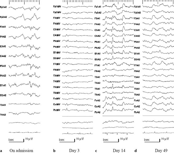
Cerebrospinal fluid analysis showed normal protein concentration and cell counts. Antibodies against herpes simplex virus, varicella zoster virus, and cytomegalovirus were negative. Polymerase chain reaction analyses for herpes simplex virus and human herpesvirus-6 were negative. Brain magnetic resonance imaging (MRI) on admission demonstrated diffusion-weighted imaging (DWI) hyperintensity with mildly reduced apparent diffusion coefficient in the right medial temporal lobe, bilateral thalami, bilateral corona radiate, and right cingulate gyrus (fig. 1). No space-occupying lesion was apparent. A magnetic resonance venogram was unremarkable. Electroencephalography (EEG) showed bilateral periodic sharp waves of approximately 0.5 Hz with the right amplitude slightly higher than the left (fig. 2).
Fig. 1.

MRI time course. MRI on admission shows DWI-hyperintense lesions in the bilateral thalami and right hippocampus (arrows). On hospital day 7, when anisocoria appeared, these lesions had vanished while a new DWI-hyperintense lesion is visible in the middle of the pons. This lesion remained apparent on follow-up MRI on hospital day 33.
Fig. 2.

EEGs. a On admission: bilateral periodic sharp waves of approximately 0.5 Hz with the right amplitude slightly higher than the left. b Day 3: sharp waves with maximum amplitude at the right temporal area. c Day 14: occasional spikes in the left posterior quadrant with right hemispheric suppression. d Day 49: no apparent spikes. The recording of day 3 was averaged reference montage. Otherwise the recordings were ear reference montage.
Four days of parenteral nutrition beginning with 100 kcal/day with intravenous phenytoin (750 mg/day) and thiamine (100 mg/day), and enteral magnesium sulfate decreased the frequency and duration of seizures, and on hospital day 5, he understood simple instructions during intervals between seizures. On the morning of hospital day 7, however, a relapse of unresponsiveness lasted for a few hours, and anisocoria was observed without apparent seizure. A repeated brain MRI revealed a lesion of CPM (hyperintensity in the central pons on T2-weighted and fluid-attenuated inversion recovery sequence and hypointensity on T1-weighted sequence), while the DWI hyperintensity lesions that were present from his MRI on admission had disappeared (fig. 1). On hospital day 20, seizures were finally controlled with the addition of carbamazepine (1,200 mg/day) and correction of electrolytes. However, altered consciousness of E4VTM4 on the GCS, anisocoria, and weakness in the left lower extremities of 3/5 on manual muscle testing persisted.
EEG was not performed on hospital day 7, when unresponsiveness recurred and the CPM lesion was revealed for the first time. However, follow-up EEGs were performed on hospital days 3 and 14 (fig. 2b, c); sharp waves with maximum amplitude at the right temporal area were observed on day 3, and occasional spikes in the left posterior quadrant areas with right-hemispheric suppression were observed on day 14. Another follow-up EEG was performed on hospital day 49, which no longer showed apparent spikes.
The patient's serum sodium and serum osmolarity had not been markedly low or drastically changed since admission. However, a review of stored blood samples before the appearance of anisocoria and relapsed unresponsiveness revealed that the patient's serum phosphate had fallen from 3.3 mg/dl on admission to 0.7 mg/dl the day after (normal range: 2.4–4.4 mg/dl), following parenteral nutrition initiation (table 1). Considering that this drastic decrease in phosphate occurred just before his change in symptoms, in the setting of preexisting hypokalemia, hypomagnesemia, and thiamine deficiency, RFS was presumed to be the cause of CPM in this patient. The neurological signs attributed to the CPM lesion (i.e., mental deterioration, anisocoria, and weakness in the left lower extremities) were sustained even after the seizures were completely controlled and serum biochemistry parameters returned to normal.
Table 1.
Parenteral nutrition rate and serum biochemistry parameters (mainly electrolytes) throughout hospital stay
| Hospital day | ||||||||||||
|---|---|---|---|---|---|---|---|---|---|---|---|---|
| 1 | 2 | 3 | 4 | 5 | 7* | 8 | 12 | 14 | 20 | 21 | 28 | |
| Parenteral nutrition, kcal/day | 100 | 100 | 800 | 800 | 1,000 | 1,200 | 1,600 | 1,600 | 1,700 | 1,700 | 1,700 | 1,700 |
| Albumin (3.7–4.9 g/dl) | 3.5 | 2.7 | 2.1 | 3.1 | 2.8 | 2.8 | 2.6 | 2.5 | 2.8 | 2.9 | 2.9 | 3.4 |
| Calcium (8.5–10.5 mg/dl) | 14.2 | 10.4 | 8.6 | 8.8 | 7.7 | 7.6 | 7.6 | 6.3 | 6.5 | 8 | 7.5 | 9.4 |
| Phosphorus (2.5–4.6 mg/dl) | 3.3 | 0.7 | 1.8 | 4.9 | ||||||||
| Sodium (135–149 mmol/l) | 133 | 139 | 138 | 140 | 141 | 139 | 137 | 141 | 139 | 134 | 132 | 136 |
| Potassium (3.5–4.9 mmol/l) | 2.7 | 2.8 | 2.8 | 3.3 | 3.4 | 4.4 | 3.7 | 3.3 | 3.3 | 4 | 4.5 | 4.9 |
| Magnesium (1.8–2.4 mg/dl) | 1.2 | 0.8 | 1.3 | 1.8 | 2 | 1.9 | 2.9 | |||||
| Thiamine (20–50 ng/ml) | 13 | |||||||||||
| Plasma osmolality (275–295 mOsm/kg H2O) | 280 | 272 | ||||||||||
The day CPM was first detected on MRI.
Discussion
This patient developed CPM without rapid change of serum osmolarity, although his background was typical of CPM in that he had both malignancy and malnutrition [1, 2]. Also, the tendency for alcohol abuse may have contributed, although he was not necessarily alcoholic.
The mechanism of how hypophosphatemia causes CPM has not been fully elucidated. One hypothesis is that the lack of energy supply to glial cells because of the reduction of ATP production caused by hypophosphatemia might lead to widespread dysfunction of Na+/K+-ATPase pumps, resulting in apoptosis [4]. Another possible mechanism may be the reduction of cell-protective organic osmolytes, such as phosphocreatine and glycerophosphorylcholine, as phosphate is required for their synthesis [5].
In our patient, hyponatremia and temporal changes in sodium and osmolarity were minimal, whereas hypophosphatemia emerged within only 2 days after the initiation of parenteral nutrition. Our patient also had preexisting hypokalemia and hypomagnesemia, and such electrolyte abnormalities prior to parenteral nutrition to an undernourished patient are typical features of RFS [6]. RFS is best defined as a series of metabolic and biochemical changes that occur as a consequence of reintroduction of feeding after a period of fasting, leading to various sequelae, such as cardiac and neurological abnormalities [7]. Although RFS has no formal definition, hypophosphatemia, caused by redistribution of phosphate, is a hallmark feature of RFS [8] and can be observed as early as 1 day after starting parenteral nutrition [9]. Based on this, a starting rate of 5 kcal/kg/24 h is recommended for extremely undernourished patients [10]. Although the rate of caloric feeding of our patient was within this standard rate on the first day of feeding, it was exceeded the following day. This might have precipitated RFS, resulting in hypophosphatemia, eventually leading to CPM. Hypokalemia is also implicated in the underlying mechanism of CPM, by increasing the potential gradient against which the ATPase must operate [11]. Thus preexisting hypokalemia in our patient may also have contributed to CPM. Other than hypokalemia, our patient also had thiamine deficiency, which is suspected, though not definitely confirmed, to be a risk factor for CPM [12, 13]. However, as a significant change was observed only for phosphate before the onset of CPM, we assume that hypophosphatemia was the main cause of CPM in our patient.
Some studies during the acute phase of CPM have suggested that clinical deficits may precede the appearance of MRI abnormalities by 2 or 3 weeks [14]. In this respect, CPM might have developed before the emergence of hypophosphatemia in our patient. However, considering that anisocoria – which is likely due to the pontine lesion, as already mentioned – developed on the very morning of MRI confirmation of CPM, we assume that this was the cause of onset of CPM in our patient.
Serum calcium rapidly decreased and serum magnesium level was low before the CPM lesion was first noticed. We do not completely deny that these factors contributed to the pathogenesis of CPM in this patient, but serum magnesium or calcium per se has not been considered to be causative for CPM [15].
This patient was hospitalized originally for a new-onset convulsive seizure. The cerebrospinal fluid findings were unremarkable, the MRI abnormality on admission was transient, and he was not necessarily alcoholic, although he used to consume higher than average amounts of alcohol and had taken a glass of sake the day before hospitalization. Thus, by exclusion, we presume that hypomagnesemia caused the convulsion. We admit it is just a speculation, because we did not taper off antiepileptic drugs, which we started even after serum magnesium was normalized.
Anisocoria and weakness in the left lower extremity of 3/5 on manual muscle testing persisted. This laterality appears contradictory to the symmetric MRI findings after day 7. Anisocoria in this patient might be inexplicable, but symmetric MRI results can coincide with neurological signs with laterality [16]. As to the weakness in the left lower extremity, Todd's palsy might be another interpretation, as the EEGs showed that right-dominant suppressed brain activity was prolonged.
This case report suggests that in addition to a conservative rate of calorie administration, frequent monitoring of serum electrolytes and replenishment of low electrolytes, including phosphate, is crucial to prevent RFS, which may lead to CPM in an undernourished patient.
Statement of Ethics
The authors have no ethical conflicts to disclose.
Disclosure Statement
The authors declare no conflict of interest.
References
- 1.Lampl C, Yazdi K. Central pontine myelinolysis. Eur Neurol. 2002;47:3–10. doi: 10.1159/000047939. [DOI] [PubMed] [Google Scholar]
- 2.Lupato A, Fazio P, Fainardi E, Cesnik E, Casetta I, Granieri E. A case of asymptomatic pontine myelinolysis. Neurol Sci. 2010;31:361–364. doi: 10.1007/s10072-009-0215-7. [DOI] [PubMed] [Google Scholar]
- 3.Falcone N, Compagnoni A, Meschini C, Perrone C, Nappo A. Central pontine myelinolysis induced by hypophosphatemia following Wernicke's encephalopathy. Neurol Sci. 2004;24:407–410. doi: 10.1007/s10072-003-0197-9. [DOI] [PubMed] [Google Scholar]
- 4.Ashrafian H, Davey P. A review of the causes of central pontine myelinosis: yet another apoptotic illness? Eur J Neurol. 2001;8:103–109. doi: 10.1046/j.1468-1331.2001.00176.x. [DOI] [PubMed] [Google Scholar]
- 5.Norenberg MD. Central pontine myelinolysis: historical and mechanistic considerations. Metab Brain Dis. 2010;25:97–106. doi: 10.1007/s11011-010-9175-0. [DOI] [PubMed] [Google Scholar]
- 6.Husein B, Iqbal J, Mohammed A, Shorrock C. ‘Too much, too soon’. Gut. 2009;58:1575–1708. doi: 10.1136/gut.2008.175174. [DOI] [PubMed] [Google Scholar]
- 7.Khan LU, Ahmed J, Khan S, Macfie J: Refeeding syndrome: a literature review. Gastroenterol Res Pract 2011, DOI: 10.1155/2011/410971. [DOI] [PMC free article] [PubMed]
- 8.Amanzadeh J, Reilly RF. Hypophosphatemia: an evidence-based approach to its clinical consequences and management. Nat Clin Pract Nephrol. 2006;2:136–148. doi: 10.1038/ncpneph0124. [DOI] [PubMed] [Google Scholar]
- 9.Marik PE, Bedigian MK. Refeeding hypophosphatemia in critically ill patients in an intensive care unit. A prospective study. Arch Surg. 1996;131:1043–1047. doi: 10.1001/archsurg.1996.01430220037007. [DOI] [PubMed] [Google Scholar]
- 10.National Institute for Health and Clinical Excellence . Enteral Tube Feeding and Parenteral Nutrition. London: National Collaborating Centre for Acute Care; 2006. Nutrition Support in Adults: Oral Nutrition Support. [PubMed] [Google Scholar]
- 11.Kallakatta RN, Radhakrishnan A, Fayaz RK, Unnikrishnan JP, Kesavadas C, Sarma SP. Clinical and functional outcome and factors predicting prognosis in osmotic demyelination syndrome (central pontine and/or extrapontine myelinolysis) in 25 patients. J Neurol Neurosurg Psychiatry. 2011;82:326–331. doi: 10.1136/jnnp.2009.201764. [DOI] [PubMed] [Google Scholar]
- 12.Bergin PS, Harvey P. Wernicke's encephalopathy and central pontine myelinolysis associated with hyperemesis gravidarum. BMJ. 1992;305:517–518. doi: 10.1136/bmj.305.6852.517. [DOI] [PMC free article] [PubMed] [Google Scholar]
- 13.Kishimoto Y, Ikeda K, Murata K, Kawabe K, Hirayama T, Iwasaki Y. Rapid development of central pontine myelinolysis after recovery from Wernicke encephalopathy: a non-alcoholic case without hyponatremia. Intern Med. 2012;51:1599–1603. doi: 10.2169/internalmedicine.51.7498. [DOI] [PubMed] [Google Scholar]
- 14.Martin PJ, Young CA. Central pontine myelinolysis: clinical and MRI correlates. Postgrad Med J. 1995;71:430–432. doi: 10.1136/pgmj.71.837.430. [DOI] [PMC free article] [PubMed] [Google Scholar]
- 15.Yu J, Zheng SS, Liang TB, Shen Y, Wang WL, Ke QH. Possible causes of central pontine myelinolysis after liver transplantation. World J Gastroenterol. 2004;10:2540–2543. doi: 10.3748/wjg.v10.i17.2540. [DOI] [PMC free article] [PubMed] [Google Scholar]
- 16.Marra TR. Hemiparesis apparently due to central pontine myelinolysis following hyponatremia. Ann Neurol. 1983;14:687–688. doi: 10.1002/ana.410140614. [DOI] [PubMed] [Google Scholar]


