Abstract
We present a case of an internal carotid web, detected on duplex ultrasound and confirmed by CT angiography. To our knowledge, this is only the third reported ultrasound case in the imaging literature. This vascular abnormality can cause a clinically significant carotid stenosis and is a risk factor for recurrent embolic cerebrovascular events. Due to small size and poor awareness among radiologists, carotid webs are often under-diagnosed on non-invasive imaging modalities. Improved awareness including knowledge of salient imaging features is useful as early diagnosis leading to appropriate intervention can eliminate the risk of future cerebrovascular events.
Keywords: Carotid artery web, atypical fibromuscular dysplasia, non-invasive imaging, ultrasound, stroke
CASE REPORT
A 76-year-old Caucasian male suffered a syncopal episode upon waking up. The patient denied acute speech impairment or motor weakness but was admitted to hospital two days later on suspicion of an ankle injury. The past medical history included coronary artery bypass grafting one month ago due to myocardial infarction, hypertension, dyslipidemia, excessive alcohol consumption, pancytopenia and a 50 pack-year smoking history. There was no personal or family history of cerebrovascular events.
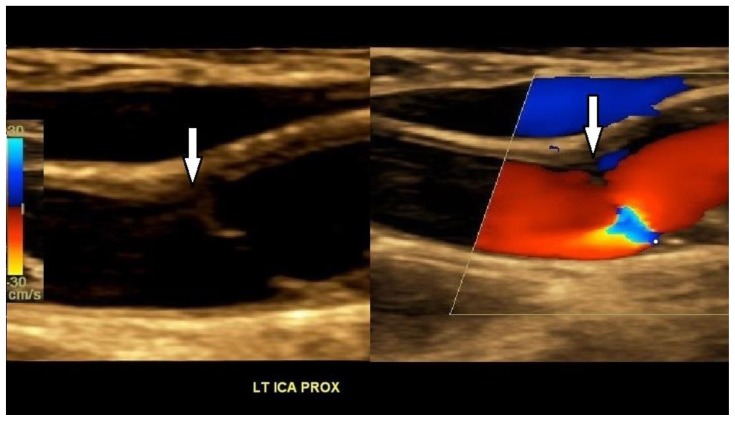
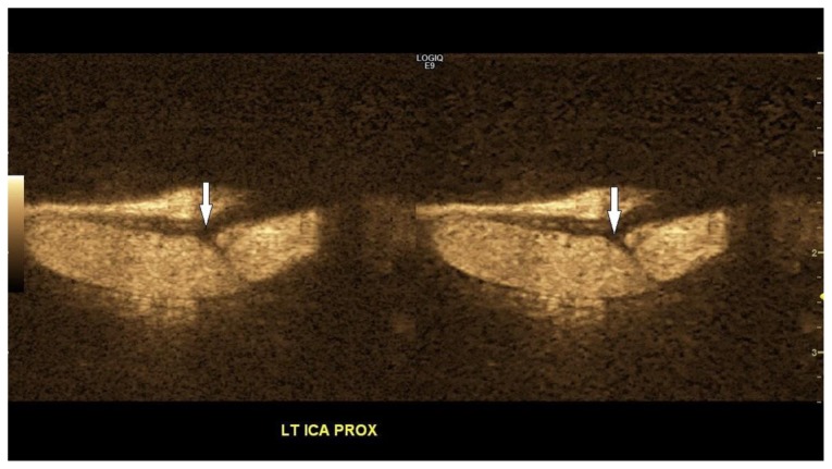
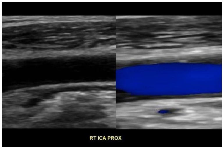
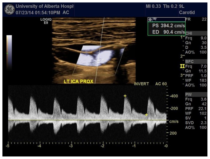
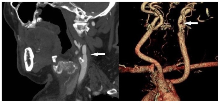
On presentation, the patient was fully orientated and afebrile with a blood pressure of 108/61 mmHg and a heart rate of 80 beats/minute. On clinical examination, a vascular bruit was detected on auscultation over the left side of the neck. The remainder of the clinical examination was essentially normal. In view of the patient’s cardiac history and risk factors for vascular occlusive disease, an echocardiogram, and a carotid ultrasound study were performed. The echocardiogram showed moderate left ventricular systolic dysfunction with an ejection fraction of 30 to 35% and akinesia of the left ventricular apex. No left ventricular thrombus or valvular abnormalities were identified. Duplex Doppler ultrasound of the carotid arteries showed a linear band of tissue (Figures 1 & 3) projecting into the lumen of the left internal carotid artery (ICA), approximately 2-cm from its origin, consistent with an incomplete carotid web, and a normal appearing right proximal ICA (Figure 2). At the level of the web in the left ICA, color Doppler demonstrated a focal aliasing artifact and a high-flow jet with a peak systolic velocity of 394 cm/s2 (Figure 4). These findings were consistent with a hemodynamically significant stenosis of > 70%. A contrast-enhanced CT angiography confirmed the ultrasound diagnosis (Figure 5). On the advice of the neurology service, the patient was treated conservatively due to his advanced age and considerable co-morbidities.
Figure 1.
A 76-year-old male with an incomplete web in the left proximal internal carotid artery. FINDINGS: Color Doppler ultrasound image shows an incomplete carotid web (arrow) in the left proximal ICA, with an aliasing artifact at the level of the web on the color Doppler image. TECHNIQUE: Color Doppler examination was performed on a GE Logiq E (xd-clear platform) equipped with a linear array transducer (9L probe) at the carotid preset, frequency = 3.6 MHz, gain = 18, wall filter = 236, pulse repetition frequency = 3.2 Hz.
Figure 3.
A 76-year-old male with an incomplete web in the left proximal internal carotid artery. FINDINGS: B-flow ultrasound image demonstrates an incomplete carotid web (arrow) in the left proximal ICA. TECHNIQUE: B mode exam was performed on a GE Logiq E (xd-clear platform) equipped with a linear array transducer (9L probe) at the carotid preset, coded harmonic imaging used, frequency = 7 MHz, gain ranging = 30 to 52, wall filter = 183, pulse repetition frequency = 10 Hz.
Figure 2.
A 76-year-old male with an incomplete web in the left proximal internal carotid artery. FINDINGS: Color Doppler ultrasound image shows a normal appearing right proximal ICA. TECHNIQUE: Color Doppler examination was performed on a GE Logiq E (xd-clear platform) equipped with a linear array transducer (9L probe) at the carotid preset, frequency = 3.6 MHz, gain = 18, wall filter = 236, pulse repetition frequency = 3.2 Hz.
Figure 4.
A 76-year-old male with an incomplete web in the left proximal internal carotid artery. FINDINGS: Spectral Doppler ultrasound of the neck demonstrates stenosis of the left proximal ICA due to the carotid web. There is a markedly elevated peak systolic velocity on spectral Doppler. TECHNIQUE: Spectral Doppler imaging was performed on a GE Logiq E (xd-clear platform) equipped with a linear array transducer (9L probe) at the carotid preset, frequency = 3.6 MHz, wall filter = 96. The pulsed wave Doppler was positioned over the vessel to be interrogated. The gain was set to 42 for optimal vessel delineation free of noise. Beam steering and angle correction were performed to acquire accurate flow measurements, and the pulse repetition frequency was set to 20.9 Hz for optimal Doppler sampling.
Figure 5.
A 76-year-old male with an incomplete web in the left proximal internal carotid artery. FINDINGS: Sagittal contrast-enhanced CT of the neck during the arterial phase and three dimensional volume rendered CT angiography of the carotid arteries show an incomplete web (arrow) in the left proximal ICA, causing >70% stenosis. TECHNIQUE: Sagittal CT of the neck, tube current = 486 mAs, tube voltage = 120 kV, slice thickness = 3 mm, intravenous administration of 75 mL Omnipaque 350 contrast material at a flow rate of 5–6 mL/s.
DISCUSSION
The detection of a carotid web is clinically important as it can cause a significant carotid stenosis and is a risk factor for recurrent embolic cerebrovascular events (1, 2). Early diagnosis is important as it impacts on the clinical management and patient outcomes. Therefore, it is essential that radiologists are aware of the imaging appearances of carotid webs.
Etiology & Demographics
A carotid web is defined as a diaphragm-like obstruction of the carotid artery. The prevalence of carotid web is unknown but is considered to be very rare (1). A carotid web, detected on duplex ultrasound (US), has previously only been reported by Kliewer et al. in 1991 and Perren et al. in 2004 (3, 4). In both cases, the carotid web was occult on contrast enhanced -computed tomography (CT) and -magnetic resonance imaging (MRI). The prevailing theory is that carotid webs are a rare form of fibromuscular dysplasia (FMD) as pathological analysis has shown abnormal fibrosis and hyperplasia in the intimal layer (1, 2, 5). FMD is an uncommon idiopathic non-inflammatory, non-atherosclerotic angiopathy that occurs in young to middle-aged individuals, and more frequently in women. It consists of abnormalities of smooth muscle and fibrous and elastic tissue that typically involves small to medium sized arteries. The involved vessel typically exhibits a ‘string of beads’ appearance on imaging. Tubular stenosis - simulating a long, smooth concentric narrowing - is a less common presentation, but one that morphologically resembles a diaphragm (6). Atypical FMD may preferentially involve one side of the vessel wall. This may manifest as a smooth or corrugated mass that project into the vessel lumen, creating the angiographic appearance of a web. It is associated with focal intimal thickening due to hyperplasia of the fibromuscular stroma (3). In our case, the presence of focal intimal thickening adjacent to the carotid web lends support to the FMD theory of origin. An atherosclerotic or developmental origin has also been proposed (7, 8).
Clinical & Imaging Findings
Diagnosis of a carotid web on US can be difficult due to the subtle nature of the entity and because of a lack of awareness among US practitioners. Therefore, meticulous sonographic technique, appropriate education and a high index of suspicion is necessary. Our patient is only the third ultrasound case in the literature, and the first with CT angiogram correlation. All cases in the literature, with the exception of one, were detected on conventional trans-catheter angiography (1). The superiority of conventional trans-catheter angiography over CT/MR angiography is related to the greater spatial and contrast resolution of the former and its ability to render a dynamic real-time vascular assessment. The low sensitivity of non-invasive imaging modalities may partly explain why these subtle vascular abnormalities are under- reported.
The prevalence of carotid webs and the likelihood of subsequent thrombus formation are unknown (1). A carotid web was first reported as a rare cause of ischemic stroke in 1967 by Ehrenfeld et al. (5). Following this initial observation, 20 other cases were reported (9). Webs remain a rare cause of recurrent embolic strokes and are often not detected on non- invasive imaging (3). CT angiogram detected only one of the carotid lesions in a case series by Morgenlander et al. (1). A conventional trans-catheter angiogram remains the gold standard, and should be performed for definitive assessment and to prevent delayed diagnosis. In individuals with a history of recurrent stroke, if the ultrasound examination is negative, there should be a low threshold to recommend trans-catheter angiography.
Treatment & Prognosis
Management of carotid webs ranges from conservative treatment in low-risk asymptomatic cases to balloon dilatation, surgical excision or stent grafting in symptomatic and high-risk cases. Conservative medical therapy is indicated for asymptomatic cases because the available data suggest a benign natural history for this disease (1). Some surgeons advocate patch angioplasty in addition to endarterectomy to prevent recurrence or regrowth of the abnormal tissue (3, 6).
Differential Diagnoses
The differential diagnoses for a carotid web include vascular dissection, post-traumatic aneurysm, and focal atherosclerotic plaque. Traumatic injury can produce tears in the intima and/or hematoma within the wall, leading to luminal stenosis and thrombosis. A carotid dissection may be traumatic or spontaneous in origin. A traumatic dissection is most commonly due to blunt trauma to the head and neck region. Spontaneous dissection is an increasingly recognized cause of cerebrovascular events in middle-aged patients. A ‘trivial’ stress (e.g. coughing, vomiting, sporting injury or cervical manipulation) may rarely precipitate a dissection in vulnerable individuals such as those with connective tissue disorders. Focal atherosclerotic plaques can occasionally simulate a web-like appearance (10). Carotid webs should be considered in the differential diagnosis of patients with a history of recurrent cerebrovascular events and individuals with no history of trauma or vascular risk factors.
In conclusion, we present a rare case of an incomplete carotid web detected on duplex US and confirmed with CT angiography. It caused a hemodynamically significant stenosis of > 70%, which manifested as a carotid bruit on auscultation. The patient was treated conservatively due to his considerable co-morbidities.
TEACHING POINT
A carotid web is a rare condition in which a band of tissue narrows the carotid lumen. Awareness of this entity is important as it can be associated with recurrent embolic stroke. Knowledge of the imaging characteristics of a carotid web is important for early diagnosis and to prevent complications such as stroke.
Table 1.
Summary table of internal carotid artery web.
| Etiology | The prevailing theory is that carotid webs are a rare form of fibromuscular dysplasia. An atherosclerotic or developmental origin has also been proposed. |
| Incidence | To our knowledge, our case is only the third reported ultrasound case in the imaging literature, and the first one with CT angiogram correlation. |
| Gender ratio | No known gender predilection |
| Age predilection | Not known |
| Risk factors | Not known |
| Treatment | Management of carotid webs ranges from conservative treatment in low-risk asymptomatic cases to balloon dilatation, surgical excision or stent grafting in symptomatic and high-risk cases. Some surgeons advocate patch angioplasty in addition to endarterectomy to prevent recurrence or regrowth of the tissue. |
| Prognosis | Conservative medical therapy is indicated for asymptomatic cases because the available data (which is sparse) suggest a benign natural history for this disease. |
| Findings on imaging | Greyscale ultrasound shows a band of tissue which projects into the vessel lumen resulting in focal narrowing of the carotid artery. Color Doppler demonstrates a high flow jet and aliasing at the carotid web, where it narrows the carotid lumen. Intravenous contrast-enhanced CT angiography of the carotid artery shows a focal filling defect corresponding to a band of tissue within the lumen of the carotid artery. |
Table 2.
Differential diagnosis table for internal carotid artery web.
| Imaging Modalities | ||
|---|---|---|
| Differential diagnoses | US | CTA |
| Web | A band of tissue that projects into the vessel lumen | A linear filling defect within the vessel lumen |
| Vascular dissection | An intimal flap within the vessel lumen | A dissection flap with true and false lumen |
| Post-traumatic aneurysm | Focal fusiform or saccular dilatation of the vessel lumen | Focal fusiform or saccular dilatation of the vessel lumen |
| Focal atherosclerotic plaque | Focal eccentric plaques +/− intimal calcifications | Plaque-like filling defect which are typically located at the arterial bifurcation |
| Thrombus | Focal eccentric foci along the vessel walls of varying echogenicities depending on the age of the blood products | Focal eccentric filling defect within the vessel lumen |
ACKNOWLEDGEMENTS
The authors would like to thank Dr. David Reich for help in preparing the CT angiographic images of the neck.
ABBREVIATIONS
- CT
computed tomography
- FMD
fibromuscular hyperplasia
- ICA
internal carotid artery
- MRI
magnetic resonance imaging
- US
ultrasound
REFERENCES
- 1.Morgenlander JC, Goldstein LB. Recurrent transient ischemic attacks and stroke in association with an internal carotid artery web. Stroke. 1991;22:94–98. doi: 10.1161/01.str.22.1.94. [DOI] [PubMed] [Google Scholar]
- 2.Wirth FP, Miller WA, Russell AP. Atypical fibromuscular hyperplasia. Report of two cases. J Neurosurg. 1981;54:685–689. doi: 10.3171/jns.1981.54.5.0685. [DOI] [PubMed] [Google Scholar]
- 3.Kliewer MA, Carroll BA. Ultrasound case of the day. Internal carotid artery web (atypical fibromuscular dysplasia) Radiographics. 1991;11:504–505. doi: 10.1148/radiographics.11.3.1852941. [DOI] [PubMed] [Google Scholar]
- 4.Perren F, Urbano L, Rossetti AO, Ruchat P, Uske A, Meuli R, Lobrinus JA, et al. Ultrasound image of a single symptomatic carotid stenosis disclosed as fibromuscular dysplasia. Neurology. 2004;62:1023–1024. doi: 10.1212/01.wnl.0000115172.99299.35. [DOI] [PubMed] [Google Scholar]
- 5.Ehrenfeld WK, Stoney RJ, Wylie EJ. Fibromuscular hyperplasia of the internal carotid artery. Arch Surg. 1967;95:284–287. doi: 10.1001/archsurg.1967.01330140122027. [DOI] [PubMed] [Google Scholar]
- 6.Finsterer J, Strassegger J, Haymerle A, Hagmuller G. Bilateral stenting of symptomatic and asymptomatic internal carotid artery stenosis due to fibromuscular dysplasia. J Neurol Neurosurg Psychiatry. 2000;69:683–686. doi: 10.1136/jnnp.69.5.683. [DOI] [PMC free article] [PubMed] [Google Scholar]
- 7.Lipchik EO, DeWeese JA, Schenk EA, Lim GH. Diaphragm-like obstructions of the human arterial tree. Radiology. 1974;113:43–46. doi: 10.1148/113.1.43. [DOI] [PubMed] [Google Scholar]
- 8.McNamara MF. The carotid web: a developmental anomaly of the brachiocephalic system. Ann Vasc Surg. 1987;1:595–597. doi: 10.1016/S0890-5096(06)61448-9. [DOI] [PubMed] [Google Scholar]
- 9.Lenck S, Labeyrie MA, Saint-Maurice JP, Tarlov N, Houdart E. Diaphragms of the carotid and vertebral arteries: an under-diagnosed cause of ischaemic stroke. Eur J Neurol. 2014;21:586–593. doi: 10.1111/ene.12343. [DOI] [PubMed] [Google Scholar]
- 10.Rodallec MH, Marteau V, Gerber S, Desmottes L, Zins M. Craniocervical arterial dissection: spectrum of imaging findings and differential diagnosis. Radiographics. 2008;28:1711–1728. doi: 10.1148/rg.286085512. [DOI] [PubMed] [Google Scholar]


