A multidisciplinary roundtable was convened on May 29, 2014, to gain insight and guidance from experts on the diagnosis and management of polycythemia vera (PV), including practical strategies, recent advances, and the emerging science. The roundtable was comprised of 10 experts in relevant fields: hematology, oncology, managed care, specialty pharmacy, translational research, and oncology nursing/nurse navigation. This supplement highlights the discussions and recommendations of the experts who participated in this meeting, with the overarching goal being to improve outcomes by enhancing the quality, delivery, and continuum of care for patients with PV.
Clinical Aspects of Polycythemia Vera
Natural history and presentation
Like myelofibrosis (MF) and essential thrombocythemia (ET), PV is a Philadelphia chromosome–negative myeloproliferative neoplasm (MPN).1 PV is characterized by clonal stem-cell proliferation of red blood cells (RBCs), white blood cells (WBCs), and platelets.2,3 Increased RBC mass results in hyperviscosity of the blood, increased risk for thrombosis, and a shortened life expectancy.4 Effective management of PV is essential, given the risk for morbidity and mortality, complexity associated with diagnosis and treatment, and overall impact on patients' quality of life (QOL).
According to the World Health Organization (WHO) classification scheme for myeloid neoplasms, PV is a BCR-ABL1–negative MPN.5 MPNs share several common features6–8:
Clonal involvement of a multipotent hematopoietic progenitor cell
Marrow hypercellularity with effective hematopoiesis (compared with ineffective hematopoiesis, as in myelodysplastic syndrome)
Extramedullary hematopoiesis; enlarged spleen and/or liver
Thrombotic and hemorrhagic diathesis
Potential evolution to MF, as well as to acute myeloid leukemia (AML).
The incidence of PV is higher among men than among women in all races and ethnicities, with rates of approximately 2.8 per 100,000 men and approximately 1.3 per 100,000 women.3 Based on several small studies, the prevalence of PV is approximately 22 cases per 100,000 population.3 PV is typically diagnosed in persons 60 to 65 years of age, and the disorder is relatively uncommon among individuals younger than 30 years. The condition is observed more often among Jews of Eastern European descent than among other European populations and Asians.3
Approximately 96% of patients with PV have a mutation of the Janus kinase 2 (JAK2) gene.9 JAK2 is involved directly in intracellular signaling in PV progenitor cells, a process that occurs after exposure to cytokines to which these cells are hypersensitive.10
The course of PV is variable. Some patients exhibit few symptoms, such that the condition is discovered only after blood work is performed during a routine medical examination. In other patients, signs, symptoms, and complications of PV arise from the high number of RBCs and platelets in the blood.3 In patients with milder symptoms, PV can persist for many years without distinct stages or clear progression.5 Other patients will evolve to post-PV MF, which occurs at a rate of up to 10% of patients every 10 years.11 Transformation to AML has been observed at a rate of up to 15% of patients with PV every 10 years.12
Symptoms of PV stem primarily from high RBC counts, which result in increased blood viscosity, and from high platelet counts, which can contribute to the formation of thrombi. Along with underlying vascular disease, which is common among older persons with PV, the risk for such clotting complications as stroke, heart attack, deep vein thrombosis, and pulmonary embolism is enhanced among persons with the disorder. Blood clots occur in about 30% of patients before a PV diagnosis is made.3 During the first 10 years after diagnosis, 40% to 60% of patients with untreated PV may develop blood clots.3
Thrombotic complications can be divided into 2 categories—microvascular and macrovascular. Microvascular complications, or microcirculatory disturbances, are caused by the formation of thrombi in small blood vessels and can result in the signs and symptoms shown in Table 1.13–15 Macrovascular complications, which are serious events caused by the development of thrombi in large arteries or veins, are often referred to as major thrombotic events.14 These major events (Table 1) are the primary cause of mortality in patients with PV, accounting for 45% of all deaths.15 Other major causes of death among individuals with PV include solid tumors (20%) and hematologic transformation to AML (13%).15
Table 1.
Thrombotic Complications in Polycythemia Vera
| Microvascular complications | Macrovascular complications |
|---|---|
| Erythromelalgia | Arterial thrombotic events
|
| Headache | |
| Dizziness | |
| Visual disturbances | |
| Paresthesia | Venous thrombotic events
|
| Transient ischemic attack |
Sources: Michiels JJ, et al. Semin Thromb Hemost. 2006;32:174–207; Falanga A, Marchetti M. Hematology Am Soc Hematol Educ Program. 2012;2012:571–581; Marchioli R, et al. J Clin Oncol. 2005;23:2224–2232.
During the roundtable, Dr Ruben A. Mesa discussed the availability of patient assessment tools that can provide hematologists with data on disease burden. One such tool is the Myeloproliferative Neoplasm Symptom Assessment Form (MPN-SAF) total symptom score, which was published in 2012.16 Because symptoms associated with PV are not always related to high blood counts, assessing patients via the use of such tools is an important clinical step. Patients can experience “PV-associated” symptoms, which are driven by higher volumes of circulating inflammatory cytokines, resulting from abnormal activation of JAK signaling.17 The most common such symptoms are fatigue (91%), pruritus (65%), early satiety (62%), concentration problems (61%), and inactivity (58%).18 Dr Mesa noted that many patients underreport these symptoms and generally appear much healthier than others seen in hematology practices, such that PV-associated symptoms often go unrecognized, specifically, symptoms that can arise from compromised blood flow, but that fall short of overt thrombosis, such as complex vascular headaches (ie, migraines with visual changes), challenges with concentration, and erythromelalgia.18
Establishing a diagnosis of Polycythemia Vera
Diagnosis of PV is made using WHO criteria, and is based on a composite assessment of clinical and laboratory features, including JAK2 mutation status and serum erythropoietin (Epo) level.19 As shown in Table 2, the presence of a JAK2 mutation and a subnormal serum Epo level confirms the diagnosis of PV.20 A subnormal serum Epo level in the absence of JAK2 V617F requires additional mutational analysis for JAK2 exon 12 mutation to identify the rare patients with PV who are JAK2 V617F negative.19 Bone marrow examination is not essential for a diagnosis; however, patients who fulfill the diagnostic criteria for PV may exhibit substantial bone marrow fibrosis.20
Table 2.
World Health Organization Diagnostic Criteria for Polycythemia Vera
| PV diagnosis requires meeting either both major criteria and 1 minor criterion or the first major criterion and 2 minor criteria: | |
| Major Criteria |
|
| Minor Criteria |
|
BM indicates bone marrow; EEC, endogenous erythroid colony; Epo, erythropoietin; Hct, hematocrit; Hb, hemoglobin; PV, polycythemia vera; RBC, red blood cell.
Reprinted with permission from Tefferi A, et al. Blood. 2007;110:1092–1097. © Copyright 2014 by American Society of Hematology.
Management of Polycythemia Vera
Although PV is a chronic, incurable disease, it can be managed effectively for long periods of time.3 Careful medical supervision and therapy are designed to reduce hematocrit and platelet concentrations to normal or near-normal value, in order to control PV-related symptoms, decrease the risk for arterial and venous thrombotic events and other complications, and avoid leukemic transformation.21,22
Patients with PV are stratified for their risk of thrombosis based on age and history of thrombosis. Those who are older than age 60 years or who have a history of thrombosis are at high risk, whereas patients younger than 60 years and with no history of thrombosis are typically classified as being at low risk.3,22
Patients with low-risk PV are usually phlebotomized and receive low-dose aspirin. These patients often report an immediate improvement in their PV symptoms, including headaches, tinnitus, and dizziness after phlebotomy.3 For many low-risk patients, phlebotomy and aspirin may be the only form of treatment required.3 In contrast, patients with high-risk PV require medical treatment to decrease their hematocrit level permanently, eliminate the need for phlebotomy, and decrease their risk for clotting. Cytoreductive chemotherapy is recommended to control RBC volume in patients in whom phlebotomy is poorly tolerated, those in whom the thrombotic risk remains high, or those whose splenomegaly continues to be symptomatic.3
Available cytoreductive medications include hydroxyurea, interferon alfa (IFN-α), and busulphan.21 Among these options, hydroxyurea is currently the treatment of choice for patients with PV who are older than 40 years of age.22,23 Hydroxyurea effectively improves myelosuppression and reduces the risk for thrombosis compared with the use of phlebotomy alone.24 Concerns about the long-term risk for secondary leukemia associated with the use of hydroxyurea, however, are relevant. After a median follow-up of more than 8 years, the Polycythemia Vera Study Group (PVSG) reported that 5.4% of patients with PV who participated in a randomized clinical trial developed leukemia after receiving hydroxyurea, compared with 1.5% of those treated with phlebotomy alone.24
Patients who are either intolerant of, or resistant to, hydroxyurea can be effectively managed with pegylated IFN-α or busulphan. Recent literature suggests a preference for IFN-α in patients who are younger than 65 years of age and busulphan in older individuals,19 although no literature or other evidence is available to validate this recommendation.19 In practice, however, the use of IFN-α is usually reserved for younger, more physically fit individuals with PV.
When discussing his approach to treatment, Dr Mesa emphasized “As we move forward, therapy for PV will be more individualized. A patient's symptom burden is an important consideration, in addition to hematocrit and spleen size. There are many nuances in terms of how the individual with PV is affected. Make no mistake: PV can clearly be fatal if an individual has a vascular event, such as myocardial infarction. Our goal in the future is cure. However, at the moment, we are talking about management of a chronic illness that has variable presentations and burdens.”
Hematologists in the roundtable panel concurred with Dr Mesa, indicating that the management of PV is not straightforward, particularly among patients who progress after initial therapy. A significant unmet need remains for individuals who continue to experience PV-associated symptoms, as well as for high-risk patients.
Michael Boxer, MD, commented, “In my experience, slightly less than one-fourth of patients with PV ‘cross the line.’ At that point, I have little to offer them. In a few patients, we have removed spleens. Interferon is generally stopped after a couple of months because of intolerable side effects, and blood counts start to cycle widely. Nothing that we use prevents patients from progressing to fibrosis and leukemia. We need new medicines that can be administered much earlier. Ideally, we need a therapy that can halt disease progression.”
John O. Mascarenhas, MD, MS, observed a similar challenge in his academic practice, stating, “I have become less sure of our approach and what I am trying to accomplish when treating individuals with PV. Of course, the patients that we see are skewed towards more advanced or complicated patients. However, even if you take time to talk with low-risk patients, you tease out symptoms that have been undiagnosed and underappreciated, even by patients themselves. Then there are the patients with thrombosis…I often talk with these patients about their fears, specifically clotting and progression to MF. I used to think that adding cytoreductive therapy reduced their risk of thrombosis, but now I do not have anything in which I feel confident. None of these therapies seems to change the natural course of this disease.”
Controversies in the Diagnosis of Polycythemia Vera
Brady Lee Stein, MD, provided deeper insight into the diagnosis of PV, beginning with the history of the disease and its classification. In 1951, a landmark paper was published by Dr William Dameshek, who speculated about the mimicry observed among myeloproliferative syndromes, including PV.25 Although he was not the first to recognize myeloproliferation, Dr Dameshek was the first to describe a unifying concept for classifying patients. He noted similar clinical and laboratory features among various myeloproliferative conditions, and was the first to hypothesize a shared pathogenesis.
The PVSG was established in 1967. This study group facilitated an understanding of the natural history of PV and the consequences, positive and negative, of the available treatments. The PVSG issued the first formal diagnostic criteria for PV, which relied heavily on demonstration of an increased RBC mass.26
Diagnostic criteria changed with identification of the JAK2 mutation—a damaged myelostimulatory factor that Dr. Dameshek predicted nearly 55 years prior to its discovery in 2005. PV is now known to result from “a signaling pathway in overdrive” that causes exuberant blood production: erythrocytosis, leukocytosis, and thrombocytosis. The JAK2 mutation is highly relevant in the diagnosis of PV, as it is present in virtually all patients.3
There are 2 variants of the JAK2 mutation: (1) the more common V617F mutation and (2) the much less common exon 12 mutation. According to WHO diagnostic guidelines, testing for the exon 12 mutation is appropriate in patients with PV who have isolated erythrocytosis and a low Epo level, but who are negative for the JAK2 V617F mutation.19 Although patients with the exon 12 mutation are phenotypically distinct, as they are more likely to have isolated erythrocytosis, the natural history of PV and rates of complications in this population are comparable to those in patients with the V617F mutation.27
The knowledge that a single mutation in JAK2 gives rise to at least 3 different disease phenotypes—PV, MF, and ET—has sparked several hypotheses regarding the evolution of these diseases, including the gene-dosage hypothesis.28 JAK2 mutation test results are typically reported in a binary fashion. A positive result can be subsequently quantified in a continuous fashion.
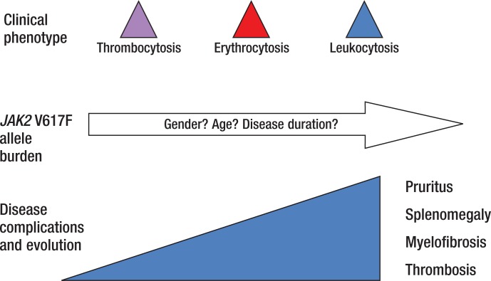
The gene-dosage hypothesis suggests a correlation between disease phenotype and the proportion of JAK2 V617F mutant alleles relative to wild-type JAK2 in hematopoietic cells, or the “allele burden.”28 As shown in Figure 1, lower allele burdens have been shown to result in thrombocytosis. As the allele burden rises, erythrocytosis and leukocytosis become more prevalent. Higher allele burdens also correlate with pruritus, splenomegaly, and MF. In the highest allele burden quartile (≥75%), data suggest that consequences of arterial thrombosis are more common.28 Although these correlations have also been observed in clinical practice, allele burden is not yet used as a prognostic parameter in the management of patients with PV.28
Figure 1. Impact of JAK2 V617F Allele Burden.
Source: Passamonti F, et al. Haematologica. 2009;94:7–10.
Upon publication of the WHO diagnostic criteria for PV in 2007,20 a “vociferous minority” of experts has been opposed to the use of hemoglobin (Hb) and hematocrit as surrogate markers for increased RBC mass. Several studies have demonstrated that these measures are flawed. In one of these studies, WHO criteria identified erythrocytosis in only 35% of men and 63% of women who had been diagnosed with PV.29 A more recent prospective study corroborated these findings, suggesting that if one relies on Hgb and hematocrit criteria as surrogates for RBC mass, the diagnosis of PV may be missed.30 Patients who are misdiagnosed are less likely to receive treatment, leaving them at risk for disease symptoms and long-term sequelae.
In critiquing the WHO criteria, Dr Stein noted that assessment of Epo levels may be valuable if the levels are abnormal but unreliable when Epo results are ambiguous. Additionally, assays of endogenous erythroid colony (EEC) growth are neither widely available nor standardized for use.
Dr Stein reminded the panelists that PV presents with a spectrum of signs and symptoms: “There are phenotypes within a phenotype.” The term “masked PV” has been operationalized to describe patients with a PV-consistent bone marrow, JAK2 mutations, and PV-like features, but low Hb levels (≤16.5 g/dL in women, ≤18.5 g/dL in men). Although the incidence of thrombosis is similar among patients with masked PV and those with overt PV, research demonstrates that patients with masked PV have significantly higher rates of progression to MF and AML, as well as higher mortality. The presence of masked PV has been identified as an independent predictor of poor survival, along with age over 65 years and a high leukocyte count.31
In light of these observations, revision of the WHO diagnostic criteria for PV may be in order. Dr Stein recalled that the British Committee for Standards in Haematology (BCSH) has switched to the use of hematocrit rather than Hb for PV diagnosis. Revised BCSH guidelines require the presence of the JAK2 mutation, as well as increased hematocrit (>48% for women, >52% for men) or increased RBC mass (>25% of predicted).32 Most hematologists who participated in the roundtable agreed that current diagnostic criteria are lacking. The majority of these physicians do not use EEC or RBC mass assays when diagnosing PV. They have consistently identified patients who clearly have PV but do not meet the strict WHO criteria.
The perceived value of information about a patient's JAK2 allele burden varied among panelists. According to 1 hematologist, “We have superimposed the importance of BCR-ABL onto JAK2. In CML [chronic myeloid leukemia], this ended up being relevant. In the MPNs, people have tried to make it relevant, but it is not really clear.” One of the physicians involved in clinical protocols for PV therapies suggested that allele burden information can add value in that context: “In a research setting when assessing a new treatment, allele burden may be a marker of molecular response. Off-protocol, however, allele burden does not enhance our understanding of the disease process.”
The panel also debated the role played by bone marrow biopsies in the diagnosis and management of patients with PV, as well as the ambiguous results that are reported by some pathologists, including hematopathologists. One of the panelists remarked, “In someone with a large spleen, extramedullary hematopoiesis, or a high white count, I want to ensure that I am not missing something. This is a low-risk procedure. For others, I discuss the fact that it is not necessary based on the fact that they meet PV criteria.” Another panel member described proactive patients who request a baseline bone marrow biopsy: “These patients are concerned about fibrosis. The idea that we have not looked for it is unsettling to them.”
JAK Signaling in Polycythemia Vera
The JAK-STAT (Signal Transducer and Activator of Transcription) pathway, with appropriate levels of JAK proteins, is crucial for normal hematopoiesis and immune function.33–35 More than 30 ligands, cytokines, and growth factors affect cell signaling through the mammalian family of Janus kinases, which includes JAK1, JAK2, JAK3, and TYK2. The JAK proteins encoded by JAK genes interact with various STAT molecules, which are signal transducers and activators of transcription, to induce key cellular responses.33–35
A variety of hematopoietic malignancies are characterized by mutations and/or translocations in JAK genes and, as a consequence, constitutively active JAK proteins35:
Myeloproliferative disorders, including PV and MF
Acute lymphoblastic leukemia
AML
Acute megakaryoblastic leukemia
T-cell precursor acute lymphoblastic leukemia.
The JAK2 V617F mutation occurs in the JH2 domain of the JAK2 gene. The valine-to-phenylalanine substitution that takes place represents a gain of function mutation and results in constitutive downstream activation of STATs, as well as the MAP kinase and PI3 kinase pathways.36
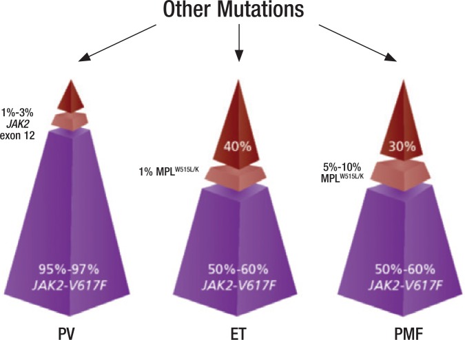
Figure 2 depicts the relative frequency of the JAK2 V617F mutation among the MPNs, including PV, ET, and Philadelphia-negative MF. As shown, the JAK2 V617F mutation is most frequently observed in PV, but is present in more than half of patients with MF, as well.37–39
Figure 2. JAK2 and Other Mutations in Patients with Myeloproliferative Neoplasms.
PV indicates polycythemia vera; ET, essential thrombocythemia; PMF, Philadelphia-negative myeloproliferative neoplasm.
Sources: Spivak JL. Blood. 2002;100:4272–4290; Levine RL, et al. Nat Rev Cancer. 2007;7:673–683; Oh ST. Ther Adv Hematol. 2011;2:11-19; Vannucchi AM, et al. Haematologica. 2008;93:972–976.
Recently, mutations in the CALR gene, which encodes calreticulin, have been identified in the majority of patients with ET or primary MF with nonmutated JAK2 and MPL.40 Calreticulin has a myriad of functions related to calcium homeostasis, as well as proper protein folding within the endoplasmic reticulum. The JAK-STAT pathway appears to be activated in all MPNs, regardless of founding driver mutations. However, these mutations may affect symptomatology and patient outcomes. Recent data suggest that evaluation of JAK2, MPL, and CALR mutation status may be important for both diagnosis and prognostication among patients with MPNs.40 According to Dr. Mascarenhas, although testing for the CALR mutation is commercially available in many laboratories, it does not yet represent a therapeutic target beyond the clinical trial setting.
Another recent development is the discovery of high expression of heat shock protein 70 (HSP70), a chaperone protein much like HSP90. This protein is upregulated preferentially in PV, compared with ET and healthy controls. HSP70 may represent a potential therapeutic target in MPNs, particularly PV.41
Acute Events
The acute events associated with PV, including thrombotic events and secondary cancers, can affect a person's overall survival, as well as disease-related symptoms that impair QOL. Laura Michaelis, MD initiated this discussion by reviewing the results of a comprehensive study conducted by an Italian PV study group, which documented overall mortality associated with PV as 2.9% per year.42 Thrombotic events and hematologic or nonhematologic cancers had similar effects on mortality in this study, which was published in 1995.42 Since that time, strategies to prevent thrombosis and alleviate symptoms in patients with PV have evolved. In 2013, the Italian Cytoreductive Therapy in Polycythemia Vera (CYTO-PV) Collaborative Group reported mortality rates of 1.6% in patients with PV whose hematocrit levels were maintained at ≤45% and 3.3% in those with PV whose hematocrit levels were maintained between 45% and 50%.43 Rates of vascular events were 2.7% in the “low hematocrit” group versus 9.8% in the “high hematocrit” group.43
Approximately two-thirds of thrombotic events experienced by patients with PV are arterial, including myocardial infarction and stroke; the remaining one-third are venous, including pulmonary embolism, splanchnic vein thrombosis, and cerebral venous sinus thrombosis. Dr Michaelis noted that in younger patients with PV, clots in the splanchnic, cerebral, or portal sinuses (including Budd-Chiari syndrome) are of particular concern in light of their potential morbidity.44,45
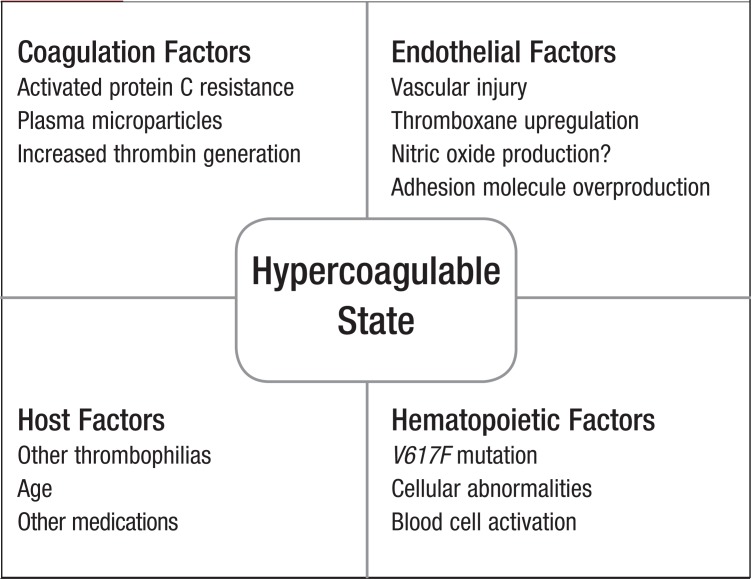
Strategies for the prevention of thrombotic events are determined after considering the multiple factors that can influence a patient's hypercoagulable state (Figure 3). Phlebotomy or cytoreduction are recommended for Hgb and hematocrit control, and antiplatelet therapy is used to prevent arterial events. Most patients take low-dose aspirin once daily, whereas those who are sensitive to aspirin are given clopidogrel. Aggressive control of cardiovascular risk factors, including blood pressure, lipids, smoking, weight, and physical fitness, is also relevant.
Figure 3. Factors Affecting Hypercoagulability in Patients.
For patients who experience an arterial or venous thrombotic event, preventive therapy is revisited. Recommendations for patient management following a thrombotic event are summarized in Table 3.
Table 3.
Recommendations for Secondary Prevention of Thrombotic Events in Patients with Polycythemia Vera
| Arterial Events | |
| Stroke and antiplatelet agents | Current guidelines recommend clopidogrel, aspirin, or extended-release dipyridamole firstline, but these agents have not been tested in patients with PV. For most patients, there is no increased benefit associated with the use of clopidogrel combined with aspirin Assess other CV risk factors: diabetes mellitus, hypertension, cholesterol |
| Cardiac and antiplatelet agents | Aspirin 150 to 325 mg daily may be combined with clopidogrel for 12 months Assess other CV risk factors: diabetes mellitus, hypertension, cholesterol |
| Venous Events | |
| Deep vein thrombosis/pulmonary embolism | Initial therapy with LMWH is recommended, but duration of therapy is unclear Data are available to guide decision-making regarding LMWH use, aspirin continuation, warfarin transition, and use of oral anticoagulants |
CV indicates cardiovascular; LMWH, low-molecular-weight heparin; PV, polycythemia vera.
In all patients who have experienced arterial or venous thrombotic events, treatment with cytoreductive agents is appropriate. Dr Michaelis indicated that she prefers IFN-α relative to hydroxyurea in younger patients with PV, particularly those who plan to have children. Other indications for the use of cytoreductive agents include poor tolerance to phlebotomy, rapidly increasing platelet counts, and progressive leukocytosis.
Management of PV-associated symptoms, including pruritus and erythromelalgia (neurovascular pain disorder), is increasingly being recognized as critical for patients. Dr Michaelis observed, “Once you start listening to patients, even though we all think about PV as the ‘safe MPN,’ these symptoms are really life-altering. Some of them can be quite severe, especially erythromelalgia.” Migraines, skin rashes, leg swelling, and burning pain in the hands are also highly debilitating. Aspirin, cytoreduction, and gabapentin can be offered to patients to help minimize these symptoms, whereas antihistamines, light therapy, aprepitant, and antidepressants may help with pruritus.
Cytoreductive Agents
In the United States, hydroxyurea is considered the standard of care for initial treatment of PV with a cytoreductive agent, despite the fact that this drug is “lacking an evidence base.” After reviewing the efficacy and safety data from clinical trials of hydroxyurea for the treatment of PV (Table 4),24,46,47 Dr. Brady Lee Stein noted, “Most data for use of hydroxyurea as a front-line cytoreductive are extrapolated from randomized, controlled trials in essential thrombocythemia…. Hydroxyurea is a relatively efficient way to control counts, it is easy to administer, and, for most patients, it is tolerable. For these reasons, hydroxyurea emerges as the front-line strategy.”
Table 4.
Studies on the Efficacy of Hydroxyurea for the Treatment of Patients with Polycythemia Vera
| Investigator/year | Number of patients, follow-up | Intervention | Comparator | Rate of thrombotic events |
|---|---|---|---|---|
| Fruchtman SM et al, 199724 | 51 patients, 795 weeks | Hydroxyurea (prospective) | Phlebotomy (134 historical controls) | 9.8% vs 32.8%, respectively |
| Najean Y et al, 199746 | 292 patients under 65 years of age, 17 years | Hydroxyurea (randomized) | Pipobroman | NSD |
| Kiladjian J-J et al, 201147 | 285 patients under 65 years of age, 16.3 years | Hydroxyurea (randomized) | Pipobroman | NSD |
NSD indicates no significant difference.
Long-term consequences of hydroxyurea use remain an area of controversy. Dr Stein summarized the available data, as shown in Table 5.12,24,47,48 Twenty-year follow-up data from a study comparing hydroxyurea and pipobroman—an agent that is not approved for use in the United States—show high rates of second malignancies with the use of both agents. It is unclear, however, whether the rate with hydroxyurea exceeds the rate of second malignancies that would be observed in untreated patients with PV. Dr Stein concluded, “There are no hard data or controlled evidence to implicate hydroxyurea as an agent that increases leukemia rates beyond what we see spontaneously with PV…. However, age plays heavily in my decision-making as I start a cytoreductive. I am concerned about prescribing hydroxyurea for long periods of time in younger patients.”
Table 5.
Studies on Potential Long-Term Consequences of Hydroxyurea Use in Patients with Polycythemia Vera
| Investigator/year | Number of patients, follow-up | Intervention | Comparator | Rate of AML and MDS transformation |
|---|---|---|---|---|
| Fruchtman SM et al, 199724 | 51 patients, 795 weeks | Hydroxyurea (prospective) | Phlebotomy | 6.0% vs 1.5%a |
| Finazzi G et al, 200512 | 1638 patients, 2.8 years (4393 person-years) | Retrospective | No association with the use of single-agent hydroxyurea | |
| Kiladjian J-J et al, 201147 | 285 patients under 65 years of age, 16.3 years | Hydroxyurea (randomized) | Pipobroman | 10 years: 6.6% vs 13% 15 years: 16.5% vs 34.1% 20 years: 24.2% vs 52.1% (P = .004) “Pipobroman is leukemogenic and is unsuitable for first-line therapy [in PV].” |
| Tefferi A et al, 201348 | 1545 patients, 6.9 years | Retrospective | No association with single-agent hydroxyurea | |
No significant difference.
AML indicates acute myeloid leukemia; MDS, myelodysplastic syndromes; PV, polycythemia vera.
Studies evaluating PV-associated symptom control with hydroxyurea demonstrate that this agent fares poorly. Dr Stein summarized 2 studies that showed no significant difference in symptom burden, as assessed using the MPN-SAF, in patients treated with hydroxyurea compared with untreated individuals.49,50 A third study of a large cohort of German patients with PV revealed that pruritus is a significant problem that is not improved by PV-directed treatment. Among 301 of 441 (68%) patients who experienced pruritus, 44 (15%) characterized the condition as “unbearable.”51 Patients with pruritus reported reduced global health status, as well as higher levels of fatigue, pain, and dyspnea. Only 24% of patients received pruritus-specific treatment, mostly antihistamines, which ameliorated symptoms in about half of the group. In only 6% of patients, PV-directed therapy, including phlebotomy or cytoreduction, resolved pruritus symptoms.51 Data such as these underscore the need for improved treatments to help alleviate common and problematic symptoms associated with PV.
An emerging issue in the treatment of PV is hydroxyurea resistance or intolerance. To address this concern, criteria defining resistance and intolerance were published by a consensus panel in 2010, primarily as guidance for clinical trials of novel therapies for PV.52
-
According to the European LeukemiaNet (ELN), hydroxyurea resistance is characterized by any of the following52:
Need for phlebotomy to keep hematocrit <45% after 3 months of hydroxyurea at a dose of 2 g/day
Platelets >400 × 109/L and WBCs >10 × 109/L after 3 months of hydroxyurea at a dose of 2 g/day
Failure to reduce splenomegaly by 50%, or failure to relieve symptoms of splenomegaly after 3 months of hydroxyurea at a dose of 2 g/day
ELN defines hydroxyurea intolerance as cytopenias, including neutropenia (absolute neutrophil count <1 × 10⁹/L), thrombocytopenia (platelet count <100 × 109/L), or anemia (Hb <10 g/dL), or the presence of mucocutaneous manifestations, such as leg ulcers, gastrointestinal symptoms, pneumonitis, or fever.52
In a study aimed at assessing the prognostic value of these ELN criteria for response, resistance, and intolerance to hydroxyurea, records from a large series of patients with PV were retrospectively reviewed. Resistance and intolerance to hydroxyurea were reported in 11% and 13% of participants, respectively.53 In this analysis, patients with hydroxyurea resistance were more likely to die (P <.001), and were also more likely to experience transformation to MF or AML (P <.001), affirming the belief that resistance to hydroxyurea is an important adverse prognostic factor. Intolerance to hydroxyurea, as defined by the ELN, did not show any association with subsequent survival or risk for hematologic transformation.53
Treatment with IFN-α may be an option for patients with resistance or intolerance to hydroxyurea, as well as for younger, newly diagnosed individuals with PV. Several phase 2 studies demonstrate the efficacy of this agent in controlling blood counts, as well as in achieving hematologic complete responses and complete molecular responses.54–58 In a minority of patients with PV, JAK2 has been eradicated, typically after 12 months of treatment with IFN-α.
Findings like these, as well as documented safety and tolerability of IFN-α, have led to renewed interest in early use of IFN-α as an alternative to hydroxyurea for patients with PV.59 Dr Stein expressed caution, however: “As we talk about renewed enthusiasm for IFN-α, we also have to be practical. Hydroxyurea is used so often because it is easy to administer. Administration of IFN-α, both for providers and patients, is not so easy. Its cost is higher, and it can be very difficult to obtain outside of a clinical trial. There is also a stigma associated with IFN-α. When we use it for other diseases, tolerability has been much worse…. If we use a modified, gradual dosing protocol [for PV], starting low and gradually increasing, tolerability is much greater, at least in my patient population.”
Novel Therapies
Ruxolitinib, a selective inhibitor of JAK1 and JAK2, was approved by the US Food and Drug Administration in November 2011 for the treatment of patients with intermediate- or high-risk MF, including primary MF, post-PV MF, and post-ET MF.60 In light of the high rate of JAK2 mutations in patients with PV, clinical trials were undertaken to evaluate the use of ruxolitinib in these patients.
Phase 2 data for ruxolitinib in patients with PV who were refractory to, or intolerant of, hydroxyurea demonstrated that ruxolitinib has long-term clinical activity, including durable response rates.61 Among 34 patients in this phase 2 trial, 97% achieved a response (defined as hematocrit <45% without phlebotomy) by week 24. Ruxolitinib use was also associated with rapid and sustained improvements in PV-associated symptoms, including pruritus, night sweats, and bone pain. Researchers in this trial observed spleen size reduction, phlebotomy independence, and improvements in blood counts and PV-related symptoms over a median follow-up of 21 months. Treatment with ruxolitinib also reduced elevated levels of inflammatory cytokines and granulocyte activation. Thrombocytopenia and anemia were the most common adverse events. Grade 3 thrombocytopenia and grade 3 anemia, which occurred in 3 patients each (9%; 1 patient experienced both), were managed with dose modification.61
On the basis of these findings, a global phase 3 registration trial for ruxolitinib use in patients with PV was initiated. This trial, known as RESPONSE (Randomized, open-label, multicenter phase 3 study of Efficacy and Safety in POlycythemia vera subjects who are resistant to or intolerant of hydroxyurea: JAK iNhibitor INC424 tablets verSus bEst available care), compared ruxolitinib with best available therapy (BAT) in approximately 200 patients with advanced PV. Study findings were presented during the annual meeting of the American Society of Clinical Oncology in June 2014.62
In this phase 3 study, patients with phlebotomy-dependent PV and splenomegaly (spleen volume of ≥450 cm) who were resistant to, or intolerant of, hydroxyurea were randomized to ruxolitinib (n = 110) or BAT (n = 112). The primary end point of the study was a composite that included achievement of both hematocrit control (<45%) and spleen response (≥ 35% reduction from baseline in spleen volume by magnetic resonance imaging) at week 32. After week 32, patients who were randomized to BAT could cross over to ruxolitinib.
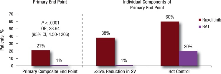
At 32 weeks, 77% of patients randomized to ruxolitinib met at least 1 component of the primary end point, and 21% met both components. Only 1% of the patients receiving BAT achieved the primary end point (Figure 4).62 The majority (91%) of ruxolitinib-treated patients who achieved the primary end point had a confirmed response at week 48, and the probability of maintaining a primary response for 1 year was 94%. The rate of thromboembolic events was lower in the ruxolitinib group, with only 1 event (portal vein thrombosis) reported through week 32, compared with 6 events among patients receiving BAT. The RESPONSE trial investigators concluded that in patients with PV who had an inadequate response to, or were intolerant of, hydroxyurea, ruxolitinib was superior to BAT in controlling hematocrit without phlebotomy, normalizing blood cell counts, reducing spleen volume, and improving PV-associated symptoms, including pruritus, fatigue, and night sweats.62
Figure 4. Primary Response with Ruxolitinib versus Best Available Therapy in the RESPONSE Trial.
BAT indicates best available therapy; CI, comfidence interval; Hct, hematocrit; OR. odds ratio; SV, spleen volume.
Source: Verstovsek S, et al. J Clin Oncol (ASCO Meeting Proceedings). 2014;32(suppl 5s): Abstract 7026.
Ruxolitinib was generally well tolerated in the RESPONSE trial, with 85% of ruxolitinib-treated patients continuing to receive treatment after a median follow-up of 81 weeks. Most adverse events were grade 1/2, and few patients developed grade 3/4 cytopenias. The rate of herpes zoster infection was higher in the ruxolitinib treatment group compared with the BAT arm. On the basis of the RESPONSE study findings in patients with PV, global regulatory filings are under way. If approved, ruxolitinib will be the first JAK1/JAK2 inhibitor available for patients with PV.
Although ruxolitinib is the farthest along in clinical development for the treatment of PV, 3 other inhibitors of JAK1 and JAK2 are in clinical development for hematologic conditions, including MF, PV, and ET (Table 6). Histone deacetylase inhibitors, including vorinostat and givinostat, are also being studied in patients with PV. Vikas Gupta, MD, summarized the results of an Italian study in which 44 patients with PV who were unresponsive to maximum tolerated doses of hydroxyurea were randomized to givinostat (either 50 or 100 mg daily) combined with hydroxyurea.63 ELN response criteria were used to assess the primary end point after 12 weeks of treatment. Complete or partial response was reported in 55% and 50% of patients receiving givinostat 50 mg or givinostat 100 mg, respectively. Control of pruritus was reported in 64% and 67% of patients in the 50 and 100-mg groups, respectively. A total of 8 patients (18%) discontinued treatment, 4 in each arm. Grade 3 adverse events were reported in 1 patient in each treatment arm. The combination of givinostat and hydroxyurea was deemed safe and clinically effective in hydroxyurea-unresponsive patients with PV.63
Table 6.
JAK1/JAK2 Inhibitors in Development for the Treatment of Patients with Polycythemia Vera
| Agent (additional names) | Targeted JAK | Indication | Stage of development |
|---|---|---|---|
| Ruxolitinib | 1, 2 | MF PV ET Psoriasis (topical) |
FDA approved Phase 3 Phase 2 Phase 2 |
| LY2784544 | 2 | MF PV ET |
Phase 1/2 |
| Momelotinib (CYT387) | 1, 2 | MF PV ET |
Phase 3 Phase 2 |
| Pacritinib (SB1518) | 2 | MF Other hematologic malignancies |
Phase 3 Phase 1/2 |
| Fedratinib (SAR302503, TG101348) | 1, 2 | MF PV ET Other malignancies |
Discontinued because of reports of Wernicke's encephalopathy |
ET indicates essential thrombocythemia; FDA, US Food and Drug Administration; JAK, Janus kinase; MF, myelofibrosis; PV, polycythemia vera.
As they discussed the RESPONSE trial data for ruxolitinib use in patients with PV, panelists identified subgroups of persons with PV for whom the unmet need remains high. In these clinical circumstances, it may be appropriate to consider ruxolitinib usage, presuming the drug is approved for the treatment of PV. The following groups of patients were included as possible candidates for ruxolitinib use:
Patients who are resistant to, or intolerant of, hydroxyurea and have high-risk disease
Patients who are taking low-dose hydroxyurea and cannot tolerate higher doses of the agent because of its effects on blood counts
Patients who experience a thrombotic event while taking hydroxyurea
Patients with problematic PV-associated symptoms, such as pruritus
Patients with signs of fibrosis in the bone marrow (“early MF”)
“Low-risk” patients with PV who are receiving aspirin and phlebotomy.
Perspective of Community Hematologists: Referrals to Academic Centers
In his presentation on the community hematologist's perspective on managing PV, Dr Boxer posed practical questions that researchers and other MPN experts continue to explore:
What therapy is best for patients with PV when hydroxyurea is ineffective?
What therapy is best for patients who cannot tolerate IFN-α?
What therapy is best for patients whose complete blood counts vary widely?
What therapy is best when splenectomy is no longer an option?
What therapy is best for patients whose pruritus is unresponsive to standard therapies?
Can any of the available agents or products in development lower the JAK2 allele burden?
Which is more important—JAK1 or JAK2 inhibition?
Is it appropriate to perform diagnostic testing, including mutation analysis? How should treatment change for patients with a JAK2 exon 12 mutation? CALR mutation? MPL mutation?
Academic centers of excellence that specialize in the management of patients with MPNs are an important resource for community clinicians. According to Dr Boxer, “Places that are true centers of excellence in myeloproliferation and that cooperate with community physicians can achieve a lot. We are more than willing to send patients for studies and help manage these patients in the community.”
Key considerations for referral of patients with PV to an academic center of excellence include the patient's risk category, symptom burden, disease complications, and whether he or she is a candidate for clinical trial enrollment.
Counseling and Monitoring Patients: The Role of Oncology Nurses and Nurse Navigators
Oncology nurses and nurse navigators play an important role as members of a multidisciplinary healthcare team managing patients with PV. Emily A. Knight, RN, BSN, OCN, summarized her responsibilities as a nurse affiliated with an academic center of excellence focused on MPNs. As the primary point of contact for patients with PV and their families, she coordinates appointments, medication authorizations, and laboratory test results. She also serves as a key resource for patient education about symptoms, the disease process, treatment options, medication dosing and side effects, and treatment adherence. When describing her interactions with patients, Ms Knight highlighted the importance of open dialogue and comprehensive knowledge of the disease: “When patients trust you, they are more willing to disclose symptoms or issues that they are having. Because we see a large number of patients with MPN, it is easy for me to triage their needs. Nurses in the community only see a few patients with PV.…Education is so important for those nurses; they need to know what to look for and what questions to ask when working with these patients.”
Deborah Christensen, RN, HNB-BC, a nurse navigator, then described her role in “steering” patients through the healthcare system and addressing barriers to care. The Academy of Oncology Nurse & Patient Navigators defines a nurse navigator as a “clinically trained individual who is responsible for the identification and removal of barriers to timely and appropriate treatment.” Nurse navigators are charged with the proactive, personalized guidance of patients throughout their care journey from diagnosis through survivorship.
Ms Christensen emphasized the importance of nurses and nurse navigators in ensuring patient adherence to oral medications, and described her center's approach to patient education. She explained, “We have established a 4-week course—an oral therapy support class—for our patients who are taking oral cancer medications. The nurse navigator and our social workers focus on adherence issues. A pharmacist talks about safe handling. Our financial resource advocate and pharmacy liaison also meet with them. Patients tell us that the course has been extra helpful for them; they have so many concerns when starting a new medication.”
The Role of Specialty Pharmacy
As new oral therapies are approved for use in patients with PV, specialty pharmacies will play an important role in facilitating drug access and supporting patients. Atheer A. Kaddis, PharmD, of Diplomat Specialty Pharmacy, in Flint, Michigan, provided an overview of specialty pharmacy services, focusing on expedition of prior authorization (PA) by patients' health plans, copayment coordination, and distribution of charitable funds. He noted, “More than half of patients [taking specialty drugs] get some sort of funding, whether it is a copay card or charitable funding, to get started on therapy and to stay on therapy. Having these programs available is so important.”
Additionally, because they interact regularly with patients receiving drug treatment, pharmacists affiliated with specialty pharmacies address disease symptoms and medication side effects, and facilitate compliance and adherence. Most specialty pharmacies provide the following services:
Direct-to-patient delivery of medications
Medication adherence calls and management of prescription refills
Assessment of disease symptoms and possible side effects of medications
Prevention of drug–drug interactions
PA support
Financial assistance with out-of-pocket cost-sharing
Referral to patient assistance programs
Patient education on disease and drug therapy
QOL assessments.
Dr Kaddis also discussed increasing trends toward “partial fills”—that is, dispensing oral or injectable medications, such as IFN-α, in 14-day increments, rather than 30-day increments. This strategy allows the patient and the physician to determine appropriateness and tolerability of treatment, while minimizing out-of-pocket expenses for these costly agents. Dr Kaddis observed, “Our list of partial-fill drugs exceeds 30 drugs, and includes oral agents for hepatitis C, cancer, and cystic fibrosis. Some plans provide 2 fills using a 14-day supply and then switch to a 30-day supply. Other health plans have gone to continuous 14-day supplies.”
For oncologists, oncology nurses, nurse navigators, and patients, specialty pharmacy services can save significant time and resources by managing initial health plan PA requirements, as well as educating patients about medication copay support programs, drug delivery and handling, dosing requirements, and potential side effects.
Health Plans and New Medications
Ken Schaecher, MD, of SelectHealth, an integrated health delivery system based in Utah, summarized key principles to use when assessing the value of medications used for patients with PV. As rare diseases, PV and other MPNs are not highly managed by his plan. If new and costly therapies become available, however, specific aspects of their supporting data will be assessed by the plan's medical and pharmacy committees when rendering formulary and coinsurance decisions.
When determining the status of a new medication for use in patients with PV, efficacy of the medication is essential, particularly when compared with current standards of care. Ideally, phase 3 trials would elucidate such comparisons, but these data are often unavailable. Key efficacy measures encompass overall survival, progression-free survival, and response rates, including potential cost offsets associated with these benefits.
Focusing on PV, Dr Schaecher predicted the following: “If ruxolitinib gets approval for PV, it is relatively straightforward. When you see the studies that have been done, it is easy to predict that it will be covered. How it is going to get covered may be the point of discussion.” In this context, he noted the specific importance of QOL data: “Typically, quality-of-life data are not valued much by health plans that are making determinations about formulary positioning. This is not fair in PV. So much of what this therapy offers and what this condition is about is QOL. It will be important for providers to understand that. Manufacturers, when talking to payers, should translate those QOL data into economic end points. We do not want to know just that patients are happier. We want to know that happier patients save money for the plan.…If patients with PV are itching constantly and are so tired that they cannot work, that needs to be emphasized. In the scheme of things, the drug's QOL benefits now equal ‘value.’”
Health plans are organized in a variety of ways. Dr Schaecher reminded the panelists that his plan, which is an integrated system, considers total medical costs in a holistic fashion. Other plans focus on medication management only, because they do not have relationships with facilities. “The Blues do not have access to patients' medical records, but we do because we are part of an integrated system. How often does PV transform to MF? What are the costs of MF? Can these costs be prevented? We care about those questions because our providers are all aligned.”
References
- 1.Tefferi A, Vardiman JW. Classification and diagnosis of myeloproliferative neoplasms: the 2008 World Health Organization criteria and point-of-care diagnostic algorithms. Leukemia. 2008; 22:14–22. [DOI] [PubMed] [Google Scholar]
- 2.Spivak JL. Polycythemia vera: myths, mechanisms, and management. Blood. 2002; 100:4272–4290. [DOI] [PubMed] [Google Scholar]
- 3.Polycythemia vera facts. FS13. Leukemia & Lymphoma Society Web site. www.lls.org/content/nationalcontent/resourcecenter/freeeducationmaterials/mpd/pdf/polycythemiavera.pdf. June 2012. Accessed June 30, 2014.
- 4.Kumar C, Purandare AV, Lee FY, Lorenzi MV. Kinase drug discovery approaches in chronic myeloproliferative disorders. Oncogene. 2009; 28:2305–2313. [DOI] [PubMed] [Google Scholar]
- 5.Barbui T, Finazzi MC, Finazzi G. Front-line therapy in polycythemia vera and essential thrombocythemia. Blood Rev. 2012; 26:205–211. [DOI] [PubMed] [Google Scholar]
- 6.Mesa RA, Li C-Y, Ketterling RP, Schroeder GS, Knudson RA, Tefferi A. Leukemic transformation in myelofibrosis with myeloid metaplasia: a single-institution experience with 91 cases. Blood. 2005; 105:973–977. [DOI] [PubMed] [Google Scholar]
- 7.Tam CS, Abruzzo LV, Lin KI, et al. The role of cytogenetic abnormalities as a prognostic marker in primary myelofibrosis: applicability at the time of diagnosis and later during disease course. Blood. 2009; 113:4171–4178. [DOI] [PMC free article] [PubMed] [Google Scholar]
- 8.Kennedy JA, Atenafu EG, Messner HA, et al. Treatment outcomes following leukemic transformation in Philadelphia-negative myeloproliferative neoplasms. Blood. 2013; 121:2725–2733. [DOI] [PubMed] [Google Scholar]
- 9.Passamonti F, Maffioli M, Caramazza D, Cazzola M. Myeloproliferative neoplasms: from JAK2 mutations discovery to JAK2 inhibitor therapies. Oncotarget. 2011; 2:485–490. [DOI] [PMC free article] [PubMed] [Google Scholar]
- 10.Vannucchi AM, Antonioli E, Guglielmelli P, Pardanani A, Tefferi A. Clinical correlates of JAK2V617F presence or allele burden in myeloproliferative neoplasms: a critical reappraisal. Leukemia. 2008; 22:1299–1307. [DOI] [PubMed] [Google Scholar]
- 11.Tefferi A. Essential thrombocythemia, polycythemia vera, and myelofibrosis: current management and the prospect of targeted therapy. Am J Hematol. 2008; 83:491–497. [DOI] [PubMed] [Google Scholar]
- 12.Finazzi G, Caruso V, Marchioli R, et al. ; for the ECLAP Investigators. Acute leukemia in polycythemia vera: an analysis of 1638 patients enrolled in a prospective observational study. Blood. 2005; 105:2664–2670. [DOI] [PubMed] [Google Scholar]
- 13.Michiels JJ, Berneman Z, Van Bockstaele D, van der Planken M, De Raeve H, Schroyens W. Clinical and laboratory features, pathobiology of platelet-mediated thrombosis and bleeding complications, and the molecular etiology of essential thrombocythemia and polycythemia vera: therapeutic implications. Semin Thromb Hemost. 2006; 32:174–207. [DOI] [PubMed] [Google Scholar]
- 14.Falanga A, Marchetti M. Thrombotic disease in the myeloproliferative neoplasms. Hematology Am Soc Hematol Educ Program. 2012; 2012:571–581. [DOI] [PubMed] [Google Scholar]
- 15.Marchioli R, Finazzi G, Landolfi R, et al. Vascular and neoplastic risk in a large cohort of patients with polycythemia vera. J Clin Oncol. 2005; 23:2224–2232. [DOI] [PubMed] [Google Scholar]
- 16.Emanuel RM, Dueck AC, Geyer HL, et al. Myeloproliferative Neoplasm (MPN) Symptom Assessment Form total symptom score: prospective international assessment of an abbreviated symptom burden scoring system among patients with MPNs. J Clin Oncol. 2012; 30:4098–4103. [DOI] [PMC free article] [PubMed] [Google Scholar]
- 17.Hasselbalch HC. The role of cytokines in the initiation and progression of myelofibrosis. Cytokine Growth Factor Rev. 2013; 24:133–145. [DOI] [PubMed] [Google Scholar]
- 18.Scherber R, Dueck AC, Johansson P, et al. The Myeloproliferative Neoplasm Symptom Assessment Form (MPN-SAF): international prospective validation and reliability trial in 402 patients. Blood. 2011; 118:401–408. [DOI] [PubMed] [Google Scholar]
- 19.Tefferi A. Annual Clinical Updates in Hematological Malignancies: a continuing medical education series: polycythemia vera and essential thrombocythemia: 2011 update on diagnosis, risk-stratification, and management. Am J Hematol. 2011; 86:292–301. [DOI] [PubMed] [Google Scholar]
- 20.Tefferi A, Thiele J, Orazi A, et al. Proposals and rationale for revision of the World Health Organization diagnostic criteria for polycythemia vera, essential thrombocythemia, and primary myelofibrosis: recommendations from an ad hoc international expert panel. Blood. 2007; 110:1092–1097. [DOI] [PubMed] [Google Scholar]
- 21.Barbui T, Barosi G, Birgegard G, et al. Philadelphia-negative classical myeloproliferative neoplasms: critical concepts and management recommendations from European LeukemiaNet. J Clin Oncol. 2011; 29:761–770. [DOI] [PMC free article] [PubMed] [Google Scholar]
- 22.Finazzi G, Barbui T. Evidence and expertise in the management of polycythemia vera and essential thrombocythemia. Leukemia. 2008; 22:1494–1502. [DOI] [PubMed] [Google Scholar]
- 23.Weinfeld A, Swolin B, Westin J. Acute leukemia after hydroxyurea therapy in polycythemia vera and allied disorders: prospective study of efficacy and leukaemogenicity with therapeutic implications. Eur J Haematol. 1994; 52:134–139. [DOI] [PubMed] [Google Scholar]
- 24.Fruchtman SM, Mack K, Kaplan ME, Peterson P, Berk PD, Wasserman LR. From efficacy to safety: a Polycythemia Vera Study group report on hydroxyurea in patients with polycythemia vera. Semin Hematol. 1997; 34:17–23. [PubMed] [Google Scholar]
- 25.Dameshek W. Some speculations on the myeloproliferative syndromes. Blood. 1951; 6:372–375. [PubMed] [Google Scholar]
- 26.Berk PD, Goldberg JD, Donovan PB, Fruchtman SM, Berlin NI, Wasserman LR. Therapeutic recommendations in polycythemia vera based on Polycythemia Vera Study Group protocols. Semin Hematol. 1986; 23:132–143. [PubMed] [Google Scholar]
- 27.Passamonti F, Elena C, Schnittger S, et al. Molecular and clinical features of the myeloproliferative neoplasm associated with JAK2 exon 12 mutations. Blood. 2011; 117:2813–2816. [DOI] [PubMed] [Google Scholar]
- 28.Passamonti F, Rumi E. Clinical relevance of JAK2 (V617F) mutant allele burden. Haematologica. 2009; 94:7–10. [DOI] [PMC free article] [PubMed] [Google Scholar]
- 29.Johansson PL, Safai-Kutti S, Kutti J. An elevated venous haemoglobin concentration cannot be used as a surrogate marker for absolute erythrocytosis: a study of patients with polycythaemia vera and apparent polycythaemia. Br J Haematol. 2005; 129:701–705. [DOI] [PubMed] [Google Scholar]
- 30.Silver RT, Chow W, Orazi A, Arles SP, Goldsmith SJ. Evaluation of WHO criteria for diagnosis of polycythemia vera: a prospective analysis. Blood. 2013; 122:1881–1886. [DOI] [PubMed] [Google Scholar]
- 31.Barbui T, Thiele J, Gisslinger H, et al. Masked polycythemia vera (mPV): results of an international study. Am J Hematol. 2014; 89:52–54. [DOI] [PubMed] [Google Scholar]
- 32.McMullin MF, Reilly JT, Campbell P, et al. Amendment to the guideline for diagnosis and investigation of polycythaemia/erythrocytosis. Br J Haematol. 2007; 138:821–822. [DOI] [PubMed] [Google Scholar]
- 33.Quintás-Cardama A, Vaddi K, Liu P, et al. Preclinical characterization of the selective JAK1/2 inhibitor INCB018424: therapeutic implications for the treatment of myeloproliferative neoplasms. Blood. 2010; 115:3109–3117. [DOI] [PMC free article] [PubMed] [Google Scholar]
- 34.Delhommeau F, Jeziorowska D, Marzac C, Casadevall N. Molecular aspects of myeloproliferative neoplasms. Int J Hematol. 2010; 91:165–173. [DOI] [PubMed] [Google Scholar]
- 35.Vainchenker W, Dusa A, Constantinescu SN. JAKs in pathology: role of Janus kinases in hematopoietic malignancies and immunodeficiencies. Semin Cell Dev Biol. 2008; 19:385–393. [DOI] [PubMed] [Google Scholar]
- 36.Silvennoinen O, Ungureanu D, Niranjan Y, Hammaren H, Bandaranayake R, Hubbard SR. New insights into the structure and function of the pseudokinase domain in JAK2. Biochem Soc Trans. 2013; 41:1002–1007. [DOI] [PubMed] [Google Scholar]
- 37.Levine RL, Pardanani A, Tefferi A, Gilliland DG. Role of JAK2 in the pathogenesis and therapy of myeloproliferative disorders. Nat Rev Cancer. 2007; 7:673–683. [DOI] [PubMed] [Google Scholar]
- 38.Oh ST. When the brakes are lost: LNK dysfunction in mice, men, and myeloproliferative neoplasms. Ther Adv Hematol. 2011; 2:11–19. [DOI] [PMC free article] [PubMed] [Google Scholar]
- 39.Vannucchi AM, Guglielmelli P. Molecular pathophysiology of Philadelphia-negative myeloproliferative disorders: beyond JAK2 and MPL mutations. Haematologica. 2008; 93:972–976. [DOI] [PubMed] [Google Scholar]
- 40.Klampfl T, Gisslinger H, Harutyunyan AS, et al. Somatic mutations of calreticulin in myeloproliferative neoplasms. N Engl J Med. 2013; 369:2379–2390. [DOI] [PubMed] [Google Scholar]
- 41.Gallardo M, Barrio S, Fernandez M, et al. Proteomic analysis reveals heat shock protein 70 has a key role in polycythemia vera. Mol Cancer. 2013; 12:142. [DOI] [PMC free article] [PubMed] [Google Scholar]
- 42.Gruppo Italiano Studio Policitemia. Polycythemia vera: the natural history of 1213 patients followed for 20 years. Ann Intern Med. 1995; 123:656–664. [DOI] [PubMed] [Google Scholar]
- 43.Marchioli R, Finazzi G, Specchia G, et al. ; CYTO-PV Collaborative Group. Cardiovascular events and intensity of treatment in polycythemia vera. N Engl J Med. 2013; 368:22–33. [DOI] [PubMed] [Google Scholar]
- 44.Falanga A, Marchetti M. Thrombosis in myeloproliferative neoplasms. Semin Thromb Hemost. 2014; 40:348–358. [DOI] [PubMed] [Google Scholar]
- 45.Casini A, Fontana P, Lecompte TP. Thrombotic complications of myeloproliferative neoplasms: risk assessment and risk-guided management. J Thromb Haemost. 2013; 11:1215–1227. [DOI] [PubMed] [Google Scholar]
- 46.Najean Y, Rain J-D. Treatment of polycythemia vera: the use of hydroxyurea and pipobroman in 292 patients under the age of 65 years. Blood. 1997; 90:3370–3377. [PubMed] [Google Scholar]
- 47.Kiladjian J-J, Chevret S, Dosquet C, Chomienne C, Rain J-D. Treatment of polycythemia vera with hydroxyurea and pipobroman: final results of a randomized trial initiated in 1980. J Clin Oncol. 2011; 29:3907–3913. [DOI] [PubMed] [Google Scholar]
- 48.Tefferi A, Rumi E, Finazzi G, et al. Survival and prognosis among 1545 patients with contemporary polycythemia vera: an international study. Leukemia. 2013; 27:1874–1881. [DOI] [PMC free article] [PubMed] [Google Scholar]
- 49.Johansson P, Mesa R, Scherber R, et al. Association between quality of life and clinical parameters in patients with myeloproliferative neoplasms. Leuk Lymphoma. 2012; 53:441–444. [DOI] [PubMed] [Google Scholar]
- 50.Scherber R, Dueck A, Knight E, et al. An international assessment of standard medical therapy on symptom burden among MPN populations: preliminary findings of the MEASURE trial. Haematologica. 2014; 99(supp 1): Abstract P1033. [Google Scholar]
- 51.Siegel FP, Tauscher J, Petrides PE. Aquagenic pruritus in polycythemia vera: characteristics and influence on quality of life in 441 patients. Am J Hematol. 2013; 88:665–669. [DOI] [PubMed] [Google Scholar]
- 52.Barosi G, Birgegard G, Finazzi G, et al. A unified definition of clinical resistance and intolerance to hydroxycarbamide in polycythaemia vera and primary myelofibrosis: results of a European LeukemiaNet (ELN) consensus process. Br J Haematol. 2010; 148:961–963. [DOI] [PubMed] [Google Scholar]
- 53.Alvarez-Larrán A, Pereira A, Cervantes F, et al. Assessment and prognostic value of the European LeukemiaNet criteria for clinicohematologic response, resistance, and intolerance to hydroxyurea in polycythemia vera. Blood. 2012; 119:1363–1369. [DOI] [PubMed] [Google Scholar]
- 54.Kiladjian J-J, Cassinat B, Chevret S, et al. Pegylated interferon-alfa-2a induces complete hematologic and molecular responses with low toxicity in polycythemia vera. Blood. 2008; 112:3065–3072. [DOI] [PubMed] [Google Scholar]
- 55.Turlure P, Cambier N, Roussel M, et al. Complete hematological, molecular and histological remissions without cytoreductive treatment lasting after peg-IFN α-2a therapy in PV: long term results of a phase 2 trial. Blood (ASH Annual Meeting Abstracts). 2011; 118: Abstract 280. [Google Scholar]
- 56.Quintás-Cardama A, Abdel-Wahab O, Manshouri T, et al. Molecular analysis of patients with polycythemia vera or essential thrombocythemia receiving pegylated interferon α-2a. Blood. 2013; 122:893–901. [DOI] [PMC free article] [PubMed] [Google Scholar]
- 57.Stauffer Larsen T, Iversen KF, Hansen E, et al. Long term molecular responses in a cohort of Danish patients with essential thrombocythemia, polycythemia vera and myelofibrosis treated with recombinant interferon alpha. Leuk Res. 2013; 37:1041–1045. [DOI] [PubMed] [Google Scholar]
- 58.Gowin K, Thapaliya P, Samuelson J, et al. Experience with pegylated interferon α-2a in advanced myeloproliferative neoplasms in an international cohort of 118 patients. Haematologica. 2012; 97:1570–1573. [DOI] [PMC free article] [PubMed] [Google Scholar]
- 59.Silver RT, Kiladjian JJ, Hasselbalch HC. Interferon and the treatment of polycythemia vera, essential thrombocythemia and myelofibrosis. Expert Rev Hematol. 2013; 6:49–58. [DOI] [PubMed] [Google Scholar]
- 60.US Food and Drug Administration (FDA). Drug Approvals and Databases. Ruxolitinib. www.fda.gov/AboutFDA/CentersOffices/OfficeofMedicalProductsandTobacco/CDER/ucm280155.htm. Accessed July 3, 2014.
- 61.Verstovsek S, Passamonti F, Rambaldi A, et al. A phase 2 study of ruxolitinib, an oral JAK1 and JAK2 inhibitor, in patients with advanced polycythemia vera who are refractory or intolerant to hydroxyurea. Cancer. 2014; 120:513–520. [DOI] [PMC free article] [PubMed] [Google Scholar]
- 62.Verstovsek S, Kiladjian J-J, Griesshammer M, et al. Results of a prospective, randomized, open-label phase 3 study of ruxolitinib (RUX) in polycythemia vera (PV) patients resistant to or intolerant of hydroxyruea (HU): the RESPONSE trial. J Clin Oncol (ASCO Meeting Proceedings). 2014; 32(suppl 5s): Abstract 7026. [Google Scholar]
- 63.Finazzi G, Vannucchi AM, Martinelli V, et al. A phase II study of Givinostat in combination with hydroxycarbamide in patients with polycythaemia vera unresponsive to hydroxycarbamide monotherapy. Br J Haematol. 2013; 161:688–694. [DOI] [PubMed] [Google Scholar]


