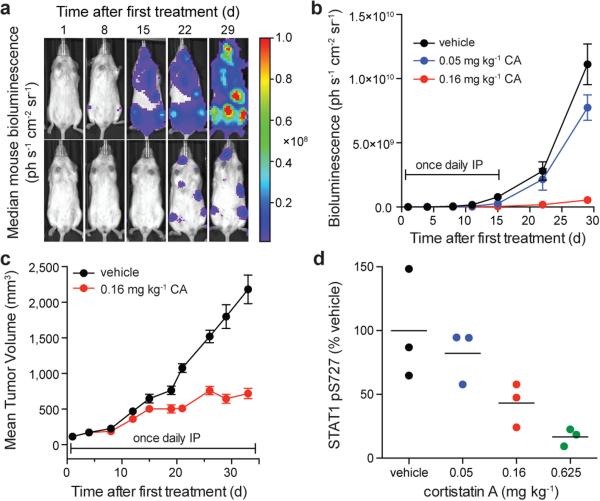
Figure 4. CA inhibits AML progression and CDK8 in vivo.
(a) Bioluminescent images of mice bearing MV4;11 leukaemia cells. Mouse with median bioluminescence shown, treatment as in (b). Color scale 1.00×10⁶ to 1.00×10⁸. (b) Mean ± s.e.m., n=11 mice; p < 0.0001 for both doses on day 33 vs. vehicle, two-way ANOVA. (c) Mice harbouring SET-2 AML xenograft tumours and treated as indicated. Mean ± s.e.m., n=10 mice; 71% tumour growth inhibition on day 33, p < 0.0001, two-tailed t-test. (d) Densitometric analysis of STAT1 pS727 in NK cells isolated from the spleen of C57BL/6 mice treated with CA or vehicle (n=3 mice), STAT1 pS727 normalized to actin, p = 0.011 for 0.625 mg kg−1, one-way ANOVA, experiment performed once.

