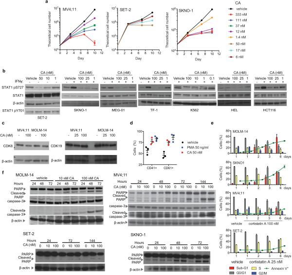
Extended Data Fig. 4. Antiproliferative activity of CA and I-BET151.
(a) Plots showing antiproliferative activity of CA over time for selected sensitive cell lines and concentrations (mean ± s.e.m., n=3 biological replicates, one of two experiments shown). (b) Immunoblots showing that CA inhibits CDK8-dependent IFN-γ-stimulated STAT1 S727 phosphorylation equally well in cells sensitive or insensitive to the antiproliferative activity of CA (one of two experiments shown, full scan in Supplementary Figure 1). (c) Immunoblots showing CDK8 and CDK19 levels upon 24 h CA treatment in sensitive cell lines MV4;11 and MOLM-14 (one of two experiments shown, full scan in Supplementary Figure 1). (d) CD41 and CD61 (vehicle vs. CA, p= 0.04 and 0.005, respectively, two-tailed t-test) on SET-2 cells after 3 days of indicated treatment (mean ± s.e.m., n=3 biological replicates, one of two experiments shown). (e) DNA content and Annexin V staining of indicated cell lines upon treatment with CA (mean ± s.e.m., n=3 biological replicates, one of two experiments shown). (f) Immunoblots of CA dose- and time-dependent induction of PARP and caspase-3 cleavage for indicated cell lines (one of two experiments shown, full scan in Supplementary Figure 1).

