Abstract
Context
Unilateral body-weight exercises are commonly used to strengthen the lower limbs during rehabilitation after injury, but data comparing the loading of the limbs during these tasks are limited.
Objective
To compare joint kinetics and kinematics during 3 commonly used rehabilitation exercises.
Design
Descriptive laboratory study.
Setting
Laboratory.
Patients or Other Participants
A total of 9 men (age = 22.1 ± 1.3 years, height = 1.76 ± 0.08 m, mass = 80.1 ± 12.2 kg) participated.
Intervention(s)
Participants performed the single-legged squat, forward lunge, and reverse lunge with kinetic data captured via 2 force plates and 3-dimensional kinematic data collected using a motion-capture system.
Main Outcome Measure(s)
Peak ground reaction forces, maximum joint angles, and peak sagittal-joint moments.
Results
We observed greater eccentric and concentric peak vertical ground reaction forces during the single-legged squat than during both lunge variations (P ≤ .001). Both lunge variations demonstrated greater knee and hip angles than did the single-legged squat (P < .001), but we observed no differences between lunges (P > .05). Greater dorsiflexion occurred during the single-legged squat than during both lunge variations (P < .05), but we noted no differences between lunge variations (P = .70). Hip-joint moments were greater during the forward lunge than during the reverse lunge (P = .003) and the single-legged squat (P = .011). Knee-joint moments were greater in the single-legged squat than in the reverse lunge (P < .001) but not greater in the single-legged squat than in the forward lunge (P = .41). Ankle-joint moments were greater during the single-legged squat than during the forward lunge (P = .002) and reverse lunge (P < .001).
Conclusions
Appropriate loading progressions for the hip should begin with the single-legged squat and progress to the reverse lunge and then the forward lunge. In contrast, loading progressions for the knee and ankle should begin with the reverse lunge and progress to the forward lunge and then the single-legged squat.
Key Words: joint moment, peak force, lunge, single-legged squat, loading
Key Points
Concentric and eccentric peak vertical ground reaction forces were greater during the single-legged squat than during the reverse and forward lunges because of an increased base of support during the lunges and greater ankle- and knee-joint moments.
Hip-joint moments were greater in the forward lunge.
Peak joint angles were greater in the lunge variations than in the single-legged squat.
Practitioners can use this information to develop a progressive loading paradigm for the hip, knee, and ankle during rehabilitation after injury.
Factors determining the progression of exercise loads during rehabilitation after injury currently follow 1 of 2 general approaches: (1) a progression according to tissue-healing time frames based on histologic studies and (2) an evaluation-based protocol in which the patient passes specific criteria before progression. Both approaches have several advantages and disadvantages.1
During the first approach, progressive loading may be applied, but it is often applied based on time-since-injury criteria rather than tissue-capability criteria. The resultant lack of progressive loading based on tissue capability may provide insufficient stimulus for optimal tissue development and has been proposed to increase the likelihood of disorganized scar formation, passive muscle and joint stiffness, muscle atrophy, and prolonged rehabilitation times.2 The second approach potentially applies controlled stresses on the injured body part, which is likely to promote tissue healing that enhances the mechanical properties of the injured tissues. The problem with the second approach is that limited objective criteria of when and how to progress exercises for the magnitude of mechanical load are available within the research literature, especially when this involves the selection of different exercises.3 If the level of loading is unknown, then a logical progressive schema of tissue loading cannot be applied. Musculoskeletal modeling has described the internal forces, including a patellofemoral-joint force range from 2.5 to 7.6 times body mass and a tibiofemoral joint force range from 2.5 to 7.3 times body mass during body-weight squatting.4–17 Such force results in increased stress on the joint articular surfaces and muscle tendons and increased force production that the muscles require to arrest movement, especially during the eccentric phase. However, no researchers have compared loading among rehabilitation exercises.
Given the necessity to control the body and resultant forces during maneuvers that occur during sporting activities, multijoint exercise using body weight and some additional external load is arguably the most common and most important type of resistance training that athletes use to increase strength and power and to rehabilitate and prevent injury. Therefore, quantifying and comparing the loads on joints and other soft tissues during such exercises are imperative to provide clear criteria of when an exercise will supply suitable loading to stimulate positive tissue changes.2 Quantifying the intensity during normal resistance-training exercise, such as machines or free weights, appears to be relatively easy to understand for the absolute level of load applied to the tissue. This is likely to be true during single-joint exercises, such as knee extension; however, during multi-joint exercises, the relative contribution of each joint and the muscles that generate movement need to be considered if loading is to be estimated for any particular structure or joint.
Whereas the tissues must adapt to the forces that are applied to the joints and surrounding tissues during sporting activities, these forces can be magnified if the athlete exhibits poor dynamic alignment.18,19 Poor dynamic alignment has been defined as an inability to control the trunk, pelvis, hip, knee, and foot in the frontal, sagittal, or transverse plane.20–23 Similar dysfunctional movements have been observed during sporting activities and are commonly screened during movements such as single-legged squats, lunges, and landing tasks.20–23 The asymmetric loading of tissues and increased joint moments created by abnormal dynamic movement patterns increase stress on the tissues, potentially leading to pathologic conditions.18 Poor dynamic-alignment movement control, especially during unilateral tasks, has been linked with patellofemoral joint pain,18,20 anterior cruciate ligament injury,19,21 and general lower limb injury.24 Therefore, the purpose of our study was to quantify and compare peak vertical ground reaction forces (vGRFs); ankle-, knee-, and hip-joint angles; and sagittal-plane joint moments and knee-valgus angles during 3 common unilateral exercises (single-legged squat, forward lunge, reverse lunge) to permit a better understanding of the demands these exercises place on the lower body. We selected these variables because several have been associated with a variety of lower limb injuries.18,23–25 This information will provide clinicians appropriate evidence-based data among exercises to identify which are most suitable within the healing constraints of the tissue at that moment to specifically and safely load the tissue to its maximal potential. We hypothesized that peak vGRFs and external joint moments during both the concentric and eccentric phases of the single-legged squat would be greater than during either lunge variation because of an increased base of support during the lunges. We also hypothesized that this would result in greater peak joint angles in the lunge variations than in the single-legged squat. An additional aim was to assess the reliability of the movement patterns during these exercises to determine if individual performances were replicated among repetitions.
METHODS
Participants
Nine healthy, recreationally active men (age = 22.1 ± 1.3 years, height = 1.76 ± 0.08 m, mass = 80.1 ± 12.2 kg) volunteered to participate. We defined recreationally active as performing structured exercise ≥ 45 minutes, 3 times or more each week. Participants reported no history of lower leg pain or pathologic condition. All participants provided written informed consent, and the study was approved by the University of Salford Institutional Review Board.
Instrumentation
Three-dimensional kinematic data were collected using a 6-camera motion-capture system (OQUS; Qualisys AB, Gothenburg, Sweden) at a sampling frequency of 250 Hz. Kinetic data from two 600- × 900-mm force plates (Advanced Medical Technologies Inc, Newton, MA), which were embedded in the floor and sampled at 2500 Hz, were integrated and simultaneously collected with the kinematic data acquired through Track Manager software (Qualisys AB).
Twenty-four 14-mm retroreflective markers were placed on anatomical landmarks of the lower extremity to define the foot, shank, thigh, and pelvis. Markers were placed bilaterally over the iliac crests; anterior-superior iliac spines; posterior-superior iliac spines; greater trochanters; medial and lateral femoral condyles; medial and lateral malleoli; calcanei; and first, second, and fifth metatarsal heads. In addition, rigid cluster plates, each of which had 4 markers, were securely attached bilaterally on the shank and thigh. Before the exercises were performed, a static calibration trial was recorded. Participants were instructed to stand in an anatomically neutral position to define anatomical markers relative to the dynamic clusters, similarly to the calibrated anatomical-systems technique.26 A CODA pelvis model (Charnwood Dynamics Ltd, Leicestershire, United Kingdom) was created to estimate hip-joint centers based on the work of Bell et al.27,28 Knee- and ankle-joint centers were defined as the midpoints between the lateral and medial epicondyle markers of the femur and the lateral and medial malleolus markers, respectively. Joint kinematics were calculated using a Cardan-Euler method in which the sequence of rotations was X (sagittal plane), Y (frontal plane), Z (vertical plane). In accordance with Decker et al,29 0° at the hip and knee in the sagittal plane was defined as an erect, anatomical stance; 0° at the ankle was defined based on the ankle angle within the static neutral calibration trial, in which a virtual foot was created and used. Positive angle values indicated hip flexion, knee flexion, and ankle dorsiflexion.
Procedures
All participants reported that they were familiar with all activities performed, as they regularly incorporated them into their warm-ups and training regimes. However, proper technique and procedures were explained and demonstrated to them before testing. Participants were allowed to practice each exercise, performing 10 repetitions on both limbs as part of a warm-up. During testing, they were required to perform 5 good trials per limb for each exercise (30 good trials in total). Trial repetitions were separated by a rest period of approximately 1 minute, with exercises performed in a counterbalanced order. Trials in which participants lost balance or did not adhere to the requirements were excluded, with a further 1-minute rest before the next trial. Identical test procedures were repeated for the contralateral limb. Participants were required to perform 5 repetitions of each exercise on each limb. All exercises were performed at a standardized cadence, with a 3-second eccentric phase and a 2-second concentric phase.
Single-Legged Squat
We instructed participants to stand on the selected test limb with their upper extremities crossed and their contralateral limbs positioned in approximately 45° of knee flexion. They squatted down as far as possible and returned to a single-legged stance while maintaining balance (Figure 1A).
Figure 1.
Example of lower limb exercises. A, Single-legged squat. B, Forward lunge. C, Reverse lunge.
Forward Lunge
Participants stood in an upright position on 1 force plate with their upper extremities crossed, stepped forward onto the second force plate with the selected test limb, lunged to a full depth while keeping the whole foot in contact with the force plate, and returned to the start position (Figure 1B).
Reverse Lunge
Participants stood in an upright position on 1 force plate with their upper extremities crossed, stepped back on the contralateral limb onto the second force plate, lunged down as far as possible, and returned to the start position. We required them to maintain balance and keep the selected test foot on the floor throughout the entire reverse lunge (Figure 1C).
Data Processing and Analysis
When digitized within Qualisys Track Manager, data were exported to Visual 3D (Visual 3D Inc, Rockville, MD) for processing and analysis. Motion and force data were filtered with a fourth-order, low-pass Butterworth filter with cutoff frequencies of 5 and 25 Hz, respectively, before further analysis. We analyzed 3 trials per limb per exercise, with the 3 trials selected based on the movement durations that were closest to the predetermined cadence: 3-second eccentric phase followed by 2-second concentric phase. Anthropometric properties, including segment variables, were derived from the anthropometric data of Dempster30 within Visual 3D. Using inverse dynamics31 in Visual 3D, we calculated hip–, knee–, and ankle–sagittal-plane joint moments with kinematic, ground reaction force, and anthropometric data. All joint moments were normalized to body mass and represented as external moments.
During single-legged stance, the onset of activity was defined when knee flexion increased 5° above the initial knee-flexion angle. Conversely, the offset of activity was defined when knee flexion reached 5° less than the final knee-flexion angle in single-legged stance. Within both the forward- and reverse-lunge exercises, foot contact and foot off were identified when the force plate achieved a threshold value ≥ 20 N and ≤ 20 N, respectively. During analysis, each exercise was divided into 2 phases: eccentric and concentric. Eccentric single-legged squat was defined as the time from onset of activity to maximum knee flexion, and concentric single-legged squat was defined as the time from maximum knee flexion to offset of activity. Eccentric forward lunge was defined as the time from foot contact to maximum knee flexion of the test limb, and concentric forward lunge was defined as the time from maximum knee flexion to foot off of the test limb. Eccentric reverse lunge was defined as the time from foot contact of the contralateral limb to maximum knee flexion of the test limb, and concentric reverse lunge was defined as the time from maximum knee flexion of the test limb to foot off of the contralateral limb. For the lunges, all data were analyzed for the lead limb (front limb; Figure 1A and C).
Each variable was exported into Excel (version 2007; Microsoft Corporation, Redmond, WA), where we conducted further analysis to determine dependent variables, such as peak joint angles, peak joint external moments, and peak ground reaction forces.
Statistical Analyses
Dependent t tests were used to determine any differences between right and left limbs, with results demonstrating no differences between limbs (P > .05; Tables 1 and 2); therefore, data from both limbs were pooled for further analysis.
Table 1.
Joint Angles and Moments Between Lower Extremitiesa for Each Exercise
| Joint |
Exercise |
Angle, ° |
Moment, Nm/kg |
||||
| Left Extremity |
Right Extremity |
Cohen d |
Left Extremity |
Right Extremity |
Cohen d |
||
| Hip | Single-legged squat | 79.82 ± 11.20 | 84.04 ± 13.29 | 0.53 | 1.79 ± 0.27 | 1.90 ± 0.45 | 0.29 |
| Forward lunge | 93.08 ± 6.46 | 95.24 ± 8.26 | 0.29 | 2.06 ± 0.32 | 2.12 ± 0.35 | 0.18 | |
| Reverse lunge | 98.01 ± 5.56 | 101.49 ± 7.78 | 0.34 | 1.59 ± 0.43 | 1.77 ± 0.44 | 0.41 | |
| Knee | Single-legged squat | 94.34 ± 11.59 | 92.25 ± 8.20 | 0.46 | 1.14 ± 0.15 | 1.22 ± 0.14 | 0.55 |
| Forward lunge | 63.54 ± 6.07 | 61.90 ± 6.75 | 0.26 | 1.40 ± 0.13 | 1.36 ± 0.22 | 0.22 | |
| Reverse lunge | 66.61 ± 8.05 | 63.69 ± 7.83 | 0.21 | 1.50 ± 0.25 | 1.41 ± 0.20 | 0.40 | |
| Ankle | Single-legged squat | 28.21 ± 6.24 | 32.77 ± 4.70 | 0.48 | 0.72 ± 0.28 | 0.71 ± 0.32 | 0.03 |
| Forward lunge | 22.71 ± 4.65 | 25.05 ± 4.95 | 0.49 | 0.99 ± 0.16 | 1.02 ± 0.20 | 0.17 | |
| Reverse lunge | 20.83 ± 5.77 | 23.62 ± 5.79 | 0.83 | 1.16 ± 0.24 | 1.18 ± 0.18 | 0.10 | |
Indicates no differences between lower extremities for any variable (P > .05).
Table 2.
Vertical Ground Reaction Forces Between Lower Extremitiesa for Each Exercise
| Phase |
Exercise |
Vertical Ground Reaction Forces, N |
||
| Left Extremity |
Right Extremity |
Cohen d |
||
| Concentric | Single-legged squat | 906.74 ± 142.68 | 691.64 ± 103.43 | 0.21 |
| Forward lunge | 764.70 ± 118.49 | 742.56 ± 111.57 | 0.19 | |
| Reverse lunge | 669.28 ± 105.82 | 902.27 ± 139.12 | 0.03 | |
| Eccentric | Single-legged squat | 876.18 ± 148.24 | 663.42 ± 110.79 | 0.17 |
| Forward lunge | 701.50 ± 128.85 | 688.45 ± 128.29 | 0.10 | |
| Reverse lunge | 646.95 ± 81.38 | 863.28 ± 137.39 | 0.09 | |
Indicates no differences between lower extremities for any variable (P > .05).
We calculated intraclass correlation coefficients (ICCs) (3,1) to determine the repeatability among trials for each dependent variable for each exercise and interpreted using the recommendations of Cortina,32 with more than 0.80 considered highly reliable. Effect sizes were determined using the Cohen d method and interpreted based on the recommendations of Rhea33 as trivial (<0.35), small (0.35–0.8), moderate (0.8–1.5), or large (>1.5). After this, mean data from each dependent variable from the 3 repetitions of each exercise were analyzed for differences using a repeated-measures analysis of variance (ANOVA), with Bonferroni post hoc analysis identifying where differences occurred. We set the α level a priori at .05. All statistical analyses were performed using SPSS statistical software (version 20; IBM Corporation, Armonk, NY).
RESULTS
Intraclass correlation coefficients demonstrated high within-sessions reliability for maximum joint angles (ICC [3,1] ≥ 0.890), maximum knee-valgus angles (ICC [3,1] ≥ 0.857), and peak joint moments (ICC [3,1] ≥ 0.864) for each joint and exercise, excluding the knee-joint moment during the reverse lunge, which demonstrated moderate reliability (ICC [3,1] = 0.714; Table 3). Peak vGRFs during the concentric (ICC [3,1] ≥ 0.890) and eccentric (ICC [3,1] ≥ 0.970) phases were also reliable (Table 4).
Table 3.
Reliability (Intraclass Correlation Coefficient [3,1]) of Joint Angles and Joint Moments for Each Exercise
| Variable Exercise |
Joint Moment |
|||
| Hip |
Knee |
Ankle |
Knee Valgus |
|
| Maximum joint angle | ||||
| Single-legged squat | 0.970 | 0.971 | 0.967 | 0.897 |
| Forward lunge | 0.967 | 0.991 | 0.975 | 0.857 |
| Reverse lunge | 0.949 | 0.890 | 0.970 | 0.891 |
| Joint moment | ||||
| Single-legged squat | 0.967 | 0.938 | 0.893 | NA |
| Forward lunge | 0.952 | 0.928 | 0.864 | NA |
| Reverse lunge | 0.958 | 0.714 | 0.903 | NA |
Abbreviation: NA, not applicable.
Table 4.
Reliability (Intraclass Correlation Coefficient [3,1]) of Peak Vertical Ground Reaction Forces for Each Exercise by Phase
| Exercise |
Phase |
|
| Concentric |
Eccentric |
|
| Single-legged squat | 0.991 | 0.989 |
| Forward lunge | 0.971 | 0.992 |
| Reverse lunge | 0.890 | 0.970 |
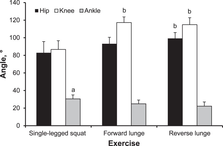
Repeated-measures ANOVA revealed a difference in peak hip angle among exercises (F2,51 = 13.22, P < .001, power = 0.996). The reverse lunge resulted in the largest hip angle (flexion; 99.25° ± 6.67°; 95% confidence interval [CI] = 94.69°, 103.80°), which was greater than during the single-legged squat (82.91° ± 12.74°; 95% CI = 78.36°, 87.47°; P < .001, d = 1.60) but not different than during the forward lunge (93.16° ± 7.36°; 95% CI = 88.60°, 97.71°; P = .19, d = 0.86; Figure 2).
Figure 2.
Comparison of peak joint angles among exercises. a Indicates greater than the forward lunge and reverse lunge (P < .05). b Indicates greater than the single-legged squat (P < .001).
We observed a difference in peak knee angle (flexion) among exercises (F2,51 = 79.26, P < .001, power = 0.996). The greatest knee angle was demonstrated in the forward lunge (117.28° ± 6.41°; 95% CI = 113.46°, 121.12°), which was larger than during the single-legged squat (86.71° ± 9.90°; 95% CI = 82.87°, 90.54°; P < .001, d = 3.67) but not different than during the reverse lunge (114.85° ± 7.94°; 95% CI = 111.00°, 118.66°; P > .99, d = 0.34). The reverse lunge also resulted in a greater knee angle than during the single-legged squat (P < .001, d = 3.14; Figure 2).
A difference in peak ankle angle was observed among exercises (F2,51 = 7.37, P = .002, power = 0.926). We noted a greater ankle angle (dorsiflexion) during the single-legged squat (30.49° ± 4.47°; 95% CI = 27.37°, 33.62°) than during the forward lunge (24.88° ± 4.30°; 95% CI = 21.75°, 27.99°; P = .041, d = 1.28) and the reverse lunge (22.22° ± 4.78°; 95% CI = 19.11°, 25.35°; P = .01, d = 1.79). No differences existed between the forward and reverse lunges (P = .70, d = 0.59; Figure 2).
We found no difference in knee-valgus angles across exercises (F2,51 = 0.45, P > .05).
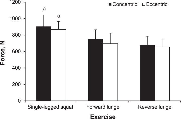
Peak eccentric vGRF (F2,51 = 16.01, P < .001, power = 0.999) and peak concentric vGRF (F2,51 = 18.12, P < .001, power = 1.000) were different across exercises. Greater peak eccentric and concentric vGRFs were observed during the single-legged squat (867.18 ± 98.05 N; 95% CI = 810.65, 923.71 N; and 904.51 ± 140.88 N; 95% CI = 848.99, 955.63 N, respectively) than during the forward lunge (694.98 ± 128.57 N; 95% CI = 638.45, 751.51 N; P < .001, d = 1.19; and 753.63 ± 110.03 N; 95% CI = 700.31, 806.95 N; P = .001, d = 1.51, respectively) and the reverse lunge (655.19 ± 96.09 N; 95% CI = 598.65, 711.72 N; P < .001, d = 2.18; and 680.46 ± 104.63 N; 95% CI = 627.14, 733.78 N; P < .001, d = 1.81, respectively). No differences were identified between the forward and reverse lunges (P > .05; Figure 3).
Figure 3.
Comparison of peak eccentric and concentric vertical ground reaction forces among exercises. a Indicates greater than the forward lunge and reverse lunge (P ≤ .001).
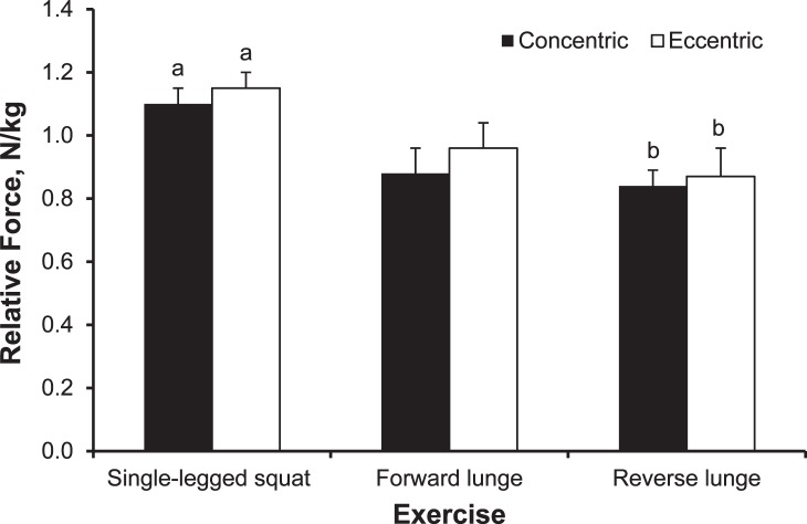
When normalized for body mass, peak relative eccentric and concentric vGRFs were different across exercises (F2,51 = 111.04, P < .001, power = 1.00; and F2,51 = 89.21, P < .001, power = 1.00, respectively). We observed greater peak relative eccentric and concentric vGRFs during the single-legged squat (1.15 ± 0.05 N/kg; 95% CI = 1.12, 1.18 N/kg; and 1.10 ± 0.05 N/kg; 95% CI = 1.08, 1.13 N/kg, respectively) than during the forward lunge (0.96 ± 0.08 N/kg; 95% CI = 0.93, 0.99 N/kg; P < .001, d = 2.85; and 0.88 ± 0.08 N/kg; 95% CI = 0.86, 0.91 N/kg; P < .001, d = 3.30, respectively) and the reverse lunge (0.87 ± 0.09 N/kg; 95% CI = 0.84, 0.90 N/kg; P < .001, d = 3.85; and 0.84 ± 0.05 N/kg; 95% CI = 0.81, 0.86 N/kg; P < .001, d = 5.00, respectively). The reverse lunge also elicited lower peak relative eccentric and concentric vGRFs than the forward lunge (P < .001; Figure 4).
Figure 4.
Comparison of relative peak eccentric and concentric vertical ground reaction forces among exercises. a Indicates greater than the forward lunge and reverse lunge (P < .001). b Indicates less than the forward lunge (P < .001).
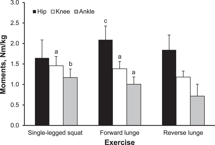
Repeated-measures ANOVA revealed a difference in hip-joint moments across exercises (F2,34 = 11.77, P < .001, power = 0.977). Greater hip-joint moment was noted in the forward lunge (2.09 ± 0.34 Nm/kg; 95% CI = 1.92, 2.26 Nm/kg) than in the reverse lunge (1.84 ± 0.36 Nm/kg; 95% CI = 1.65, 2.03 Nm/kg; P = .003, d = 0.71) and the single-legged squat (1.65 ± 0.44 Nm/kg; 95% CI = 1.42, 1.88 Nm/kg; P = .01, d = 1.12; Figure 5). We found no differences in hip-joint moment between the reverse lunge and single-legged squat (P = .19).
Figure 5.
Comparison of peak joint moments among exercises. a Indicates greater than the reverse lunge (P ≤ .001). b Indicates greater than the forward lunge and reverse lunge (P ≤ .002). c Indicates greater than the single-legged squat and reverse lunge (P < .05).
Knee-joint moments were different among exercises (F2,34 = 24.28, P < .001, power = 1.00). Knee-joint moment was greater during the single-legged squat (1.46 ± 0.22 Nm/kg; 95% CI = 1.36, 1.58 Nm/kg) than during the reverse lunge (1.18 ± 0.15 Nm/kg; 95% CI = 1.10, 1.26 Nm/kg; P < .001, d = 1.49) but not greater than during the forward lunge (1.38 ± 0.18 Nm/kg; 95% CI =1.29, 1.47 Nm/kg; P = .41, d = 0.40; Figure 5). In addition, the forward lunge demonstrated a greater knee-joint moment than the reverse lunge (P = .001, d = 1.21).
Ankle-joint dorsiflexion moments demonstrated differences among exercises (F2,34 = 37.16, P < .001, power = 1.00). We observed greater ankle-joint dorsiflexion moments in the single-legged squat (1.17 ± 0.21 Nm/kg; 95% CI = 1.06, 1.28 Nm/kg) than in the forward lunge (1.00 ± 0.18 Nm/kg; 95% CI = 0.91, 1.08 Nm/kg; P = .002, d = 0.87) and reverse lunge (0.72 ± 0.29 Nm/kg; 95% CI = 0.58, 0.86 Nm/kg; P < .001, d = 1.78; Figure 5). The forward lunge also resulted in greater ankle-joint dorsiflexion moments than the reverse lunge (P < .001, d = 1.16).
DISCUSSION
The purpose of our study was to compare hip, knee, and ankle kinematic and kinetic variables during several commonly prescribed rehabilitation and general training exercises to support recommendations on loading progressions for each relevant joint. Practitioners can use this information to make decisions about the relative loading of each joint (hip, knee, and ankle) during the specific exercise and develop a progressive-loading (progressively increasing stress on the relevant structures) paradigm. As hypothesized, both concentric and eccentric peak vGRFs were greater during the single-legged squat than during either lunge variation because of an increased base of support during the lunges along with greater knee-joint and ankle-joint moments. Whereas the forward and reverse lunges had similar magnitudes of ground reaction force, the anterior-posterior components may have been increased in these exercises relative to those in the single-legged squat. Chappell et al34 reported that anterior-posterior forces during stop-jump tasks were related to proximal knee shear forces, but van den Bogert and McLean35 disputed these findings. In contrast, we observed that hip-joint moments were greatest in the forward lunge. In addition, as hypothesized, greater peak joint angles were observed in the lunge variations than in the single-legged squat.
The first attribute of the 3 exercises we assessed was the reliability of movement patterns. All exercises demonstrated excellent reliability for all variables tested, showing that participants can perform these exercises consistently. Having consistent loading and movement patterns for each exercise provides the practitioner with greater confidence that the exercises will produce consistent levels of tissue stress.
In relation to the kinematics of the hip joint, the reverse lunge created the greatest hip-flexion angle, which was larger than during a single-legged squat but not different than during a forward lunge. However, this result is likely a product of the greater knee-flexion angles in the lunge exercises than in the single-legged squat, resulting in an increased flexion angle between the thigh and trunk. We observed a greater hip-extensor moment during the forward lunge than during the reverse lunge or single-legged squat, with no difference between the hip moment of the latter 2 exercises. Therefore, if the client has limited range of hip movement or is at risk of a pathologic condition, such as hip impingement, it would appear sensible to choose the single-legged squat over the forward and reverse lunges, as it requires less range of motion at the hip. If the clinician wants to improve hip-extensor strength, the forward lunge will be more challenging than the reverse lunge or single-legged squat. Conversely, if the client has noticeably weak hip extensors or injury to those structures, the opposite would apply.
At the knee joint, the 2 lunge exercises demonstrated greater knee-flexion angles than in the single-legged squat. No difference in the range of knee flexion was observed between lunges. The single-legged squat generated the greatest knee-extensor moment but this was not different than the forward lunge. Both exercises produced greater moments than the reverse lunge. If the clinician wants to challenge the knee extensors, then either the single-legged squat or the forward lunge would be more suitable than the reverse lunge. Patellofemoral-joint compressive forces increase with increased ranges of knee flexion6,36; therefore, the forward-lunge exercise, with its increased range of motion and knee-extensor moments, is likely to be the most stressful of the 3 exercises for the patellofemoral joint. Similarly, tibiofemoral-joint compressive load and shear are increased progressively with increased knee-flexion angles10,36; therefore, for individuals with degeneration of this joint (osteoarthritis), the forward lunge is again likely to be a less favorable choice of exercise. The reduced range of knee flexion during the single-legged squat coupled with similar knee moments and decreased hip moments (potentially relatively decreased hamstrings coactivation) may expose structures such as the anterior cruciate ligament to increased load because of relatively increased shear force.37
The 2 lunging exercises generated less ankle-dorsiflexion range than was required for the single-legged squat. The single-legged squat produced a greater ankle–plantar-flexion moment than both forward- and reverse-lunge exercises, and the forward lunge produced less than the reverse lunge. For individuals with a condition such as Achilles tendinopathy, the single-legged squat appears to be the most stressful exercise, and the forward lunge appears to be the least stressful and, therefore, a less favorable choice for these individuals.
We identified lower limb kinematic and kinetic variables while participants performed the single-legged squat, forward lunge, and reverse lunge, but we did not examine muscle activity during these tasks, which is a limitation. Distefano et al,38 Boren et al,39 and Begalle et al40 reported relative levels of muscle (electromyographic) activity during single-legged squatting and lunging, but they did not study the kinematics and kinetics of these exercises. By combining their work and ours, the reader is likely to have a more complete picture of these exercises related to the loads applied and the muscles involved. We undertook this work only on healthy, physically active individuals. Future study is required to evaluate if these findings are consistent across other populations and injured groups.
CONCLUSIONS
When clinicians rehabilitate patients, applying controlled stresses to the injured body part is likely to enhance the mechanical properties of the injured tissues, promoting healing. However, limited objective data related to the loading characteristics of different rehabilitation exercises are available. Our study produced kinetic and kinematic data for 3 commonly used lower limb exercises, which may help clinicians make more informed decisions when loading or unloading particular structures is desirable during injury rehabilitation to appropriately progress loading and facilitate appropriate healing and tissue recovery.
REFERENCES
- 1.Creighton DW, Shrier I, Shultz R, Meeuwisse WH, Matheson GO. Return-to-play in sport: a decision-based model. Clin J Sport Med. 2010;20(5):379–385. doi: 10.1097/JSM.0b013e3181f3c0fe. [DOI] [PubMed] [Google Scholar]
- 2.Khan KM, Scott A. Mechanotherapy: how physical therapists' prescription of exercise promotes tissue repair. Br J Sports Med. 2009;43(4):247–252. doi: 10.1136/bjsm.2008.054239. [DOI] [PMC free article] [PubMed] [Google Scholar]
- 3.Myer GD, Paterno MV, Ford KR, Quatman CE, Hewett TE. Rehabilitation after anterior cruciate ligament reconstruction: criteria-based progression through the return-to-sport phase. J Orthop Sports Phys Ther. 2006;36(6):385–402. doi: 10.2519/jospt.2006.2222. [DOI] [PubMed] [Google Scholar]
- 4.Wallace DA, Salem GJ, Salinas R, Powers CM. Patellofemoral joint kinetics while squatting with and without an external load. J Orthop Sports Phys Ther. 2002;32(4):141–148. doi: 10.2519/jospt.2002.32.4.141. [DOI] [PubMed] [Google Scholar]
- 5.Sharma A, Leszko F, Komistek RD, Scuderi GR, Cates HE, Liu F. In vivo patellofemoral forces in high flexion total knee arthroplasty. J Biomech. 2008;41(3):642–648. doi: 10.1016/j.jbiomech.2007.09.027. [DOI] [PubMed] [Google Scholar]
- 6.Reilly DT, Martens M. Experimental analysis of the quadriceps muscle force and patello-femoral joint reaction force for various activities. Acta Orthop Scand. 1972;43(2):126–137. doi: 10.3109/17453677208991251. [DOI] [PubMed] [Google Scholar]
- 7.Collins JJ. Antagonistic-synergistic muscle action at the knee during competitive weightlifting. Med Biol Eng Comput. 1994;32(2):168–174. doi: 10.1007/BF02518914. [DOI] [PubMed] [Google Scholar]
- 8.Dahlkvist NJ, Mayo P, Seedhom BB. Forces during squatting and rising from a deep squat. Eng Med. 1982;11(2):69–76. doi: 10.1243/emed_jour_1982_011_019_02. [DOI] [PubMed] [Google Scholar]
- 9.Escamilla RF, Fleisig GS, Zheng N, Barrentine SW, Wilk KE, Andrews JR. Biomechanics of the knee during closed kinetic chain and open kinetic chain exercises. Med Sci Sports Exerc. 1998;30(4):556–569. doi: 10.1097/00005768-199804000-00014. [DOI] [PubMed] [Google Scholar]
- 10.Wilk KE, Escamilla RF, Fleisig GS, Barrentine SW, Andrews JR, Boyd ML. A comparison of tibiofemoral joint forces and electromyographic activity during open and closed kinetic chain exercises. Am J Sports Med. 1996;24(4):518–527. doi: 10.1177/036354659602400418. [DOI] [PubMed] [Google Scholar]
- 11.Nagura T, Matsumoto H, Kiriyama Y, Chaudhari A, Andriacchi TP. Tibiofemoral joint contact force in deep knee flexion and its consideration in knee osteoarthritis and joint replacement. J Appl Biomech. 2006;22(4):305–313. doi: 10.1123/jab.22.4.305. [DOI] [PubMed] [Google Scholar]
- 12.Nisell R. Mechanics of the knee: a study of joint and muscle load with clinical applications. Acta Orthop Scand Suppl. 1985;56(S216):1–42. [PubMed] [Google Scholar]
- 13.Salem GJ, Powers CM. Patellofemoral joint kinetics during squatting in collegiate women athletes. Clin Biomech (Bristol, Avon) 2001;16(5):424–430. doi: 10.1016/s0268-0033(01)00017-1. [DOI] [PubMed] [Google Scholar]
- 14.Shelburne KB, Pandy MG. Determinants of cruciate-ligament loading during rehabilitation exercise. Clin Biomech (Bristol, Avon) 1998;13(6):403–413. doi: 10.1016/s0268-0033(98)00094-1. [DOI] [PubMed] [Google Scholar]
- 15.Smith SM, Cockburn RA, Hemmerich A, Li RM, Wyss UP. Tibiofemoral joint contact forces and knee kinematics during squatting. Gait Posture. 2008;27(3):376–386. doi: 10.1016/j.gaitpost.2007.05.004. [DOI] [PubMed] [Google Scholar]
- 16.Thambyah A. How critical are the tibiofemoral joint reaction forces during frequent squatting in Asian populations? Knee. 2008;15(4):286–294. doi: 10.1016/j.knee.2008.04.006. [DOI] [PubMed] [Google Scholar]
- 17.Toutoungi DE, Lu TW, Leardini A, Catani F, O'Connor JJ. Cruciate ligament forces in the human knee during rehabilitation exercises. Clin Biomech (Bristol, Avon) 2000;15(3):176–187. doi: 10.1016/s0268-0033(99)00063-7. [DOI] [PubMed] [Google Scholar]
- 18.Souza RB, Powers CM. Differences in hip kinematics, muscle strength, and muscle activation between subjects with and without patellofemoral pain. J Orthop Sports Phys Ther. 2009;39(1):12–19. doi: 10.2519/jospt.2009.2885. [DOI] [PubMed] [Google Scholar]
- 19.Hewett TE, Myer GD, Ford KR, et al. Biomechanical measures of neuromuscular control and valgus loading of the knee predict anterior cruciate ligament injury risk in female athletes: a prospective study. Am J Sports Med. 2005;33(4):492–501. doi: 10.1177/0363546504269591. [DOI] [PubMed] [Google Scholar]
- 20.Earl J, Hertel J, Denegar CR. Patterns of dynamic malalignment, muscle activation, joint motion, and patellofemoral pain syndrome. J Sport Rehabil. 2005;14(3):216–234. [Google Scholar]
- 21.Noyes FR, Barber-Westin SD, Fleckenstein C, Walsh C, West J. The drop-jump screening test: difference in lower limb control by gender and effect of neuromuscular training in female athletes. Am J Sports Med. 2005;33(2):197–207. doi: 10.1177/0363546504266484. [DOI] [PubMed] [Google Scholar]
- 22.Sugimoto D, Myer GD, McKeon JM, Hewett TE. Evaluation of the effectiveness of neuromuscular training to reduce anterior cruciate ligament injury in female athletes: a critical review of relative risk reduction and numbers-needed-to-treat analyses. Br J Sports Med. 2012;46(14):979–988. doi: 10.1136/bjsports-2011-090895. [DOI] [PMC free article] [PubMed] [Google Scholar]
- 23.Hewett TE, Lindenfeld TN, Riccobene JV, Noyes FR. The effect of neuromuscular training on the incidence of knee injury in female athletes: a prospective study. Am J Sports Med. 1999;27(6):699–706. doi: 10.1177/03635465990270060301. [DOI] [PubMed] [Google Scholar]
- 24.Leetun DT, Ireland ML, Willson JD, Ballantyne BT, Davis IM. Core stability measures as risk factors for lower extremity injury in athletes. Med Sci Sports Exerc. 2004;36(6):926–934. doi: 10.1249/01.mss.0000128145.75199.c3. [DOI] [PubMed] [Google Scholar]
- 25.Myer GD, Ford KR, Brent JL, Hewett TE. An integrated approach to change the outcome part I: neuromuscular screening methods to identify high ACL injury risk athletes. J Strength Cond Res. 2012;26(8):2265–2271. doi: 10.1519/JSC.0b013e31825c2b8f. [DOI] [PMC free article] [PubMed] [Google Scholar]
- 26.Cappozzo A, Catani F, Croce UD, Leardini A. Position and orientation in space of bones during movement: anatomical frame definition and determination. Clin Biomech (Bristol, Avon) 1995;10(4):171–178. doi: 10.1016/0268-0033(95)91394-t. [DOI] [PubMed] [Google Scholar]
- 27.Bell AL, Pedersen DR, Brand RA. A comparison of the accuracy of several hip center location prediction methods. J Biomech. 1990;23(6):617–621. doi: 10.1016/0021-9290(90)90054-7. [DOI] [PubMed] [Google Scholar]
- 28.Bell AL, Brand RA, Pederson DR. Prediction of hip joint center location from external landmarks. Hum Mov Sci. 1989;8(1):3–16. [Google Scholar]
- 29.Decker MJ, Torry MR, Wyland DJ, Sterett WI. Richard Steadman J. Gender differences in lower extremity kinematics, kinetics and energy absorption during landing. Clin Biomech (Bristol, Avon) 2003;18(7):662–669. doi: 10.1016/s0268-0033(03)00090-1. [DOI] [PubMed] [Google Scholar]
- 30.Dempster WT. Space Requirements of the Seated Operator: Geometrical, Kinematic, and Mechanical Aspects of the Body With Special Reference to the Limbs. US Air Force technical report 55-159. Wright-Patterson Air Force Base, OH: US Air Force;; 1955. [Google Scholar]
- 31.Winter DA. Biomechanics and Motor Control of Human Movement. 2nd ed. New York, NY: John Wiley & Sons;; 1990. pp. 107–138. [Google Scholar]
- 32.Cortina JM. What is coefficient alpha? An examination of theory and applications. J Appl Psychol. 1993;78(1):98–104. [Google Scholar]
- 33.Rhea MR. Determining the magnitude of treatment effects in strength training research through the use of the effect size. J Strength Cond Res. 2004;18(4):918–920. doi: 10.1519/14403.1. [DOI] [PubMed] [Google Scholar]
- 34.Chappell JD, Herman DC, Knight BS, Kirkendall DT, Garrett WE, Yu B. Effect of fatigue on knee kinetics and kinematics in stop-jump tasks. Am J Sports Med. 2005;33(7):1022–1029. doi: 10.1177/0363546504273047. [DOI] [PubMed] [Google Scholar]
- 35.van den Bogert AJ, McLean SG. Comment: effect of fatigue on knee kinetics and kinematics in stop-jump tasks. Am J Sports Med. 2006;34(2):312–315. doi: 10.1177/0363546505283276. [DOI] [PubMed] [Google Scholar]
- 36.Escamilla RF. Knee biomechanics of the dynamic squat exercise. Med Sci Sports Exerc. 2001;33(1):127–141. doi: 10.1097/00005768-200101000-00020. [DOI] [PubMed] [Google Scholar]
- 37.Blackburn JT, Norcross MF. The effects of isometric and isotonic training on hamstring stiffness and anterior cruciate ligament loading mechanisms. J Electromyogr Kinesiol. 2014;24(1):98–103. doi: 10.1016/j.jelekin.2013.10.010. [DOI] [PubMed] [Google Scholar]
- 38.Distefano LJ, Blackburn T, Marshall SW, Padua DA. Gluteal muscle activation during common therapeutic exercises. J Orthop Sports Phys Ther. 2009;39(7):532–540. doi: 10.2519/jospt.2009.2796. [DOI] [PubMed] [Google Scholar]
- 39.Boren K, Conrey C, Le Coquic J, Paprocki L, Voight M, Robinson TK. Electromyographic analysis of gluteus medius and gluteus maximus during rehabilitation exercises. Int J Sports Phys Ther. 2011;6(3):206–223. [PMC free article] [PubMed] [Google Scholar]
- 40.Begalle RL, DiStefano LJ, Blackburn T, Padua DA. Quadriceps and hamstrings coactivation during common therapeutic exercises. J Athl Train. 2012;47(4):396–405. doi: 10.4085/1062-6050-47.4.01. [DOI] [PMC free article] [PubMed] [Google Scholar]


