Abstract
Based on recent molecular data, it has been suggested that Sporothrix globosa is the main causal agent of sporotrichosis in China. The objective of this study was to compare the morphology, growth characteristics, patterns of carbon source usage, and susceptibility to antifungal agents among Sporothrix strains. A total of 15 clinical strains confirmed to be S. globosa, from three different regions of China, and 11 ex-type strains from the CBS-KNAW biodiversity center were obtained. The elongated conidia of S. pallida, S. variecibatus, S. schenckii, and S. schenckii luriei were clearly different from the subglobose and globose conidia of S. globosa strains. S. schenckii is able to assimilate sucrose, raffinose, and ribitol. Susceptibility profiles of these Sporothrix species were evaluated by measuring minimum inhibitory concentrations (MICs). Fluconazole, itraconazole, terbinafine, and amphotericin B showed good activity against most S. globosa clinical isolates from China. Potassium iodide also showed a low MIC against S. pallida, while fluconazole showed a high MIC for S. mexicana, S. humicola, S. globosa, S. schenckii, and S. inflata; these strains might be considered tolerant. The species showed differences in susceptibility to antifungal drugs and should therefore be properly identified during diagnosis prior to designing therapeutic strategies.
Keywords: Sporothrix globosa, Phenotypic characters, Antifungal susceptibility
1. Introduction
Sporotrichosis is a chronic subcutaneous mycosis that can be acquired by contact with contaminated objects or organic material (O'Reilly and Altman, 2006). It also can be acquired through zoonotic transmission (Oliveira et al., 2011; Silva-Vergara et al., 2012). The incidence of sporotrichosis is increasing in China, particularly in the northeastern region of the country (Zhang and Lin, 2008). Sporothrix globosa was identified as the causal agent of sporotrichosis in many of these cases (Tan et al., 2013; Yu et al., 2013; Liu et al., 2014; Zhou et al., 2014). Four species of the Sporothrix complex have been reported as causal agents of sporotrichosis: S. brasiliensis, S. globosa, S. schenckii s. str., and S. schenckii luriei (Rodrigues et al., 2014). S. pallida and S. mexicana are mainly environmental but may also be potentially pathogenic to humans and other mammals. It is not clear if these species have different physiological features that determine their pathogenicity or if they differ in their susceptibility to antifungals.
Species in the S. schenckii complex have been shown to differ in their calmodulin (CAL), internal transcribed spacer (ITS), and a fragment of the β-tubulin gene sequences (Marimon et al., 2007). Few studies have focused on the morphological and physiological variation among strains that comprise the S. schenckii complex. Recent studies have shown that morphologically similar species in this group could differ in their physiological features (Marimon et al., 2007; Rodrigues et al., 2014). Colony characteristics, growth rates, and carbon assimilation tests have been shown to be useful for distinguishing species of Sporothrix (Marimon et al., 2007; de Meyer et al., 2008). Moreover, the distribution of sessile conidia and their morphological variation have been suggested to be the key features distinguishing species within the Sporothrix complex. In this study, using type strains of different species of Sporothrix, we compared colony and microscopic morphologies, assimilation of carbon sources, and antifungal susceptibilities. The variation in these characteristics among different Sporothrix species, as reported here, provides new insights into their pathogenicity mechanisms and suggests different susceptibilities to antifungals, thereby providing new avenues for clinical therapeutic strategies.
2. Materials and methods
2.1. Strains
Fifteen strains of Sporothrix from clinical sources in China were included in the study. Isolates consisted of 15 strains that were morphologically and genetically identified as S. globosa (Liu et al., 2014) and 11 ex-type strains, including S. brunneoviolacea (n=1), S. dimorphospora (n=1), S. globosa (n=1), S. humicola (n=1), S. inflata (n=1), S. lignivora (n=1), S. mexicana (n=1), S. pallida (n=1), S. variecibatus (n=1), S. schenckii (n=1), and S. schenckii luriei (n=1) (Table 1). Isolates were stored at room temperature on slant cultures containing 2% (0.02 g/ml) potato dextrose agar. Cultures were stored at 4 °C in distilled water for long-term preservation.
Table 1.
Sources of isolates used in this study and analysis
| Strain | Name | Source | Country of origin |
| LC2454 | S. globosa | Clincal strain | Beijing, China |
| LC2460 | S. globosa | Clincal strain | Beijing, China |
| LC2466 | S. globosa | Clincal strain | Beijing, China |
| LC2467 | S. globosa | Clincal strain | Beijing, China |
| LC2469 | S. globosa | Clincal strain | Beijing, China |
| LC2432 | S. globosa | Clincal strain | Chongqing, China |
| LC2433 | S. globosa | Clincal strain | Chongqing, China |
| LC2445 | S. globosa | Clincal strain | Chongqing, China |
| LC2447 | S. globosa | Clincal strain | Chongqing, China |
| LC2480 | S. globosa | Clincal strain | Chongqing, China |
| LC2404 | S. globosa | Clincal strain | Jilin, China |
| LC2405 | S. globosa | Clincal strain | Jilin, China |
| LC2411 | S. globosa | Clincal strain | Jilin, China |
| LC2413 | S. globosa | Clincal strain | Jilin, China |
| LC2419 | S. globosa | Clincal strain | Jilin, China |
| CBS 120341 | S. mexicana | Environmental | Mexcio |
| CBS 124561 | S. brunneoviolacea | Soil | Spain |
| CBS 118129 | S. humicola | Soil | South Africa |
| CBS 239.68 | S. inflata | Wheat-field soil | Germany |
| CBS 131.56 | S. pallida | Stemonitis fusca | Japan |
| CBS 121961 | S. variecibatus | Trichopoda sp. from Protea repens | South Africa |
| CBS 119148 | S. lignivora | Utility poles | South Africa |
| CBS 120340 | S. globosa | Clincal strain | Spain |
| CBS 553.74 | S. dimorphospora | Soil | Canada |
| CBS 359.36 | S. schenckii | Clincal strain | Unknown |
| CBS 937.72 | S. schenckii luriei | Clincal strain | South Africa |
2.2. Morphological characteristics
Mycelial discs (2 mm diameter) of each strain were transferred to three replicate plates containing 2% (0.02 g/ml) potato dextrose agar, and the plates were incubated at 25 °C. Colony size, shape, and pigment production were recorded after 7, 14, 21, and 28 d of incubation. Colony diameter was measured daily for 7 d and growth rate was calculated as the 7-d average of mean weekly growth (mm/week). Growth rates were subjected to an analysis of variance (P<0.05) with Duncan’s multiple range test by using SPSS software (Version 16.0). After 7 d, the shape and size of the appressoria formed across the underside of the cover slip were studied (Marimon et al., 2007).
2.3. Physiological studies
Carbon assimilation was tested in liquid medium according to Marimon et al. (2007). Variation in the ability to grow on sucrose, raffinose, and maltose was tested in all strains. The tests were conducted in 96-well microplates, containing either no carbon source (negative control) or glucose (positive control). The inocula were adjusted to a final concentration of from 2×10⁵ to 2×10⁶ colony-forming unit (CFU)/ml. Microplates were read (530 nm) after 5 d of incubation at 25 °C, as previously described (Marimon et al., 2007).
2.4. Antifungal susceptibility testing
The following antifungals were tested for their activity against Sporothrix strains: fluconazole (FLC), itraconazole (ITC), terbinafine (TRB), amphotericin B (AMB), and potassium iodide (KI). Antifungal susceptibility tests were performed on the isolates using the methods recommended by the Clinical Laboratory and Standards Institute standard document M38-A2 (Kohler et al., 2004; Trilles et al., 2005; Marimon et al., 2008; Oliveira et al., 2011). Ninety-six-well microplates containing 0.01 ml of RPMI-1640 nutrient broth in each well were incubated at 25 °C. Nutrient broth RPMI-1640 was amended with antifungal drug concentrations to final concentrations that spanned 0.50 to 500.00 μg/ml (KI), 0.125 to 64.00 μg/ml (FLC), and 0.03 to 16.00 μg/ml (ITC, TRB, and AMB). The concentration of cells used to inoculate assays was adjusted to a final concentration that ranged from 1×10⁴ to 5×10⁴ CFU/ml. Candida parapsilosis (ATCC 22019) was included as a control for each antifungal test (Marimon et al., 2008). Growth was monitored spectrophotometrically at 530 nm during the growth period and growth in the presence of antifungals was compared with growth in the absence of antifungals to establish minimum inhibitory concentrations (MICs) (Espinel-Ingroff et al., 2002). The MICs for AMB, ITC, and KI were defined as the lowest concentration showing 100% growth inhibition. MICs for TRB were defined as the lowest concentrations that showed 80% inhibition of growth, and for FLC as the lowest concentration that produced 50% growth inhibition (Trilles et al., 2005; Marimon et al., 2008; Oliveira et al., 2011; Rodrigues et al., 2014). The tests were carried out three times and an MIC based on these replicates was calculated. Carbon assimilation was tested in liquid medium according to Marimon et al. (2007). Variation in the ability to grow on sucrose, raffinose, and maltose was tested in all strains. The tests were conducted in 96-well microplates, containing either no carbon source (negative control) or glucose (positive control). The inocula were adjusted to a final concentration of from 2×10⁵ to 2×10⁶ CFU/ml. Microplates were read (530 nm) after 5 d of incubation at 25 °C, as previously described (Marimon et al., 2007).
3. Results
3.1. Morphological characteristics
3.1.1 Colony characters
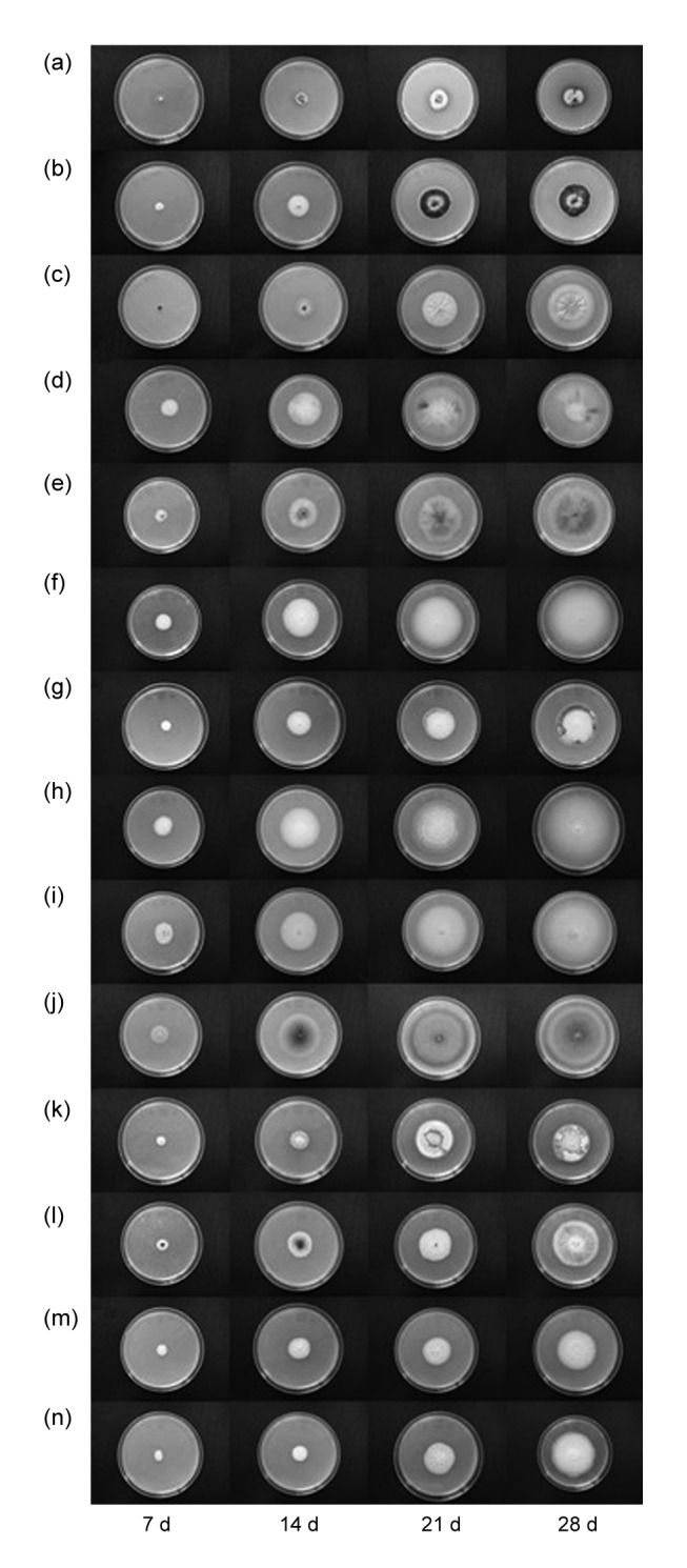
Images of Sporothrix isolate colonies are depicted in Fig. 1. All strains grew well on potato dextrose agar at 25 °C. Colonies of clinical isolates from China produced cream-colored, smooth or verrucous, moist colonies with occasional aerial mycelia. Over time, these colonies matured into black leathery colonies with a wrinkled and folded surface. Colonies of S. schenckii, S. schenckii luriei, S. pallida, and S. humicola produced white aerial mycelium, with white colonies in the center. These colonies remained white to cream-colored over time.
Fig. 1.

Colony morphology of isolates grown on potato dextrose agar at 25 °C following incubation for 7, 14, 21, and 28 d
(a) S. globosa (Beijing, China); (b) S. globosa (Jilin, China); (c) S. globosa (Chongqing, China); (d) S. mexicana (CBS 120341); (e) S. brunneoviolacea (CBS 124561); (f) S. humicola (CBS 118129); (g) S. inflata (CBS 239.68); (h) S. pallida (CBS 31.56); (i) S. variecibatus (CBS 121961); (j) S. lignivora (CBS 119148); (k) S. globosa (CBS 120340); (l) S. dimorphospora (CBS 553.74); (m) S. schenckii (CBS 359.36); (n) S. schenckii luriei (CBS 937.72)
3.1.2 Conidial morphology
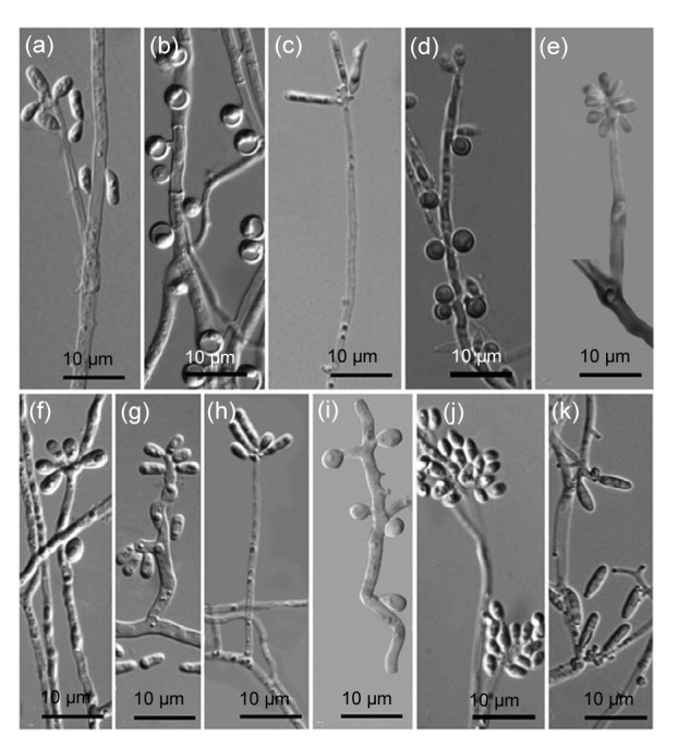
The morphology of conidia in S. schenckii isolates differed from that of the S. globosa isolates examined. The conidia of S. globosa isolates were septate hyphae with terminal clavate obovoid structures. The conidia of S. schenckii isolates were ovate, hyaline, and thin-walled conidia in sympodial conidiophores, with oval to oblong phialides. The type strain of S. inflata produced globose, sessile, brown conidia. The type strains of S. mexicana, S. variecibatus, S. lignivora, and S. pallida produced oval conidia and hyphae with terminal obovoid conidia. The type strain of S. brunneoviolacea showed hyaline, globose to smooth and thin-walled guttulate conidia.The conidia of ex-type S. humicola were solitary, straight, and hyaline. The type strain of S. dimorphosporas showed subglobose to obovoid sessile conidia in a conidiogenous terminal structure. The type strain of S. schenckii luriei produced septate, oblong, and hyaline conidia that were slightly irregular to irregular in shape (Fig. 2).
Fig. 2.

Morphology of the conidia of the species in Sporothrix schenckii sensu lato when grown on potato dextrose agar at 25 °C for 7 d
(a) S. mexicana (CBS 120341); (b) S. brunneoviolacea (CBS 124561); (c) S. humicola (CBS 118129); (d) S. inflata (CBS 239.68); (e) S. pallida (CBS 131.56); (f) S. variecibatus (CBS 121961); (g) S. lignivora (CBS 119148); (h) S. globosa (CBS 120340); (i) S. dimorphospora (CBS 553.74); (j) S. schenckii (CBS 359.36); (k) S. schenckii luriei (CBS 937.72)
3.1.3 Growth rate
Duncan’s multiple range test showed no significant differences in the growth rates among 15 S. globosa clinical strains from China (P=0.45). However, there was a statistical difference in growth rates among the different species that comprise the complex. Moreover, statistical analysis revealed a faster growth rate of strains isolated from the natural environment compared with those obtained from clinical specimens (P<0.05). The maximum growth rate of S. globosa strains was (12.33±5.29) mm/week and the minimum growth rate was (8.41±3.02) mm/week; these were not significantly different (P>0.05). S. mexicana type strains grew the fastest with a rate of (19.93±2.15) mm/week. S. schenckii and S. globosa type strains had growth rates of (8.68±1.56) mm/week and (9.77±2.86) mm/week, respectively. The colony diameters for different isolates at the same temperature are summarized in Table 2.
Table 2.
Summary of morphological data obtained from Sporothrix isolates
| Fungus isolate | Diameter (mm) |
Growth rate (mm/week) | |||
| 7 d | 14 d | 21 d | 28 d | ||
| LC2454 | 10.15±4.83 | 21.33±1.36 | 26.83±1.72 | 38.33±3.20 | 8.41±3.02 |
| LC2460 | 10.65±4.84 | 20.50±1.51 | 32.00±3.03 | 39.83±3.31 | 9.17±4.05 |
| LC2466 | 11.66±1.21 | 21.16±1.47 | 30.33±1.36 | 40.66±2.94 | 9.05±1.84 |
| LC2467 | 11.66±1.03 | 21.00±1.54 | 33.16±2.02 | 42.66±3.66 | 10.44±3.68 |
| LC2469 | 12.16±1.16 | 21.16±1.72 | 32.66±2.33 | 44.33±3.93 | 10.72±2.63 |
| LC2432 | 9.98±4.48 | 21.16±2.31 | 31.83±2.99 | 46.10±1.16 | 11.83±4.34 |
| LC2433 | 8.16±6.26 | 22.63±2.16 | 34.33±3.20 | 44.33±1.50 | 11.11±3.06 |
| LC2445 | 8.28±5.76 | 22.66±3.93 | 35.50±2.07 | 43.00±3.57 | 11.38±4.52 |
| LC2447 | 9.98±4.57 | 21.50±1.37 | 30.33±2.58 | 44.66±2.16 | 11.11±3.37 |
| LC2480 | 9.63±4.35 | 21.50±2.73 | 31.66±2.92 | 44.50±2.25 | 11.22±4.09 |
| LC2404 | 9.96±4.63 | 20.16±1.16 | 31.00±3.09 | 39.50±3.01 | 9.55±2.70 |
| LC2405 | 10.15±4.74 | 20.50±1.08 | 31.50±2.66 | 39.33±2.42 | 9.50±4.24 |
| LC2411 | 9.80±4.86 | 20.33±2.33 | 31.33±3.38 | 47.66±1.36 | 12.33±5.29 |
| LC2415 | 9.96±4.55 | 20.00±2.28 | 31.50±2.25 | 46.00±3.46 | 11.05±2.97 |
| LC2419 | 9.46±6.67 | 21.00±0.89 | 32.16±2.31 | 47.83±3.18 | 11.88±4.01 |
| S. mexicana | 25.70±2.98 | 46.00±2.13 | 63.63±2.13 | 85.50±0.83 | 19.93±2.15 |
| S. brunneoviolacea | 16.83±2.50 | 33.16±1.72 | 52.66±2.25 | 64.50±0.57 | 15.85±3.65 |
| S. humicola | 27.25±4.52 | 47.25±3.19 | 59.66±2.73 | 71.33±1.21 | 14.74±4.38 |
| S. inflata | 13.75±1.28 | 24.66±2.50 | 39.00±1.78 | 45.66±3.72 | 10.63±6.77 |
| S. pallida | 28.62±3.50 | 51.00±4.14 | 62.33±2.58 | 71.50±1.51 | 14.29±6.46 |
| S. variecibatus | 29.33±5.20 | 48.22±5.08 | 65.16±1.16 | 74.50±0.54 | 15.05±5.16 |
| S. lignivora | 29.66±1.62 | 58.50±6.41 | 76.83±7.47 | 77.50±0.54 | 18.03±10.3 |
| S. globosa | 17.01±4.69 | 24.50±1.97 | 33.83±1.47 | 46.33±1.50 | 9.77±2.86 |
| S. dimorphospora | 15.50±2.07 | 30.66±1.86 | 42.66±3.01 | 55.00±1.09 | 13.20±2.05 |
| S. schenckii | 17.20±3.35 | 27.35±2.25 | 34.16±1.83 | 43.00±1.78 | 8.68±1.56 |
| S. schenckii luriei | 14.25±3.84 | 24.83±5.07 | 35.33±1.03 | 55.66±1.63 | 13.78±5.73 |
3.2. Physiological studies
The type strain of S. lignivora (CBS 119148) was unable to assimilate sucrose, unlike all of the other strains. All isolates were able to assimilate dextrose and ribitol with the exception of S. dimorphospora, S. inflata, S. lignivora, and S. mexicana. With respect to carbon source utilization, the clinical isolates from China were similar to the type strain of S. globosa (Table 3).
Table 3.
Carbon assimilation associated with Sporothrix isolates used in this study
| Strain | Sucrose | Raffinose | Ribitol |
| S. globosa (n=15) | + | − | + |
| S. mexicana | + | + | − |
| S. brunneoviolacea | + | + | + |
| S. humicola | + | − | + |
| S. inflata | + | − | − |
| S. pallida | + | − | + |
| S. variecibatus | + | − | + |
| S. lignivora | − | − | − |
| S. globosa | + | − | + |
| S. dimorphospora | + | − | − |
| S. schenckii | + | + | + |
| S. schenckii luriei | + | + | + |
Number of tests showing positive (+)/negative (−) as carbon source (+: able to grow sufficiently; −: unable to grow sufficiently)
3.3. Antifungal susceptibility test
Table 4 summarizes the MIC ranges for the Sporothrix isolates. Fifteen clinical isolates of S. globosa were subjected to antifungal susceptibility testing. These isolates showed a low MIC to KI (MIC range, 31.25–250.00 μg/ml), FLC (MIC range, 0.50– 4.00 μg/ml), ITC (MIC range, 0.25–1.00 μg/ml), TRB (MIC range, 0.06–1.00 μg/ml), and AMB (MIC range, 0.13–0.50 μg/ml) (Table 4). ITC, TRB, and AMB had the lowest MICs against S. schenckii with values of 2.00, 1.00, and 1.00 μg/ml, respectively, while FLC showed no activity (MIC>64.00 μg/ml). We detected moderate activity for ITC and TRB against the S. globosa type strain, with MICs of 1.00 μg/ml, while KI and FLC showed a low activity indicating that they are not effective against this strain. FLC was not effective against S. mexicana, S. humicola, S. inflata, or S. schenckii luriei (MIC>64.00 μg/ml).
Table 4.
Minimum inhibitory concentration (MIC) of Sporothrix isolates
| Strain | Name | MIC (μg/ml) | ||||
| KI | FLC | ITC | TRB | AMB | ||
| LC2454, LC2460, LC2466, LC2467, LC2469 | > S. globosa | 31.25–62.50 | 0.50–2.00 | 0.50–1.00 | 0.06–1.00 | 0.13–0.50 |
| LC2404, LC2405, LC2411, LC2413, LC2419 | > S. globosa | 62.50–125.00 | 2.00–4.00 | 0.50–1.00 | 0.25–1.00 | 0.13–0.50 |
| LC2432, LC2433, LC2445, LC2447, LC2480 | > S. globosa | 62.50–250.00 | 1.00–4.00 | 0.25–1.00 | 0.13–1.00 | 0.25–0.50 |
| CBS 120341 | > S. mexicana | 62.50 | >64.00 | 4.00 | 0.25 | 8.00 |
| CBS 124561 | > S. brunneoviolacea | 62.50 | 4.00 | 1.00 | 2.00 | 1.00 |
| CBS 118129 | > S. humicola | 31.25 | >64.00 | 0.06 | 2.00 | 8.00 |
| CBS 239.68 | > S. inflata | 31.25 | >64.00 | 1.00 | 0.06 | 0.25 |
| CBS 131.56 | > S. pallida | 7.80 | 4.00 | 0.13 | 0.25 | >16.00 |
| CBS 121961 | > S. variecibatus | 31.25 | 4.00 | 1.00 | 0.06 | 1.00 |
| CBS 119148 | > S. lignivora | 62.50 | 1.00 | >16.00 | 1.00 | 8.00 |
| CBS 120340 | > S. globosa | >500.00 | >64.00 | 1.00 | 1.00 | 4.00 |
| CBS 553.74 | > S. dimorphospora | 25.65 | 1.00 | 4.00 | 1.00 | 4.00 |
| CBS 359.36 | > S. schenckii | 250.00 | >64.00 | 2.00 | 1.00 | 1.00 |
| CBS 937.72 | > S. schenckii luriei | 31.25 | >64.00 | 1.00 | 1.00 | 4.00 |
KI: potassium iodide; FLC: fluconazole; ITC: itraconazole; TRB: terbinafine; AMB: amphotericin B
KI was the most effective antifungal agent against S. pallida (MIC=7.80 μg/ml). The MICs of FLU showed moderate activity against clinical isolates of S. globose from China compared with S. schenckii, S. globosa, and S. schenckii luriei. TRB was the most active drug for all the strains tested. AMB was slightly less effective against S. schenckii luriei and S. globosa compared with S. globosa clinical isolates from China.
For ITC, a strain with an MIC of >4.00 μg/ml was considered tolerant, while a strain with an MIC in the range of 1.00–2.00 μg/ml was considered susceptible. Our data showed that ITC was effective against S. globosa clinical isolates from China, but was ineffective for S. schenckii, S. globosa, and S. schenckii luriei.
4. Discussion
Different strains comprising the S. schenckii complex exhibited variation in colony characteristics. The type strain of S. globosa showed terminal clavate obovoid conidia. However, we were unable to observe any dark colony characters, and we presume that the isolate had degenerated, thereby losing its ability to produce these dark mycelia (Marimon et al., 2007). Moreover, pathogenic Sporothrix species differed in growth rates and physiological characteristics when compared with environmental isolates, suggesting that these strains have adapted specific, albeit yet to be defined, traits that enable their pathogenicity.
ITC, when administered at a dose of 200 mg orally daily for 3–6 months, is the first antifungal treatment for cutaneous and lymphocutaneous sporotrichosis recommended by the Clinical Practice Guidelines for the Management of Sporotrichosis (de Lima Barros et al., 2004; Silva-Vergara et al., 2012; Rodrigues et al., 2014). In this study, ITC showed good activity against all species, except for S. lignivora, consistent with the results of Kohler et al. (2004). Moreover, TRB exhibited high activity against all the strains tested, as demonstrated by other authors (Marimon et al., 2008).
In this study, we found that S. globosa isolates from China were more susceptible to antifungals than the type strain of S. globosa. The study included strains isolated from the three different regions of China. Even in this limited sample we found variation in morphological characteristics and susceptibilities to antifungal agents. Therefore, our findings reinforce the importance of identifying S. schenckii sensu stricto and of evaluating its antifungal susceptibilities to determine the optimum therapeutic option for each case of sporotrichosis. Thus, it is imperative that species causing infection be identified during diagnosis and that treatment be prescribed accordingly. The results of this study provide fundamental data to assist in the selection of antifungal agents with enhanced activities against selected strains.
Acknowledgements
Dr. Lei CAI from the State Key Laboratory of Mycology, Chinese Academy of Sciences (Beijing, China) is acknowledged for providing lab space and facilities to carry out this work, and for providing valuable comments on this manuscript. The authors thank the Dermatology Department of the First Hospital of Jilin University, the Beijing University First Hospital, and the Southwest Hospital of the Third Military Medical University for supplying the clinical strains used in this study. The authors thank the CBS-KNAW biodiversity center for supplying the ex-type strains used in this study.
Footnotes
Project supported by the National Natural Science Foundation of China (No. 31270062)
Compliance with ethics guidelines: Ming-dan ZHAO, Xun ZHOU, Ting-ting LIU, and Zhibang YANG declare that they have no conflict of interest.
This article does not contain any studies with human or animal subjects performed by any of the authors.
References
- 1.de Lima Barros MB, de Oliveira Schubach A, do Valle ACF, et al. Cat-transmitted sporotrichosis epidemic in Rio de Janeiro, Brazil: description of a series of cases. Clin Infect Dis. 2004;38(4):529–535. doi: 10.1086/381200. [DOI] [PubMed] [Google Scholar]
- 2.de Meyer EM, de Beer ZW, Summerbell RC, et al. Taxonomy and phylogeny of new wood- and soil-inhabiting Sporothrix species in the Ophiostoma stenoceras-Sporothrix schenckii complex. Mycologia. 2008;100(4):647–661. doi: 10.3852/07-157R. [DOI] [PubMed] [Google Scholar]
- 3.Espinel-Ingroff A, Chaturvedi V, Fothergill A, et al. Optimal testing conditions for determining MICs and minimum fungicidal concentrations of new and established antifungal agents for uncommon molds: NCCLS collaborative study. J Clin Microbiol. 2002;40(10):3776–3781. doi: 10.1128/JCM.40.10.3776-3781.2002. [DOI] [PMC free article] [PubMed] [Google Scholar]
- 4.Kohler LM, Monteiro PC, Hahn RC, et al. In vitro susceptibilities of isolates of Sporothrix schenckii to itraconazole and terbinafine. J Clin Microbiol. 2004;42(9):4319–4320. doi: 10.1128/JCM.42.9.4319-4320.2004. [DOI] [PMC free article] [PubMed] [Google Scholar]
- 5.Liu TT, Zhang K, Zhou X. Molecular identification of Sporothrix clinical isolates in China. J Zhejiang Univ Sci B (Biomed & Biotechnol) 2014;15(1):100–108. doi: 10.1631/jzus.B1300136. [DOI] [PMC free article] [PubMed] [Google Scholar]
- 6.Marimon R, Cano J, Gené J, et al. Sporothrix brasiliensis, S. globosa, and S. mexicana, three new Sporothrix species of clinical interest. J Clin Microbiol. 2007;45(10):3198–3206. doi: 10.1128/JCM.00808-07. [DOI] [PMC free article] [PubMed] [Google Scholar]
- 7.Marimon R, Serena C, Gené J, et al. In vitro antifungal susceptibilities of five species of Sporothrix . Antimicrob Agents Chemother. 2008;52(2):732–734. doi: 10.1128/AAC.01012-07. [DOI] [PMC free article] [PubMed] [Google Scholar]
- 8.Oliveira DC, Lopes PG, Spader TB, et al. Antifungal susceptibilities of Sporothrix albicans, S. brasiliensis, and S. luriei of the S. schenckii complex identified in Brazil. J Clin Microbiol. 2011;49(8):3047–3049. doi: 10.1128/JCM.00255-11. [DOI] [PMC free article] [PubMed] [Google Scholar]
- 9.O'Reilly LC, Altman SA. Macrorestriction analysis of clinical and environmental isolates of Sporothrix schenckii . J Clin Microbiol. 2006;44(7):2547–2552. doi: 10.1128/JCM.00078-06. [DOI] [PMC free article] [PubMed] [Google Scholar]
- 10.Rodrigues AM, de Hoog GS, de Cássia Pires D, et al. Genetic diversity and antifungal susceptibility profiles in causative agents of sporotrichosis. BMC Infect Dis. 2014;14(1):219. doi: 10.1186/1471-2334-14-219. [DOI] [PMC free article] [PubMed] [Google Scholar]
- 11.Silva-Vergara ML, de Camargo ZP, Silva PF, et al. Disseminated Sporothrix brasiliensis infection with endocardial and ocular involvement in an HIV-infected patient. Am J Trop Med Hyg. 2012;86(3):477–480. doi: 10.4269/ajtmh.2012.11-0441. [DOI] [PMC free article] [PubMed] [Google Scholar]
- 12.Tan JW, Liu W, Wan Z, et al. Reclassification of 33 clinical strains of Sporothrix from northern China based on phenotypic and molecular characters. Mycosystema. 2013;32(2):161–167. (in Chinese) [Google Scholar]
- 13.Trilles L, Fernández-Torres B, dos Santos Lazéra M, et al. In vitro antifungal susceptibilities of Sporothrix schenckii in two growth phases. Antimicrob Agents Chemother. 2005;49(9):3952–3954. doi: 10.1128/AAC.49.9.3952-3954.2005. [DOI] [PMC free article] [PubMed] [Google Scholar]
- 14.Yu X, Wan Z, Zhang Z, et al. Phenotypic and molecular identification of Sporothrix isolates of clinical origin in Northeast China. Mycopathologia. 2013;176(1-2):67–74. doi: 10.1007/s11046-013-9668-6. [DOI] [PMC free article] [PubMed] [Google Scholar]
- 15.Zhang JD, Lin JP. Clinical analysis of 316 cases of cutaneous sporotrichosis. Chin J Mycol. 2008;3(4):207–210. (in Chinese) [Google Scholar]
- 16.Zhou X, Rodrigues AM, Feng P, et al. Global ITS diversity in the Sporothrix schenckii complex. Fungal Divers. 2014;66(1):153–165. doi: 10.1007/s13225-013-0220-2. [DOI] [Google Scholar]


