

Highlights
-
•
The physical fitness of the patient for general anaesthesia was not very good, and the patient developed cardiac arrhythmia during the operation, obliging the surgeon to work quickly, possibly leading a thermal overshooting injury to the CBD that caused stricture.
-
•
The patient lived in a city with simple medical facilities, explaining why he did the surgery in another city, the ERCP was performed in a 3rd city, and the final management was performed in a 4th city. Because of the war circumstances, we do not have a city with full medical services in Iraq, placing a burden on the patient as well as on the medical staff to complete the job properly. All of these complicating situations are due to the poor living conditions and security situations in Iraq.
-
•
Significant medical complications may be resolved by simple procedures and minimal surgical interventions using simple tools that may save the life of the patient.
Keywords: Biloma, Endoscopic retrograde cholangiopancreatography, Sphincterotomy, Percutaneous drainage
Abstract
Background
Biliary leak can occur as a complication of biliary surgery, endoscopic retrograde cholangiopancreatography manipulations and endoscopic biliary sphincterotomy. Consequently, bile may collect in the abdominal cavity, a condition called biloma. Rarely, it may reach a massive size.
Case presentation
A 72-year-old man presented with gastric upset with gradual abdominal distension reaching a large size due to intra-abdominal bile collection (biloma) after endoscopic retrograde cholangiopancreatography plus endoscopic biliary sphincterotomy and stenting for post laparoscopic cholecystectomy common bile duct stricture. This huge biloma was treated by percutaneous insertion of a tube drain for a few days, evacuating the collection successfully without recurrence.
Discussion
This patient might sustain injury to the common bile duct either by the guide wire or stent, or the injury occurred at the angle between the common bile duct and duodenum during sphincterotomy of the ampulla. Although any of these rents may lead to a bile leak, causing a huge biloma, they could be successfully treated by percutaneous drainage.
Conclusions
(1) Following endoscopic retrograde cholangiopancreatography, a patient’s complaints should not be ignored. (2) A massive biloma can occur due to such procedures. (3) Conservative treatment with minimal invasive technique can prove to be effective.
1. Introduction
A biloma is defined as an encapsulated collection of bile outside the biliary tree. It is mainly caused by iatrogenic injury (surgery, percutaneous trans-hepatic interventions, endoscopic retrograde cholangiopancreatography “ERCP”) or abdominal trauma involving the biliary system; very rarely, it may occur spontaneously [1], [2], [3].
Gould and Patel used the term biloma for the first time in 1979 to describe a loculated collection outside the biliary tree but had extended to include both intra and extra hepatic collections of bile. Leaking bile, by virtue of the detergent and tissue destroying action of bile acids, elicits low-grade inflammation, resulting in a thin capsule or adhesions, thereby forming an isolated collection called biloma [3]. Rarely, it may reach a huge size.
Since its introduction in 1968, ERCP has become a commonly performed endoscopic procedure [4]. The diagnostic and therapeutic utility of ERCP has been well demonstrated for various disorders, including the management of choledocholithiasis, the diagnosis and management of biliary and pancreatic neoplasms, and the postoperative management of biliary perioperative complications [5], [6], [7]. The evolution of the role of ERCP has occurred simultaneously with that of other diagnostic and therapeutic modalities, most notably magnetic resonance imaging (MRI)/magnetic resonance cholangiopancreatography (MRCP), laparoscopic cholecystectomy (with or without intraoperative cholangiography), and endoscopic ultrasonography (EUS). For endoscopists to accurately assess the clinical appropriateness of ERCP, it is important to have a thorough understanding of the potential complications of this procedure. Numerous studies have helped to determine the expected rates of complications, potential contributing factors for these adverse events, and possible methods to improve the safety of ERCP. The recognition and understanding of potential complications of ERCP are vital in the acquisition of appropriately informed consent [8]. Reported complication rates vary widely in the published literature because of differences in study design, patient population, and definitions of complications. The diagnosis and management of all complications of ERCP are beyond the scope of this study.
The most frequent complications of ERCP and endoscopic biliary sphincterotomy (ES) are pancreatitis, cholangitis, haemorrhage, CBD and duodenal perforation. Several less common adverse events have also been described, including cardiopulmonary complications, contrast allergy, impaction of a retrieval basket and numerous other events reported in only small numbers of patients or individual case reports. These unusual adverse events, which may be difficult to manage, can be associated with significant morbidity and mortality [9], [10].
Perforation rates with ERCP range from 0.1% to 0.6% [11], [12], [13], [14], [15].
Three distinct types of perforation have been described: (1) guidewire-induced perforation, (2) periampullary perforation during ES, and (3) luminal perforation at a site remote from the papilla [16]. Risk factors for perforation determined in a large retrospective study included the performance of ES, Billroth II anatomy, intramural injection of contrast, prolonged duration of the procedure, biliary stricture dilation, and sphincter of Oddi dysfunction (SOD) [14], [15]. However, in a more recent multicentre prospective study, only malignancy and precut access were associated with an increased risk of perforation [14]. Prompt recognition of periampullary perforation and treatment with aggressive biliary and duodenal drainage (using nasobiliary and nasogastric tubes) coupled with broad spectrum antibiotics can result in clinical resolution without the need for operative intervention in as many as 86% of patients [17]. The management of perforation will depend on many factors, such as the site and location, clinical status, and radiographic imaging. Early identification and expeditious management of a perforation have been shown to decrease associated morbidity and mortality [18]. Perforations related to endoscopy are best approached in collaboration with surgeons [19].
2. Case presentation
A 72-year-old man, a diabetic for the last 20 years, underwent a difficult laparoscopic cholecystectomy for calculus cholecystitis with a smooth post-operative period until the 4th month after the surgery, when the patient developed an attack of acute cholangitis, upper abdominal pain, a fever of 39.5 °C, rigor and mild jaundice (total serum bilirubin = 3.5 mg/dl, direct = 2.5 mg/dl, indirect = 1 mg/dl), a fasting blood sugar = 162 mg/dl, and a WBC count = 13000/cm m. Ultrasonography (US) revealed mild intrahepatic biliary tree dilatation with mild CBD dilation of 12-mm calibre, with a small stone of a 5-mm diameter in its lower part.
With broad-spectrum antibiotic coverage and blood sugar control, the patient underwent ERCP, which showed a dilated intra and extra hepatic biliary tree with moderate stricture of the CBD at the level of cystic duct insertion and a small stone in the lower part of the CBD (Fig. 1), ES for the ampulla of Vater and sphincter of Oddi done, extraction of the stone was performed using an extraction balloon, and insertion of a plastic stent in the CBD passing the stricture was performed without the need for CBD dilatation.
Fig. 1.

ERCP showing a post cholecystectomy stricture at the level of the cystic duct.
The patient improved, and the discomfort, fever, jaundice and leukocytosis disappeared within a few days.
Two weeks later, the patient started to complain of abdominal discomfort, epigastric heaviness and fullness, anorexia, nausea, repeated vomiting with gradual abdominal distension, reaching a huge size after two months (Fig. 2). Blood investigations and esophagogastroduodenoscopy (EGD) were performed and were normal.
Fig. 2.

Severe abdominal distension 2 months after ERCP, ES and CBD stenting.
US and CT scan examinations revealed a huge fluid collection with a small amount of gas in between the bowel and anterior abdominal wall (Fig. 3, Fig. 4).
Fig. 3.

CT scan of the abdomen in the transverse section showing the large fluid collection.
Fig. 4.

CT scan of the abdomen in the longitudinal section showing the large fluid collection.
Diagnostic aspiration through the abdominal wall using a syringe revealed a dirty, thick, light-green-coloured fluid, of which 4 l were aspirated by the syringe, and its direct examination by Gram staining revealed Gram-negative bacteria with acute and chronic inflammatory cells; the chemical analysis was positive for bile pigment and bile salts.
The abdomen remained distended, so we took the patient to the operating theatre, used five millilitres of local anaesthetic lidocaine solution 2% injected locally just above the umbilicus, and performed a one-centimetre transverse skin incision to introduce a Fr 20 tube drain into the cavity of the biloma through the anterior abdominal wall fixed by silk suture (Fig. 5). Approximately 11 l of the same fluid was drained (Fig. 6), the abdomen collapsed with no more fluid discharge after the third day (Fig. 5), the tube drain was removed at the seventh day, and the patient obtained relief from all of his symptoms, has been doing well and was followed for one year with no recollection or any other related complaint.
Fig. 5.
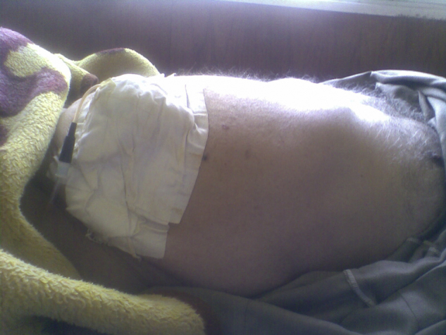
A tube drain fixed to the anterior abdominal wall.
Fig. 6.

Some of the amount of the drained fluid.
3. Discussion
Although perforation of the biliary system or duodenum is very rare after ERCP, it occurred in this case; however, we could not identify the exact site of perforation.
In ES, the cut is usually performed at the angle between the duodenum and CBD at 2 o‘clock. If this cutting is overdone, it may reach the serosa of the organs that may open to the peritoneal cavity. The most probable scenario is that the CBD was injured at the site of the stricture during insertion of the plastic stent. However, through any of these rents, the bile seeped slowly and gradually into the Morison pouch enlarging insidiously and heaping in front of the duodenum, transverse colon and greater omentum just behind the anterior abdominal wall, reaching the urinary bladder downward. Because the patient has been diabetic for the last 20 years, he did not have severe pain due to bile irritation, possibly due to neuropathy, but he was complaining of dyspepsia, epigastric discomfort, and repeated vomiting due to the pressure effect of the bile collection on the duodenum and stomach.
Because of the massive collection of bile, we expected a large perforation; however, it closed spontaneously possibly due to securing the patency of the CBD and bile flow by the insertion of the plastic stent in the CBD.
4. Conclusion
-
1
After ERCP, any complain of the patient should not be ignored, even if it is simple because it may hide a serious underlying problem that could be discovered and treated early.
-
2
However, very rare, after an ERCP, a biloma can occur that may reach a very large size causing many symptoms.
-
3
The biloma can be treated by aspiration with minimally invasive surgical interference particularly if the patient is clinically stable and with the help of US guidance.
Conflict of interest
The authors declare that they have no competing interests.
Funding
No source of funding is needed. The authors are ready to pay the publication fee.
Ethical approval
It is not needed.
Consents
Written informed consent was obtained from the patient to publish the case with its related pictures. A copy of the written consent is available for review by the Editor-in-Chief of this journal.
Authors’ contributions
Harith M. Alkhateeb: Surgery for the patient, writing of the original text, communication with the editorial team.
Thaer J. Aljanabi: Assistance in the surgery, sharing the analysis of the data, and follow up of the patient.
Khairallah H. Alazzawi: EGD and medical control of the diabetes mellitus.
Taha A. Alkarboly: ERCP, ES of the ampulla of Vater, CBD stone extraction and plastic stent insertion in the CBD.
Guarantor
Dr. Harith M. Alkhateeb.
Aknowledgments
We would like to thank the patient for allowing us to contact him for follow up and consenting for us to publish the case with its related photographs. We would like to thank the efficient ultrasonographist Dr. Bushra M. Shabeeb for her kind help in performing the follow-up US examinations on the patient. We would like to thank Zayd H. Alkhateeb for his help in the editing of the manuscript and pictures.
Contributor Information
Harith M. Alkhateeb, Email: khateeb_hm@yahoo.com.
Thaer J. Aljanabi, Email: thaere.jasim@yahoo.com.
Khairallh H. Al-azzawi, Email: jaguar_fp@yahoo.com.
Taha A. Alkarboly, Email: alkarbolytaha@gmail.com.
References
- 1.Gould L., Patel A. Ultrasound detection of extrahepatic encapsulated bile: biloma. Am. J. Roentgenol. 1979;132:1014–1015. doi: 10.2214/ajr.132.6.1014. [DOI] [PubMed] [Google Scholar]
- 2.Kannan Umashankkar, Parshad Rajinder, Regmi Suboudh Kumar. Cases J. 2009;2:8048. doi: 10.4076/1757-1626-2-8048. [DOI] [PMC free article] [PubMed] [Google Scholar]
- 3.Bas Gurhan, Okan Ismail, Sahin Mustafa, Eryilmaz Ramazan, Isik Arda. J. Med. Case Rep. 2011;5:3. doi: 10.1186/1752-1947-5-3. [DOI] [PMC free article] [PubMed] [Google Scholar]
- 4.McCune W.S., Shorb P.E., Moscovitz H. Endoscopic cannulation of the ampulla of Vater: a preliminary report. Ann. Surg. 1968;167:752–756. doi: 10.1097/00000658-196805000-00013. [DOI] [PMC free article] [PubMed] [Google Scholar]
- 5.Maple J.T., Ben-Menachem T., Anderson M.A. The role of endoscopy in the evaluation of suspected choledocholithiasis. Gastrointest. Endosc. 2010;71:1–9. doi: 10.1016/j.gie.2009.09.041. [DOI] [PubMed] [Google Scholar]
- 6.Baron T.H., Mallery J.S., Hirota W.K. The role of endoscopy in the evaluation and treatment of patients with pancreaticobiliary malignancy. Gastrointest. Endosc. 2003;58:643–649. doi: 10.1016/s0016-5107(03)01994-1. [DOI] [PubMed] [Google Scholar]
- 7.Costamagna G., Shah S.K., Tringali A. Current management of postoperative complications and benign biliary strictures. Gastrointest. Endosc. Clin. N. Am. 2003;13:635–648. doi: 10.1016/s1052-5157(03)00103-x. [DOI] [PubMed] [Google Scholar]
- 8.Zuckerman M.J., Shen B., Harrison M.E., 3rd Informed consent for GI endoscopy. Gastrointest. Endosc. 2007;66:213–218. doi: 10.1016/j.gie.2007.02.029. [DOI] [PubMed] [Google Scholar]
- 9.Kwon C.I., Song S.H., Hahm K.B., Ko K.H. Unusual complications related to endoscopic retrograde cholangiopancreatography and its endoscopic treatment. Clin. Endosc. 2013;46:251. doi: 10.5946/ce.2013.46.3.251. [DOI] [PMC free article] [PubMed] [Google Scholar]
- 10.Chavalitdhamrong D., Donepudi S., Pu L., Draganov P.V. Uncommon and rarely reported adverse events of endoscopic retrograde cholangiopancreatography. Dig. Endosc. 2014;26:15. doi: 10.1111/den.12178. [DOI] [PubMed] [Google Scholar]
- 11.Freeman M.L., Nelson D.B., Sherman S. Complications of endoscopic biliary sphincterotomy. N. Engl. J. Med. 1996;335:909–918. doi: 10.1056/NEJM199609263351301. [DOI] [PubMed] [Google Scholar]
- 12.Masci E., Toti G., Mariani A. Complications of diagnostic and therapeutic ERCP: a prospective multicenter study. Am. J. Gastroenterol. 2001;96:417–423. doi: 10.1111/j.1572-0241.2001.03594.x. [DOI] [PubMed] [Google Scholar]
- 13.Loperfido S., Angelini G., Benedetti G. Major early complications from diagnostic and therapeutic ERCP: a prospective multicenter study. Gastrointest. Endosc. 1998;48:1–10. doi: 10.1016/s0016-5107(98)70121-x. [DOI] [PubMed] [Google Scholar]
- 14.Williams E.J., Taylor S., Fairclough P. Risk factors for complication following ERCP; results of a large-scale, prospective multicenter study. Endoscopy. 2007;39:793–801. doi: 10.1055/s-2007-966723. [DOI] [PubMed] [Google Scholar]
- 15.Howard T.J., Tan T., Lehman G.A. Classification and management of perforations complicating endoscopic sphincterotomy. Surgery. 1999;126:658–663. discussion 64-5. [PubMed] [Google Scholar]
- 16.Cotton P.B., Lehman G., Vennes J. Endoscopic sphincterotomy complications and their management: an attempt at consensus. Gastrointest. Endosc. 1991;37:383–393. doi: 10.1016/s0016-5107(91)70740-2. [DOI] [PubMed] [Google Scholar]
- 17.Enns R., Eloubeidi M.A., Mergener K. ERCP-related perforations: risk factors and management. Endoscopy. 2002;34:293–298. doi: 10.1055/s-2002-23650. [DOI] [PubMed] [Google Scholar]
- 18.Lai C.H., Lau W.Y. Management of endoscopic retrograde cholangiopancreatography-related perforation. Surgeon. 2008;6:45–48. doi: 10.1016/s1479-666x(08)80094-7. [DOI] [PubMed] [Google Scholar]
- 19.Anderson Michelle. A., Fisher Laurel, Jain Rajeev. Complications of ERCP. GIE. 2012;75(3) doi: 10.1016/j.gie.2011.07.010. [DOI] [PubMed] [Google Scholar]