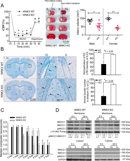
Figure 1. Genetic deletion of WNK3 in vivo decreases infarct volume and demyelination and improves neurological recovery following ischemic stroke.
A. Changes in regional cerebral blood flow (rCBF) during and after 60-min MCAO are similar in littermate wild type (WT) and WNK3 KO mice. Values are mean ± SD. *p < 0.05 vs. -15 min, #p < 0.05 vs 0 min with one-way ANOVA . Representative coronal brain sections from WT and WNK3 KO mice. Infarct volume at 24 h Rp was determined by TTC staining. Values are mean ± SEM (n = 8, 4F/4M). *p < 0.05 vs. WT. B. WNK3 KO mice exhibit less white matter injury after MCAO than WT. Left panel: Bright field images of WT (i) or WNK3 KO brain sections (ii) stained with Luxol Fast Blue (LFB) and cresyl violet. Black box: Demyelination analysis in external capsules (EC) of white matter in the CL and IL hemispheres. Right panel: higher magnification images. Arrowhead: myelinated or demyelinated axonal bundles. Arrow: Axonal track. Right panel, Summary of demyelination data. Values are Mean ± SEM (n = 3). *p < 0.05 vs. WT. C. Improved neurological behavior in WNK3 KO mice after MCAO, scored as described in Methods 27. Values are median composite values and data analyzed by non-parametric Mann-Whitney test (n= 8-9, M). *p < 0.05 vs. WT, #p < 0.05 vs. 0 day. D. Expression of pNKCC1 in cytosolic or membrane fractions of ischemic brains. Values are Mean ± SEM (n= 3-4). *p < 0.05 vs. CL.

