



Background: The mechanism coupling substrate binding to transport in neurotransmitter: sodium symporters (NSSs) is poorly understood.
Results: Site-directed fluorescence quenching spectroscopy experiments on the NSS homologue LeuT reveal a structural intermediate preceding transition to the inward-open conformation.
Conclusion: Stability of the intermediate might represent a rate-limiting barrier in the transport mechanism.
Significance: The data add to our mechanistic understanding of Na+-coupled transport across lipid bilayers.
Keywords: amino acid transport, conformational change, dopamine transporter, gating, membrane protein, monoamine transporter, neurotransmitter transport, fluorescence quenching, fluorescence spectroscopy, mechanisms of membrane transport
Abstract
Neurotransmitter:sodium symporters (NSSs) mediate reuptake of neurotransmitters from the synaptic cleft and are targets for several therapeutics and psychostimulants. The prokaryotic NSS homologue, LeuT, represents a principal structural model for Na+-coupled transport catalyzed by these proteins. Here, we used site-directed fluorescence quenching spectroscopy to identify in LeuT a substrate-induced conformational rearrangement at the inner gate conceivably leading to formation of a structural intermediate preceding transition to the inward-open conformation. The substrate-induced, Na+-dependent change required an intact primary substrate-binding site and involved increased water exposure of the cytoplasmic end of transmembrane segment 5. The findings were supported by simulations predicting disruption of an intracellular interaction network leading to a discrete rotation of transmembrane segment 5 and the adjacent intracellular loop 2. The magnitude of the spectroscopic response correlated inversely with the transport rate for different substrates, suggesting that stability of the intermediate represents an unrecognized rate-limiting barrier in the NSS transport mechanism.
Introduction
Specialized integral membrane proteins terminate neurotransmission in the CNS by catalyzing Na+-dependent clearance of neurotransmitters from the synaptic cleft (1, 2). For dopamine, norepinephrine, serotonin, glycine, and γ-aminobutyric acid (GABA), this role is fulfilled by members of the neurotransmitter/sodium symporter (NSS)2 family (also referred to as the solute carrier 6 gene family) (3). Altered function of these transporters has been implicated in both psychiatric disorders and neurological diseases (4–6). Moreover, NSSs constitute important targets for several pharmaceutical agents, including antidepressants, antiepileptics, and illicit drugs, such as cocaine and amphetamines (7).
Our understanding of how NSSs operate at the molecular level has been substantially improved by the high resolution structural information revealed by crystallization of LeuT, a prokaryotic NSS homologue from Aquifex aeolicus (8–10) and more recently of MhsT, a prokaryotic homologue from Bacillus halodurans (11), and of the dopamine transporter from Drosophila (12). The crystal structures have revealed a structural fold with 11 or 12 packed transmembrane segments of which the first 10 are arranged in a pseudosymmetric inverted repeat pattern with a primary substrate-binding site (S1) in the center and two adjacent sodium-binding sites (8, 13) (Fig. 1A). The inverted repeat fold has also been identified in symporters and antiporters with no sequence homology to NSSs (14–19), suggesting that the fold represents a structural paradigm for ion-coupled transport (20). Importantly, LeuT is widely accepted as a principal model for NSSs (21–25), which is supported by the striking structural similarity among LeuT, MhsT, and Drosophila dopamine transporter despite the evolutionary distance between the three proteins (11, 12).
FIGURE 1.

Probing small scale conformational changes at the cytosolic face of LeuT. A, two-dimensional diagram of LeuT embedded in the membrane. The cysteine (E192C) introduced for labeling with the sulfhydryl-reactive fluorophore TMR maleimide is indicated in orange (LeuT E192CTMR). Mutations introduced in the primary and secondary substrate-binding sites are show in green (Y108F, F253A, and F253L) and blue (L400S), respectively. The I359Q mutation, shown in red, introduced in the primary substrate site converts LeuT to a tryptophan transporter. The conserved residues involved in the intracellular interaction network (Arg5, Trp8, Tyr268, and Asp369) are outlined in teal. B, structure of LeuT (outward occluded) showing the position of Glu192 with TM1i and TM5i highlighted in green. Glu192 is positioned in close proximity to the conserved intracellular interaction network. C, chemical structure of TMR maleimide that was conjugated to E192C at the cytosolic end of TM5.
It is believed that NSSs mediate sodium-driven substrate translocation by a classical alternating access mechanism (26). Consistent with such a model, crystallization efforts on LeuT have outlined three canonical conformational states including an “outward-occluded,” an “outward-open,” and an “inward-open” state (8–10). Nonetheless, despite the importance of these states, the structures represent stabilizable states in crystallography and may not provide a complete picture of the translocation cycle (27). To address this and gain better insight into the structural dynamics of the transport process, biophysical techniques such as double electron-electron resonance and single molecule fluorescence resonance energy transfer (smFRET) have been applied to LeuT (28–31). Of interest, the smFRET data substantiated predictions from computational steered molecular dynamics simulations by supporting the functional role of a second high affinity substrate (S2)-binding site in LeuT situated in a vestibule extracellular to the S1 site (29, 32). To this point, however, our understanding of how substrate binding leads to dissociation of a conserved intracellular interaction network (Fig. 1A) and subsequent isomerization of the transporter to the inward-open state (21) remains incomplete.
Here, we applied site-directed fluorescence quenching spectroscopy (SDFQS) to LeuT as a highly sensitive method to investigate conformational rearrangements linked to substrate binding. Labeling with tetramethylrhodamine (TMR) of a single cysteine inserted at the cytoplasmic end of TM5 (TM5i) enabled detection of a Na+-dependent, substrate-induced increase in aqueous exposure of the fluorophore, consistent with a substrate-induced conformational change at the inner gate. Mutational data suggested the requirement for an intact S1 site, and computational simulations were able to link the enhanced aqueous exposure of the fluorophore to the spatial relation between the discrete outward movement of the N terminus and the cytoplasmic end of TM1 and specific changes in the intracellular loop 2 (IL2)-TM5i region. The data suggest a novel coupling mechanism between the primary substrate-binding site and the inner gate involving the formation of a conformational intermediate that may precede transition to the inward-open conformation.
Experimental Procedures
Construction, Expression, Purification, and Fluorescent Labeling of LeuT
Residues were mutated by the QuikChange method (Agilent Technologies) in pET16b LeuT containing a C-terminal octahistidine tag. The generated LeuT mutants were confirmed by DNA sequencing. LeuT variants were expressed in Escherichia coli C41(DE3) by cultivation in lysogeny broth medium supplemented with 0.1 mg/ml ampicillin until A600 reached ∼0.6. Expression was induced by addition of 0.1 mm isopropyl β-d-thiogalactopyranoside, and the bacterial culture was cultivated at 20 °C for another 20 h. The membrane fraction was isolated by disruption of the cells (Constant Systems homogenizer, Kennesaw, GA) and solubilized with 1% (w/v) n-dodecyl β-d-maltopyranoside (DDM; Affymetrix, Santa Clara, CA). Detergent-solubilized LeuT was immobilized on Chelating Sepharose Fast Flow resin (GE Healthcare), washed with ice-cold buffer A, and incubated with 200 μm tetramethylrhodamine-5-maleimide (TMR maleimide; Life Technologies) for 16 h at 4 °C. Resin slurry was loaded on a packing column and washed with ice-cold Buffer A (20 mm Tris-HCl (pH 7.50), 200 mm KCl, 20% (v/v) glycerol, 0.1 mm tris(2-carboxyethyl)phosphine (TCEP), and 0.05% (w/v) DDM) containing 60 mm imidazole until flow-through A541 was reduced to 0. Subsequently LeuT was eluted in ice-cold Buffer A containing 300 mm imidazole. Protein samples were desalted using HiTrap spin columns (GE Healthcare) equilibrated with Buffer A. Protein concentration was determined by measuring the TMR-corrected absorbance at 280 nm (ϵ = 1.91 cm2 mg−1). TMR labeling efficiency was calculated as the TMR:LeuT molar ratio measuring TMR absorbance at 541 nm (ϵ = 95.000 cm−1 m−1). TMR labeling- efficiency was >89% in all TMR-labeled LeuT preparations.
Radioligand Binding
Binding experiments were performed using a scintillation proximity assay (SPA) (33). In a 96-well white wall clear bottom plate, 100 ng or 1 μg of LeuT was used as indicated. [3H]Leucine (0.25 (Y108F) or 20.1 Ci mmol−1) or [3H]tryptophan (0.2 Ci mmol−1) (PerkinElmer Life Sciences) was used at the indicated concentrations together with 0.125 mg ml−1 yttrium silicate-copper His tag SPA beads (PerkinElmer Life Sciences). Binding buffer consisted of 20 mm Tris-HCl (pH 7.50), 200 mm NaCl, 20% (v/v) glycerol, 0.1 mm TCEP, and 0.05% DDM. Nonspecific background was detected in the presence of 5 mm alanine. Samples were incubated for 16 h at 4 °C, and activity was recorded on a MicroBetaTM plate counter (PerkinElmer Life Sciences) with a 1-min counting protocol. No change in sample activity was observed when incubating beyond 16 h. Data were analyzed by non-linear regression analysis and fitted to a single site hyperbolic function or sigmoidal dose-response curve using GraphPad Prism 5.0 (GraphPad Software, La Jolla, CA). Ki values were calculated as follows: Ki = IC50 × (1 + [L]/KD)−1 where L denotes the [3H]leucine concentration and KD is the dissociation constant for leucine.
Reconstitution and Uptake
Purified LeuT variants were reconstituted at a weight ratio of 1:150 (LeuT:lipid) in E. coli polar lipid extract (Avanti Lipids, Alabaster, AL) liposomes prepared as described (33) except for using a reconstitution buffer (200 mm KCl, 20 mm Tris-HCl (pH 7.50), and 0.1 mm TCEP) supplemented with 0.11% Triton X-100 to destabilize the liposomes. Control liposomes lacking LeuT were prepared in parallel. Detergent was removed by incremental addition of adsorbent Bio-Beads SM-2 (Bio-Rad) to a final concentration of ∼250 mg ml−1 followed by incubation overnight at 4 °C. The LeuT-containing proteoliposomes or control liposomes were collected by ultracentrifugation at 323,000 × g for 30 min, resuspended in reconstitution buffer to a lipid concentration of 100 mg ml−1, and flash frozen for storage at −80 °C. Prior to uptake experiments, thawed proteoliposomes were extruded through a 100-nm filter (Avanti Lipids).
Time course accumulation of 1 μm [3H]alanine (84.5 Ci mmol−1; PerkinElmer Life Sciences) was measured in a 96-well 0.22-μm PVDF filter plate (Millipore, Billerica, MA) at 22 °C in assay buffer consisting of 200 mm NaCl, 20 mm Tris-HCl (pH 7.50), and 0.1 mm TCEP. At the indicated time points, samples were rapidly filtered using a MultiScreenTM vacuum manifold apparatus (Millipore) and washed three times with ice-cold quenching buffer (200 mm KCl, 20 mm Tris-HCl (pH 7.50), and 0.1 mm TCEP) to remove free [3H]alanine. Samples were incubated with 0.1 ml of scintillation liquid (PerkinElmer Life Sciences) before activity was recorded on a MicroBeta plate counter. cpm were converted into pmol with an [3H]alanine activity standard curve (R2 = 0.9991). Total uptake was corrected for nonspecific accumulation in control liposomes and analyzed by non-linear regression using GraphPad Prism 5.0.
The maximum rates of transport were quantified by mixing 0.3 μg of reconstituted LeuT with 0–8 μm [3H]alanine (60 Ci mmol−1; American Radiolabeled Chemicals, St. Louis, MO), [3H]valine (60 Ci mmol−1; American Radiolabeled Chemicals), [3H]isoleucine (50 Ci mmol−1; American Radiolabeled Chemicals), [3H]methionine (80 Ci mmol−1; American Radiolabeled Chemicals), or [3H]leucine (125 Ci mmol−1; American Radiolabeled Chemicals) in assay buffer containing 50 mm Tris/MES (pH 8.5) and 50 mm NaCl at 22 °C. The bound fraction and nonspecific accumulation were assessed by dissipating the Na+ gradient with 25 μg ml−1 gramicidin added 1 min prior to the experiment. The reactions were terminated at t = 1 min by dilution into ice-cold stopping buffer containing 100 mm KPi (pH 6.0) and 100 mm LiCl immediately followed by rapid filtration through 0.22-μm nitrocellulose filters (Millipore). Radioactivity retained on the filters was recorded by scintillation counting, and cpm were converted into nmol using known amounts of the corresponding 3H-labeled amino acids. Specific uptake was determined as the difference between total activity and 3H-substrate binding (as determined in the presence of gramicidin), and kinetic constants were obtained by data fitting to the Michaelis-Menten function in GraphPad Prism 5.0.
Fluorescence Spectroscopy
Fluorescence-based experimentswere carried out with 0.5 μg ml−1 of the TMR-labeled LeuT variants diluted in fluorescence buffer containing 200 mm NaCl or 200 mm KCl (as specified), 20 mm Tris-HCl (pH 7.50), 0.1 mm TCEP, and 0.05% DDM unless otherwise stated. For ligand binding experiments, a small aliquot of the ligand (as specified) was added to the samples followed by incubation on an orbital shaker for 30 min at room temperature. It should be noted that we observed no change in the EC50 value for the quenching response when incubating the samples for up to 60 min. Steady-state fluorescence intensities were recorded on a FluoroMax2 (Horiba Scientific, Edison, NJ) at λem = 572 nm using an excitation source at λex = 540 nm and excitation and emission band passes of 5 nm at room temperature. Emission spectra were recorded by varying λem between 550 and 650 nm by 1-nm increments. Quencher titration was carried out by successive additions of small aliquots containing (i) 1 m potassium iodide (KI) with 10 mm Na2S2O3 or (ii) 100 mm 2,2,6,6-tetramethylpiperidine-1-oxyl (TEMPO) in the appropriate fluorescence buffers followed by recording of the fluorescence intensity. Fluorescence intensities (F) were corrected for sample dilution, normalized to the initial fluorescence intensity in the sample (F0), and analyzed by linear regression in GraphPad Prism 5.0. The degree of accessibility was obtained from the Stern-Volmer equation: F0/F = 1 + KSV × [Q] where F0/F is the normalized fluorescence quenching, [Q] is the quencher concentration, and KSV is the Stern-Volmer constant.
Fluorescence time course experiments were recorded at λem = 572 nm (10 data points per s) using an excitation source at λex = 540 nm and excitation and emission band passes of 5 nm with constant stirring in the cuvette containing 0.5 μg ml−1 TMR-labeled LeuT in buffer consisting of 200 mm NaCl, 20 mm Tris-HCl (pH 7.50), 0.1 mm TCEP, and 0.05% DDM plus 200 mm KI or KCl (control). Data were normalized to the average steady-state fluorescence prior to leucine addition with control conditions (200 mm KCl) subtracted.
Molecular Dynamics Simulations
Based on our established simulation protocols and molecular system, we carried out the MD simulations of LeuT using NAMD (34) as described previously (32). Briefly, all-atom simulations of LeuT immersed in an explicit 1-palmitoyl-2-oleoyl-sn-glycero-3-phosphocholine lipid bilayer were carried out with the CHARMM22/CMAP and CHARMM36/lipid force fields (35, 36). In the isothermal-isobaric (NPT) ensemble, constant temperature (310 K) was maintained with Langevin dynamics, and 1-atm constant pressure was achieved with the hybrid Nosé-Hoover Langevin piston method (37) applied to an anisotropic flexible periodic cell with orthogonal pressure components computed independently. The particle mesh Ewald method was used to evaluate long range electrostatic effects. A time step of 2 fs was used for the unbiased MD simulations, and 1fs was used for the targeted molecular dynamics (tMD) simulations.
In tMD simulations (38), a harmonic potential, VtMD
 0.5·ktMD ·(RMSD(t) − RMSDtarget(t))2, is added to the MD force field to bias a starting conformation toward a target by gradually reducing an instantaneous target RMSD(t) at simulation time t in response to a user-defined reference value RMSDtarget(t) (ktMD is a force constant). In this study, we implemented a “staircase” variation of tMD (stMD) (39) in which RMSDtarget(t) is reduced from RMSD(0) in “staircases”, i.e. alternating steps of targeted motion (“move”) and constrained equilibration (“pause”); this procedure allowed us to compute averages along transition “pauses” of constant RMSDtarget(t) values. For each move step, the slope of RMSDtarget(t) is −RMSD(0)/nsΔts where ns is the number of staircases along the transition and Δts the simulation time of each stMD step so that, at the end of each stMD run with total simulation time Γ
ns×(Δtm+Δtp), RMSDtarget(Γ) is zero.
0.5·ktMD ·(RMSD(t) − RMSDtarget(t))2, is added to the MD force field to bias a starting conformation toward a target by gradually reducing an instantaneous target RMSD(t) at simulation time t in response to a user-defined reference value RMSDtarget(t) (ktMD is a force constant). In this study, we implemented a “staircase” variation of tMD (stMD) (39) in which RMSDtarget(t) is reduced from RMSD(0) in “staircases”, i.e. alternating steps of targeted motion (“move”) and constrained equilibration (“pause”); this procedure allowed us to compute averages along transition “pauses” of constant RMSDtarget(t) values. For each move step, the slope of RMSDtarget(t) is −RMSD(0)/nsΔts where ns is the number of staircases along the transition and Δts the simulation time of each stMD step so that, at the end of each stMD run with total simulation time Γ
ns×(Δtm+Δtp), RMSDtarget(Γ) is zero.
In this study, the stMD simulations were performed on the Cα atoms of the N terminus and the cytoplasmic segments of TMs1, 3, 6, and 8 of LeuT, i.e. residues 1–21, 91–113, 261–267, and 343–368, with Γ = 50 ns, ktMD = 0.25 kcal/mol/Å2/Cα atom, Δts = 0.5 ns, and ns = 50. For the execution protocol for the tMD simulations, we devised an stMD wrapper script that runs the tMD module provided in NAMD.
The initial conformation of LeuT-WT for the tMD was a selected frame of the equilibration of the substrate-bound crystal structure of LeuT (Protein Data Bank code 2A65); the targeted conformation is based on an inward-facing conformation resulting from our previous steered MD study (32) with the N terminus dissociated from the TM domain. Based on the same initial conformation of LeuT-WT, LeuT E192CTMR was constructed by covalently attaching TMR at position 192 after mutating it from Glu to Cys using the covalent docking protocol of Schrödinger Suite (2012; Schrödinger LLC, New York, NY). The parameters of TMR were generated from Multipurpose Atom-Typer for CHARMM (MATCH) server (40).
Data Analysis
All experiments were repeated at least three times unless otherwise indicated. Data points are given as means ± S.E. or means with 95% confidence intervals (when derived from a log scale). Data fits of kinetic analyses were carried out using non-linear regression algorithms in GraphPad Prism. Statistical analyses were performed using Student's t test or one-way analysis of variance followed by Bonferroni post hoc test as appropriate.
Results
Detection of Substrate-induced Conformational Changes by Collisional Quenching
To understand the coupling between substrate binding and conformational rearrangements at the inner gate, we used LeuT as a model system in SDFQS experiments. SDFQS is a highly sensitive method that has been successfully applied to reveal conformational rearrangements in other membrane proteins, such as the β2-adrenergic receptor and the lactose permease of E. coli (41–45). Note that LeuT contains no endogenous cysteines, which enables straightforward site-selective incorporation of thiol-reactive fluorophores into targeted cysteine mutants (Fig. 1A).
In the transport cycle, the transition from the inward-occluded to inward-open state is a key step in allowing substrate release to the intracellular side. According to a combined computational and smFRET study (28), the structural rearrangement of such a transition in NSSs involves a tilting of the intracellular portion of TM1 (TM1i) away from the helix bundle in an overall direction that is consistent with the position of TM1i in a crystal structure of LeuT in the inward-open state (Protein Data Bank code 3TT3) (9). Importantly, in the transport mechanism, such a rearrangement of TM1i must coordinate with other nearby structural elements in a controlled manner. One such element is the region that includes the IL2 and TM5i, which has been proposed to have a role in Na+ binding (46). Indeed, substituted cysteine accessibility method studies of serotonin transporter (47, 48) have suggested that conformational rearrangements take place in the IL2-TM5i region during translocation. Furthermore, compared with the crystal structure of LeuT in the inward-open state (9), the recent crystal structure of MhsT showed different features of TM5i rearrangements (11), supporting both a critical role for TM5i in the dynamic transition to the inward-open state and the possible involvement of additional conformational states (11).
To explore the involvement of this dynamic region in substrate-induced conformational rearrangements, we introduced a cysteine at position 192 (E192C) of LeuT. This position was chosen for its location at the junction between TM1i and TM5i so that it is in contact with interior residues and external solvent (Fig. 1, A and B). The construct was expressed in E. coli, purified, and conjugated with the thiol-reactive fluorophore TMR maleimide (Fig. 1, Fig. 2A). TMR maleimide specifically labeled the inserted cysteine as no fluorescence was detected in purified WT LeuT after treatment with 200 μm TMR maleimide (data not shown).
FIGURE 2.
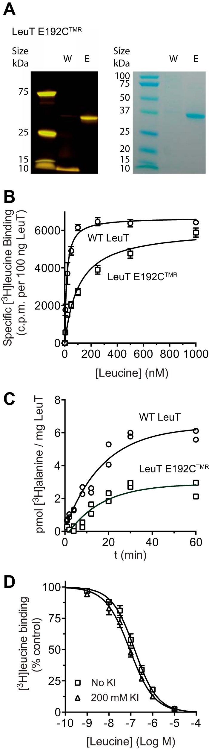
Purified and fluorescently labeled LeuT E192C (LeuT E192CTMR) retains WT-like substrate binding activity. A, representative SDS-PAGE showing the purity of LeuT E192CTMR eluted from nickel immobilized-metal affinity chromatography. Left, fluorescence scan showing specific TMR labeling of eluted LeuT E192C (“E”) and that unconjugated TMR is removed in the wash step (“W”). Right, the same gel stained with Coomassie Blue. B, saturation binding of [3H]leucine in 200 mm NaCl assessed in the SPA for WT LeuT (circles) and LeuT E192CTMR (squares). Data are fitted to a single site model, and equilibrium binding constants are shown in Table 1. Data points are means ± S.E. (error bars) from four independent experiments performed in duplicates. C, specific accumulation of 1 μm [3H]alanine in liposomes reconstituted with WT LeuT (circles) or LeuT E192CTMR (squares). Data points are duplicate determinations from a representative experiment. D, competition SPA binding of 10 nm [3H]leucine to LeuT E192CTMR with leucine in the absence (squares) or presence of 200 mm KI (triangles). Data points are means ± S.E. (error bars) from two independent experiments performed in at least duplicates.
As assessed in a SPA (33), the labeled mutant (LeuT E192CTMR) maintained high affinity [3H]leucine binding (KD = 108 ± 14 nm, mean ± S.E., n = 4), although the affinity was moderately decreased compared with WT LeuT (KD = 15 ± 2 nm, mean ± S.E., n = 4) (Fig. 2B and Table 1). We did not observe a difference in the total number of binding sites (Bmax) (Fig. 2B and Table 1), indicating no change in substrate binding stoichiometry. Furthermore, as determined after reconstitution into proteoliposomes and using [3H]alanine as substrate, LeuT E192CTMR maintained its ability to transport, although the capacity was partially reduced compared with WT LeuT (Fig. 2C). These results show that despite the mutation and derivatization with TMR the transporter exhibits functional substrate binding and transport.
TABLE 1.
Radioligand binding constants for LeuT E192CTMR variants
Values were calculated from SPA saturation binding experiments in the presence of 200 mm NaCl. Data are means ± S.E. from three to four independent experiments. ND, not determined.
| LeuT variant | KD | Bmax |
|---|---|---|
| nm | % of WT | |
| [3H]Leucine | ||
| LeuT E192CTMR | 108 ± 14 | 91 ± 4 |
| +L400S | 25 ± 5 | 58 ± 3 |
| +Y108F | >25,000 | ND |
| +F253A | 60 ± 10 | 50 ± 2 |
| +F253L | 94 ± 16 | 32 ± 2 |
| [3H]Tryptophan | ||
| LeuT E192CTMR | 50 ± 5 | ND |
| +I359Q | 30 ± 12 | ND |
To evaluate putative conformational changes surrounding the fluorophore, we used a collisional quenching approach. Collisional quenching requires an interaction between the quencher and fluorophore during the excited state of the fluorophore. Upon contact, the fluorophore returns to the ground state without emission of a photon. This makes it possible to assess solvent accessibility of a fluorophore by using a water-soluble quencher (49), such as iodide (I−). Indeed, increasing concentrations of KI strongly quenched the fluorescence emitted from free TMR (Fig. 3A). The ability of KI to quench TMR conjugated to the transporter (LeuT E192CTMR) was markedly reduced compared with the free fluorophore (Fig. 3B). Plotting F0/F (F0 = fluorescence in the absence of KI) against the KI concentration yielded linear Stern-Volmer plots with a slope (the Stern-Volmer constant, KSV) of 10.3 ± 0.4 m−1 (means ± S.E., n = 4) for free TMR and a slope (KSV) of ∼2.2 m−1 for TMR conjugated to the transporter (Fig. 3C and Table 2). Thus, the fluorophore is less exposed to the solvent when conjugated to the transporter, suggesting that it is partially buried in the protein structure and/or the hydrophobic environment of the detergent micelle.
FIGURE 3.

Leucine induces a Na+-dependent increase in the aqueous accessibility of TMR bound to E192C (LeuT E192CTMR). A and B, TMR maleimide fluorescence emission spectra in the presence of increasing concentrations of the hydrophilic quencher KI, normalized to maximum fluorescence intensity in the absence of KI (dashed line). A, fluorescence from unconjugated dye was quenched concentration-dependently by iodide. B, conjugation of TMR to E192C in LeuT (LeuT E192CTMR) substantially decreased iodide quenching of TMR fluorescence. C, Stern-Volmer plots of quenching data in A and B showing that F0/F (fluorescence normalized to intensity in the absence of KI) is linearly dependent on the KI concentration but that the slope (the Stern-Volmer quenching constant, KSV) is substantially lower for TMR conjugated to E192C (LeuT E192CTMR) (black circles) compared with unconjugated TMR (purple circles). D, iodide quenching of LeuT E192CTMR in 200 mm NaCl in the absence of substrate (black circles) or presence of increasing fixed concentrations of leucine (blue squares, 100 nm; blue triangles, 1 μm; blue inverted triangles, 10 μm; blue diamonds, 100 μm; blue circles, 1 mm). E, iodide quenching of LeuT E192CTMR in 200 mm KCl in the absence of substrate (black circles) or presence of increasing fixed concentrations of leucine (blue diamonds, 100 μm; blue circles, 1 mm). F, KSV values obtained from D and E plotted as a function of leucine concentration, showing that leucine in 200 mm Na+ (filled squares) induces a concentration-dependent and saturable increase in iodide quenching of LeuT E192CTMR. The response is abolished in the absence of Na+ (200 mm KCl; open squares). Data points are means ± S.E. (error bars) from four to five independent experiments.
TABLE 2.
Iodide quenching response constants for LeuT E192CTMR variants
Values were calculated from SDFQS experiments. KSV values are means ± S.E., and EC50 values are means (95% confidence interval) from three to five independent experiments.
| KSV basal | KSV leucine | EC50 leucine | |
|---|---|---|---|
| m−1 | m−1 | μm | |
| LeuT E192CTMR | 2.21 ± 0.05 | 3.43 ± 0.04 | 1.94 (1.15;3.27) |
| +L400S | 2.60 ± 0.06 | 3.66 ± 0.05 | 1.32 (0.71;2.45) |
| +Y108F | 1.80 ± 0.06 | 2.61 ± 0.07 | 52.1 (22.8;119) |
| +F253A | 2.13 ± 0.08 | 3.35 ± 0.12 | 82.8 (34.0;201) |
| +F253L | 2.47 ± 0.06 | 3.57 ± 0.08 | 21.3 (9.03;50.2) |
Incubation with increasing concentrations of leucine enhanced iodide-dependent quenching of LeuT E192CTMR as illustrated by a concentration-dependent increase in the slope of the Stern-Volmer plots (Fig. 3D). The enhanced quenching required Na+ as the response was eliminated in Na+-free buffer (200 mm KCl) (Fig. 3E). Removal of Na+ did not change the basal quenching of LeuT E192CTMR, suggesting that Na+ binding has no effect per se on fluorophore accessibility (KSV = 2.21 ± 0.05 m−1 in Na+ versus KSV = 2.27 ± 0.06 m−1 in K+, means ± S.E., n = 5). By plotting the KSV obtained in Na+ as a function of leucine concentration, we observed a saturable response with an EC50 of ∼1.9 μm and a maximal KSV of ∼3.4 m−1 (Fig. 3F and Table 2). Note that the EC50 value is larger than the KD of leucine obtained in radiotracer binding experiments (∼100 nm; Table 1). Importantly, it is unlikely that this higher value is because the binding assay and the quenching assay reflect binding to two different pools of transporters; the TMR labeling stoichiometry of purified LeuT E192C was >89% (see “Experimental Procedures”), and in our binding assay, we did not observe more than one affinity state, and there was no significant decrease in the total number of binding sites (see above). Moreover, the difference is also unlikely caused by addition of the quencher (KI) because the [3H]leucine binding properties were unchanged even in the presence of 200 mm KI (Fig. 2D).
We also assessed the rate of the leucine-induced iodide quenching response by measuring fluorescence over time with 200 mm KI added to the sample of purified of LeuT E192CTMR. As shown in Fig. 4A and consistent with movement of the fluorophore from a lower to a higher KI accessibility, addition of Leu (100 μm) elicited a marked and rapid decrease in the fluorescence signal. Importantly, the response occurred with a rate (kTMR = 504 h−1) substantially faster than the rate for the transport (see below).
FIGURE 4.

Kinetics of the LeuT E192CTMR fluorescence quenching response and effects of CMI. A, fluorescence time course experiment monitoring the rate of change in LeuT E192CTMR fluorescence upon addition of 100 μm leucine (arrow) in the presence of 200 mm KI. Data points are individual fluorescence recordings from two independent experiments normalized to the average steady-state fluorescence prior to leucine addition with control conditions (200 mm KCl) subtracted. Data are fitted to a two-phase exponential decay function. A.U., arbitrary units. B, addition of 100 μm CMI does not induce a significant increase in iodide quenching when the primary substrate site has no substrate bound. However, 100 μm CMI significantly potentiates the quenching response to leucine (1 mm) by decreasing the substrate off-rate. Note that fluorescence buffer containing Tris/MES (pH 7.50) instead of Tris-HCl was used to improve the solubility of CMI. Data points are percent change in the Stern-Volmer constant, KSV (means ± S.E. (error bars) from three to five individual measurements; ***, p < 0.001; n.s., not significant; unpaired t test).
Next, we tested the effect on LeuT E192CTMR of the tricyclic antidepressant clomipramine (CMI) that acts as a non-competitive inhibitor of LeuT by binding to the S2 site (50). CMI (100 μm) potentiated the iodide quenching response of LeuT E192CTMR to leucine (Fig. 4B), but CMI alone did not increase the iodide quenching significantly (Fig. 4B). This suggests that CMI can potentiate the LeuT E192CTMR quenching response by stabilizing the leucine-induced conformation via its binding to S2 (50). It is unlikely that the effect of CMI relates to spectral interference of the compound as CMI did not affect quenching of free TMR (data not shown).
To further substantiate the specificity of the leucine-induced change in quenching of LeuT E192CTMR, we carried out the same experimental procedure for the adjacent position, 191. For the resulting fluorescently labeled construct (LeuT I191CTMR), we observed no significant effect of either Na+ or leucine addition on iodide quenching (Fig. 5, A and B). Position 191 appears to be surrounded by a cluster of hydrophobic residues (Ile182, Leu183, Ile187, and Ile357) and is in a part of the transporter that is not expected to be exposed to a different environment when TM1 is moving away from the helical bundle.
FIGURE 5.

Specificity and directionality of the LeuT E192CTMR quenching response. A and B, KI quenching experiments on LeuT I191CTMR or LeuT E192CTMR. A and B, Stern-Volmer constants (KSV) obtained from KI quenching experiments with the purified and TMR-labeled single cysteine LeuT mutants. KSV values determined in a low sodium buffer (1 mm NaCl and 199 mm KCl), in a high sodium buffer (190 mm NaCl and 10 mm KCl), or in the presence of 1 mm leucine (in 190 mm NaCl and 10 mm KCl) are shown. Data points are means ± S.E. (error bars) from four to six independent experiments. Neither Na+ nor leucine causes a significant change in quenching of LeuT I191CTMR. In a parallel series of experiments with LeuT E192CTMR, leucine but not Na+ causes a marked increase in iodide quenching (***, p < 0.001). C, D, and E, leucine binding to LeuT E192CTMR induces a conformation where TMR is less exposed to hydrophobic quencher molecules, incorporated in the detergent micelle. C, diagram showing the hydrophilic quencher iodide (I−; yellow circles) and the chemical structure of the hydrophobic quencher molecule TEMPO (red hexagons). TEMPO localizes to the hydrophobic environment of the detergent micelle and is expected to produce a higher degree of fluorescence quenching if TMR becomes more buried. In contrast, I− exerts a higher degree of quenching when TMR exposure to the aqueous environment is increased. D, Stern-Volmer plots obtained from quenching of LeuT E192CTMR fluorescence by the hydrophobic quencher TEMPO. Stern-Volmer relationships were determined in a low sodium buffer (inverted triangles; 1 mm NaCl and 199 mm KCl), in a high sodium buffer (circles; 190 mm NaCl and 10 mm KCl), or in the presence of 1 mm leucine (squares; in 190 mm NaCl and 10 mm KCl). E, Stern-Volmer constants (KSV) obtained from the experiments shown in D. Data points are means ± S.E. (error bars) from four individual experiments (*, p < 0.05; unpaired t test).
The solvent accessibility of TMR conjugated to E192C was further assessed with the hydrophobic quencher TEMPO, which incorporates into the detergent micelle or lipid bilayer (51) and thereby is capable of quenching fluorescence from fluorophores buried from the aqueous milieu (Fig. 5C) (51). TEMPO strongly quenched the fluorescence from TMR conjugated to E192C, consistent with partial burying of the fluorophore (Fig. 5D). Changing from a Na+-free (KSV ∼ 83 m−1) to a Na+-containing buffer (KSV ∼ 82 m−1) did not cause a significant change in accessibility (p = 0.8416); however, addition of leucine markedly decreased TEMPO accessibility (KSV ∼ 62 m−1) (Fig. 5, D and E), suggesting, in full agreement with our findings using iodide as quencher, that TMR conjugated to E192C becomes more solvent-exposed upon leucine binding and thus that leucine induces a conformational change that moves the fluorophore away from a buried water-inaccessible environment.
Simulations Predict Coordinated Rearrangements between TM1i and TM5i
To predict the character of conformational rearrangements detected in the fluorescence quenching experiments, we used tMD simulations to investigate how the IL2-TM5i region would respond to coordinated rearrangements of the N terminus-TM1i (NT-TM1i) and IL2-TM5i. In the tMD simulations starting from the inward-closed state, the NT-TM1i segment was gradually pulled away from the TM domain in a direction defined in our previous steered MD study (32); this direction is along that identified as well in the inward-open structure of LeuT (Protein Data Bank code 3TT3). The tMD run was followed by extended unbiased MD (uMD) simulations to allow the system to equilibrate. In parallel, we also carried out an uMD simulation of the inward-closed state for comparison.
The distancing of NT-TM1i from the TM domain involves the breaking of three sets of interactions between NT and other TMs: (i) the interactions between NT and TM6 in the cluster of aromatic residues (Trp8-Tyr265-Tyr268), (ii) the cation-π/ionic interactions among NT, TM6, and TM8 (Arg5-Tyr268-Asp369), and (iii) an H-bond between NT and TM5i (Thr10-Glu192). As a result, the IL2-TM5i region rotated inward to occupy the space left by the displaced N-terminal residues (Fig. 6). The changes are quantified by the angle between the axes of TM1i and TM5i, revealing a substantial increase of this angle relative to the inward-closed state determined from the corresponding uMD simulations (Fig. 6b). Using the same protocol, we carried out tMD → uMD simulations for LeuT E192CTMR. Importantly, similar rearrangements were observed (Fig. 6, b, c, and d), and these rearrangements were associated with a rotation of TMR buried among the lipid headgroups in the inward-closed state toward the water milieu (Fig. 6, b, c, and d). To quantify the changes of the TMR label, we compared the solvent-accessible surface area of the tricyclic ring of TMR along the simulations and found a significant increase of solvent-accessible surface area of TMR in the conformation resulting from tMD/uMD simulations (Fig. 6e). Altogether, the simulations show how the substrate-induced change in fluorescence quenching of LeuT E192CTMR can describe a discrete outward movement of TM1i, supporting the inference from the measurements that this movement in turn leads to a rotation of IL2-TM5i that increases the solvent exposure of TMR.
FIGURE 6.

The coordinated rearrangements of TM1i and TM5i in the conformational transition predicted from the simulations. a, comparison in the WT LeuT of the inward-closed state (with TM1i and TM5i colored in green) and the modeled state (with TM1i and TM5i colored in cyan) resulting from the tMD/uMD study (see text). The conserved salt bridge between Arg5 and Asp369 is disrupted during the transition from the inward-closed to the modeled state. b, quantification of the modeled TM5i movement. The graph shows the probability distribution of the angle between TM1i and TM5i for WT LeuT (dashed line) and LeuT E192CTMR (solid line) in the inward-closed (green) and modeled state (cyan). The probability of LeuT assuming a conformation with a larger angle between TM1 and TM5 is markedly increased in the modeled state for both WT LeuT and LeuT E192CTMR. c and d, predicted conformation of TMR conjugated to E192C in the inward-closed state (c) and in the modeled state (d). The color scheme in c and d is the same as in a and b. In the modeled state, rotation of TM5i results in a movement of TMR attached to E192C (colored in magenta) from the lipid phase to the aqueous environment. e, quantification of TMR solvent exposure in the LeuT E192CTMR conformations. Probability distribution of the solvent-accessible surface area (SASA) for TMR attached to E192C, showing a clear increase in the probability of the fluorophore to have a higher degree of solvent exposure in the modeled state compared with the inward-closed state.
Conformational Coupling to the Inner Gate Depends on Substrate Binding to S1
To discern whether leucine binding to S1 and/or S2 mediated the observed conformational coupling, we first mutated Tyr108 in TM3 to Phe (LeuT E192CTMR/Y108F). This mutation is presumed to impair the binding of leucine to both S1 and S2 by disrupting a hydrogen bond to the carboxylate group of S1 (8) and promoting an outward-open conformation (9). As expected (52), LeuT E192CTMR/Y108F displayed a dramatic decrease in [3H]leucine binding affinity (KD > 25 μm; Table 1). Moreover, we observed marked changes in iodide quenching in our assay. In contrast to our findings for LeuT E192CTMR, an increase in quenching and thus in solvent exposure of TMR could only be detected at higher leucine concentrations yielding an EC50 of 52 μm (Fig. 7, A and B, and Table 2). We also observed a decrease in the baseline iodide quenching (KSV ∼ 1.80 m−1 for LeuT E192CTMR/Y108F versus ∼2.21 for LeuT E192CTMR) (Fig. 7, A and B, and Table 2).
FIGURE 7.

Conformational coupling to the inner gate depends on substrate binding to S1. A, Stern-Volmer plots of KI quenching data for the S1 substrate-binding site mutant LeuT E192CTMR/Y108F. Experiments were conducted in 200 mm NaCl in the absence of substrate (black circles) or presence of increasing fixed concentrations of leucine (blue squares, 100 nm; blue triangles, 1 μm; blue inverted triangles, 10 μm; blue diamonds, 100 μm; blue circles, 1 mm). B, KSV values obtained from A plotted as a function of leucine concentration. The dashed line shows data for LeuT E192CTMR taken from Fig. 2F. C, Stern-Volmer plots of KI quenching data for the S2 substrate-binding site mutant LeuT E192CTMR/L400S. Experiments were performed as described in A. D, KSV values obtained from C plotted as a function of leucine concentration. The dashed line shows data for LeuT E192CTMR taken from Fig. 2F. E and F, Stern-Volmer plots of KI quenching data for the S1 substrate-binding site mutants LeuT E192CTMR/F253A and LeuT E192CTMR/F253L. Experiments were performed as described in A. G and H, KSV values obtained from E and G plotted as a function of leucine concentration. The dashed line shows data for LeuT E192CTMR taken from Fig. 2F. By revealing a right shift in dose-response curves (EC50 values given in Table 1) for the S1 mutants Y108F, F253A, and F253L but not for L400S, the data support the importance of leucine binding to S1 for the conformational response. Data points are means ± S.E. (error bars) of three to four independent experiments using protein from two separate preparations. For F253L, data points are means ± S.E. (error bars) of two to three independent experiments.
To block possible substrate binding to S2 (53), we mutated Leu400 in TM10 to Ser (LeuT E192CTMR/L400S). Consistent with previous data for this mutation (32), we found only a modest change in [3H]leucine affinity for LeuT E192CTMR/L400S (KD = 24.9 ± 4.7 nm, mean ± S.E., n = 4) as compared with LeuT E192CTMR but a marked reduction in Bmax consistent with loss of binding to S2 (53) (Table 1). In the iodide quenching assay, leucine produced a concentration-dependent increase in apparent TMR accessibility in LeuT E192CTMR/L400S of the same magnitude and with a similar EC50 as that in LeuT E192CTMR (EC50 ∼ 1.3 μm) (Fig. 7, C and D, and Table 2). The baseline quenching and the maximal quenching in response to leucine were also similar although slightly elevated (Fig. 7, C and D, and Table 2). Together with the blunted response to leucine in LeuT E192CTMR/Y108F, the data suggest that the substrate-mediated conformational change in LeuT E192CTMR requires leucine binding to S1 and not S2.
To further establish the role of S1 binding, we mutated Phe253 to alanine (F253A). Phe253 is part of a hydrophobic pocket accommodating the side chain of leucine bound in S1, and it is critical for trapping S1 leucine by closing access to the extracellular milieu (8, 32). Of interest, the side chain of Phe253 rotates away in the outward-open conformation, permitting free access to S1 (9). For the resulting mutant, LeuT E192CTMR/F253A, basal TMR accessibility was unchanged (Fig. 7, E and F, and Table 2). The EC50 for the leucine response increased nonetheless 43-fold (Fig. 7, E and F, and Table 2), further substantiating a critical role of S1 leucine binding in the conformational coupling to the cytoplasmic end of TM5. However, the maximum response was unchanged, suggesting that Phe253 is not required per se for the conformational change. When Phe253 was substituted with leucine (LeuT E192CTMR/F253L), thus introducing a bulkier side chain than that of Ala, the EC50 for leucine increased only 11-fold compared with LeuT E192CTMR (Fig. 7, G and H, and Table 2). Of note, the KD values for binding of [3H]leucine to LeuT E192CTMR/F253A/L were similar to that of LeuT E192CTMR, but the Bmax values were reduced to ∼35–55% of LeuT E192CTMR (Table 1) as would be expected from previous data for this mutant (53).
Tryptophan Elicits a Response in the Quenching Assay upon Mutation of Ile359 to Gln
Tryptophan (Fig. 8A) binds to LeuT but is not a transportable substrate because the indole side chain sterically hinders the movement of Phe253 and thereby stabilizes the transporter in an outward-open conformation (10). If our quenching assay reports a functional step in the transport cycle then we surmised that tryptophan, as an inhibitor, would not elicit a response. Indeed, tryptophan elicited no change in iodide quenching (Fig. 8B). Interestingly, it has been shown that mutating Ile359 to glutamine makes LeuT capable of transporting tryptophan (54). We reasoned, therefore, that introducing a glutamine in position 359 of LeuT E192CTMR (LeuT E192CTMR/I359Q) should give rise to a tryptophan response. The resulting mutant (LeuT E192CTMR/I359Q) bound [3H]tryptophan with similar affinity as LeuT E192CTMR (Table 1), and tryptophan did elicit a clear response in the iodide quenching assay (EC50 6.34 μm (3.19; 12.6), mean (95% confidence interval], n = 4) (Fig. 8C). The baseline iodide quenching of LeuT E192CTMR-I359Q was not significantly altered compared with LeuT E192CTMR (2.18 ± 0.05 m−1, mean ± S.E., n = 5 versus 2.21 ± 0.05 m−1, mean ± S.E., n = 5), suggesting no basal change in the conformation surrounding position 192.
FIGURE 8.

The substrate-induced increase in iodide quenching of LeuT E192CTMR is inversely correlated to the maximum attainable uptake rate of the substrate. A, chemical structures of the neutral amino acids used for the experiments in B–G. B, KSV values obtained from KI quenching of LeuT E192CTMR in 200 mm NaCl plotted against the indicated concentrations of tryptophan. No significant quenching response was observed. C, KSV values obtained from KI quenching of LeuT E192CTMR/I359Q in 200 mm NaCl plotted against the indicated concentrations of tryptophan, showing that introduction of I359Q enabled a Na+-dependent tryptophan-induced quenching response. D, KSV values obtained from KI quenching of LeuT E192CTMR in 200 mm NaCl plotted against the indicated concentrations of alanine. Alanine induced a concentration-dependent increase in quenching, but the magnitude of the response was smaller than that of leucine. E, maximum KI quenching response for the indicated amino acids. The experiment was performed using a 1 mm concentration of the amino acids in 200 mm NaCl (filled bars) or 200 mm KCl (empty bars). Data points are means ± S.E. (error bars) from three to five independent experiments. Note that fluorescence buffer containing Tris/MES (pH 7.50) instead of Tris-HCl was used to improve the solubility of the tested amino acids and that similar results were obtained in Tris-HCl buffer except with a smaller range of the individual KSV values. F, uptake experiments on reconstituted LeuT using [3H]alanine (red symbols), [3H]valine (purple), [3H]isoleucine (green), [3H]methionine (yellow), and [3H]leucine (blue). Data points are specific uptake in nmol/min/mg of protein given as means ± S.E. (error bars) from three independent experiments. Kinetic constants are shown in Table 4. Aa, amino acid. G, the catalytic efficiencies (kcat/Km) for [3H]alanine, [3H]valine, [3H]isoleucine, [3H]methionine, and [3H]leucine are inversely correlated with the maximal quenching responses shown in E.
The Quenching Response Is Inversely Correlated with Substrate Efficacy
Alanine (Fig. 8A), which is a better substrate for LeuT than leucine (10, 32), also caused a concentration-dependent increase in iodide quenching of LeuT E192CTMR with an EC50 of 25.6 μm [12.1;54.0] (mean (95% confidence interval), n = 4). Surprisingly, however, the maximum quenching was lower than in response to leucine (Fig. 8D). We consequently decided to test methionine, another known LeuT substrate (10), as well as valine and isoleucine (Fig. 8A). Like alanine and leucine, a saturating concentration (1 mm) of these amino acids elicited a Na+-dependent increase in iodide quenching of LeuT E192CTMR, but the magnitudes of the responses differed (Fig. 8E). This is unlikely caused by differences in binding affinities because all tested amino acids displayed Ki values distributed between those of leucine and alanine, and thus full occupancy of the transporter should be obtained at 1 mm (Table 3). We therefore reasoned that the different quenching responses reflected the intrinsic ability of each amino acid to stabilize the substrate-bound state, which gives rise to the measured quenching efficiencies. This could provide a quantitative explanation for the observation that alanine is transported with a ∼5-fold higher Vmax than leucine (10, 32). Thus, we surmised that the Vmax for valine, isoleucine, and methionine, in that order, would be between the Vmax of alanine and leucine and proceeded to investigate the maximum uptake capacity for 3H-labeled versions of the amino acids by LeuT reconstituted in proteoliposomes. All tested amino acids were substrates for transport, but the maximum uptake rate varied considerably with alanine having the highest and leucine the lowest uptake rate (Fig. 8F and Table 4). Remarkably, by plotting catalytic efficiencies (kcat/Km) against KSV for the different amino acids, we observed an inverse linear correlation (R2 = 0.9850) between the catalytic efficiencies and the fluorescence quenching response (Fig. 8G).
TABLE 3.
[3H]Leucine displacement constants for LeuT E192CTMR
Values were calculated from SPA competition binding experiments using 100 nm [3H]leucine. Values are means with 95% confidence intervals from a representative duplicate determination.
| Amino acid | Ki |
|---|---|
| nm | |
| Alanine | 6,600 (4,300; 10,200) |
| Valine | 1,250 (920; 1,700) |
| Isoleucine | 249 (199; 312) |
| Leucine | 77 (60; 99) |
| Methionine | 1,910 (1,560; 2,350) |
| Tryptophan | 36,600 (26,100; 51,500) |
TABLE 4.
Kinetic constants for LeuT
Values were calculated from uptake experiments using the indicated 3H-labeled amino acids. Values are means ± S.E. from three independent experiments.
| Amino acid | Km | Vmax | kcat | kcat/Km |
|---|---|---|---|---|
| nm | nmol/min/mg LeuT | h−1 | nm−1 h−1 | |
| [3H]Alanine | 901 ± 140 | 6.38 ± 0.32 | 22.0 ± 1.10 | 0.024 |
| [3H]Valine | 730 ± 220 | 4.30 ± 0.38 | 14.8 ± 1.30 | 0.020 |
| [3H]Isoleucine | 749 ± 140 | 3.43 ± 0.19 | 11.8 ± 0.67 | 0.016 |
| [3H]Leucine | 392 ± 150 | 0.85 ± 0.08 | 2.92 ± 0.28 | 0.007 |
| [3H]Methionine | 643 ± 310 | 2.20 ± 0.24 | 7.59 ± 0.83 | 0.012 |
Discussion
In this study, we used SDFQS to investigate conformational changes linked to substrate binding in LeuT, a prototypic NSS protein. SDFQS measures environmental changes around a single fluorophore and can therefore be used as an extremely sensitive technique for detection of discrete conformational rearrangements (49). The technique has previously shown its strength in delineating conformational changes associated with activation of G protein-coupled receptors (42, 43). Here, we covalently coupled TMR-maleimide to a cysteine inserted at position 192 of LeuT (LeuT E192CTMR) and performed a series of collisional quenching experiments offering dynamic rearrangement data in support of a direct coupling between substrate binding and a conformational rearrangement at the intracellular end of TM5. Thus, leucine elicited a Na+-dependent increase in the accessibility of TMR to the aqueous quencher iodide while decreasing accessibility to the hydrophobic quencher (TEMPO) (51).
The leucine-induced increase in quenching displayed an EC50 value of ∼2 μm, which is higher than the affinity of leucine obtained by radioligand binding (KD ∼ 100 nm). The EC50 of alanine in the quenching assay was also higher than the affinity determined by radioligand binding, although the difference was smaller (Ki ∼ 7 μm versus EC50 ∼ 26 μm). Based on the range of results obtained with other substrates we analyzed as described, it is tempting to suggest that the observed discrepancy might indicate a range of coupling efficiencies for the various substrates in connecting the binding event with the induction of a putative transport-competent conformation. An inefficient coupling would be consistent with observations for the norepinephrine transporter that for a specific substrate required ∼1000 binding events for a single transport event to occur (55). Note also that the kinetic constant (Km) for leucine transport is ∼150 nm for WT LeuT, roughly 1 order of magnitude higher than the KD for radiotracer binding (10, 32). It is moreover important that our time course experiments showed that the change in iodide quenching in response to leucine developed rapidly (kTMR = 504 h−1) with a rate markedly faster than that for transport (∼3 h−1; Table 4), supporting that indeed the conformational change can be a critical step in the transport process. Finally, differences in substrate binding affinities and transport Km have been reported in other transporters (see e.g. Ref. 56). Previous application of SDFQS to the β2-adrenergic receptor also yielded ligand potencies in a fluorescence quenching assay that were lower than their corresponding binding affinities (41).
In our computational simulations, we observed that, in response to a discrete outward movement of TM1, the increase in aqueous solvent accessibility of TMR attached to position 192 is consistent with the experimentally observed enhanced accessibility of the TMR to iodide upon addition of substrate. Importantly, neither the simulations nor the measured substrate-induced change is likely to reflect the structural context of a full transition to the inward-open configuration. A full transition to the inward-open state is improbable in light of recent smFRET and spin labeling experiments indicating that under similar experimental conditions Na+ promotes a more inward-closed configuration, whereas leucine in high Na+ was found to have no or little effect on the distribution between the conformational states detected in these studies (28–30). Conceivably, in the state visited under these conditions, the leucine-induced change observed in the present study is not altering the net distance between the probes sufficiently to be detected. This underlines the unique sensitivity of the current approach and suggests that our data might reflect a distinct Na+- and leucine-dependent rearrangement in the IL2-TM5i region that, although not readily detectable by smFRET and spin labeling experiments, describes the formation of a heretofore unrecognized substrate-stabilized structural intermediate that may precede transition to the inward-open conformation.
The possible existence of a substrate-stabilized structural intermediate is interesting to consider in relation to the structure of the NSS member MhsT (11). This structure was solved in a substrate-bound, inward-occluded state and thus in a state that should occur subsequent to the outward-occluded state but precede the formation of the inward-open state. In the solved structure (Protein Data Bank code 4US4), MhsT has both Na1 and Na2 bound; however, a water access pathway reaching the Na2 site has formed. The formation of this path is caused by a striking structural rearrangement of the intracellular part of TM5 involving an unwinding of the helix (11). It is difficult to predict whether an unwinding of TM5 can be generalized to LeuT and other NSS proteins; however, the results in MhsT strongly support a substrate- and Na+-dependent structural rearrangement at the intracellular end of TM5 as a key step in the transport cycle that precedes transition to the inward facing state by providing a water access pathway to the Na2 site (11).
By mutating residues impairing leucine binding to S1, we observed major changes in the quenching response. Substituting Tyr108 with phenylalanine (LeuT E192CTMR/Y108F) caused a rightward shift in the leucine dose-response curve and decreased basal accessibility to TMR attached to E192C. The shift in the dose-response curve substantiated the requirement for S1 binding, whereas the decrease in basal accessibility might reflect the shift of Y108F to an outward-open state (9), a shift that concomitantly would decrease E192CTMR accessibility. Mutation of Phe253 to alanine (LeuT E192CTMR/F253A) also caused a right shift of the leucine dose-response curve, further supporting the requirement for S1 binding. Interestingly, substitution of Phe253 with leucine (LeuT E192CTMR/F253L) instead of alanine caused a more moderate right shift in the dose-response curve, most likely because the side chain is bulkier. It is tempting to speculate that the presence of Phe253 increases the coupling efficiency for each leucine binding event by prolonging the substrate residence time in S1; such a function of the highly conserved residue has recently been proposed for GABA transporter-1 (57).
Mutation of Ile359 to glutamine (I359Q) transforms tryptophan from an inhibitor of LeuT into a transported substrate (54). Hence, it was important that tryptophan did not affect iodide quenching in LeuT E192CTMR but did increase quenching in LeuT E192CTMR/I359Q. This provides strong support for the hypothesis that the substrate-induced change reports on a conformational change of functional relevance to the transport process. Of further interest, we compared the effects of a series of substrates, including alanine, valine, isoleucine, leucine, and methionine, on the quenching efficiencies and found that their maximum KSV responses showed an inverse linear correlation with the efficacy of the amino acids as substrates. An intriguing explanation for this correlation is that a larger propensity of the substrate to stabilize a structural intermediate imposes a rate-limiting effect on the catalytic process in the transporter. Interestingly, the ability of the tricyclic antidepressant CMI to potentiate the effect of leucine in the quenching assay may provide additional support for this explanation. Thus, by binding to the S2 site, CMI may increase the conformational coupling by preventing dissociation of leucine from S1; however, as an allosteric inhibitor, it may not allow further progression of the transport cycle. For transport to occur, both the binding of a substrate molecule (but not an inhibitor) in the S2 site (29, 32) and the presence of a Na+ gradient might be required. Of note, it has been hypothesized before that the lower transport rate of leucine in LeuT is caused by its high affinity for the occluded intermediate, increasing the energy barrier for transitioning to the inward facing state (10).
Summarized, our findings provide, to our knowledge, the first direct evidence for a coupling mechanism between the primary substrate-binding site (S1) and the inner gate in NSSs, suggesting formation of a conformational intermediate that may precede transition to the inward-open state. Such coupling has been proposed for LeuT on the basis of computational simulations in the frame of Shannon information theory with the magnitude of this coupling emerging as a determinant factor in transport (58). Consistent with this theoretical analysis, we show here evidence for this coupling and that the conserved residue Phe253 is critical for the concentration dependence of the effect trapping the substrate in S1. Furthermore, the striking inverse correlation between catalytic efficiencies and the degree of the conformational response provides an attractive structural explanation for why a substrate might be poorly transported despite its binding with high affinity to the transporter. It underscores the inference that, although the structural intermediate could be a key step in transport cycle, an increase in the stability of the intermediate becomes a limiting factor in the maximum attainable transport rate.
Author Contributions
C. B. B., M. B. K., and J. K. generated LeuT constructs. C. B. B. prepared, purified, and labeled LeuT variants and performed fluorescence quenching experiments with help from J. K., J. S. M., and M. B. K. K. G. helped with preparations and purifications of LeuT. C. B. B. performed radioligand binding experiments. C. B. B. and M. Q. performed functional reconstitution and radioligand uptake experiments. L. S., Z. L., and S. S. performed molecular dynamics simulations and analysis. C. B. B., M. B. K., L. S., J. K., M. Q., J. A. J., C. J. L., H. W., and U. G. were all involved in designing the experiments and interpretation of the data. C. B. B., C. J. L., and U. G. wrote the manuscript, and all authors commented on the manuscript.
Acknowledgments
The pET16b WT LeuT plasmid was kindly provided by Dr. Eric Gouaux, Vollum Institute, Oregon Health Science University. We thank Dr. Mark Sonders for helpful suggestions.
This work was supported, in whole or in part, by National Institutes of Health Grants P01 DA 12408 (to U. G. and H. W.), DA022413 (to J. A. J.), and DA17293 (to J. A. J.). This work was also supported by the Danish Council for Independent Research-Sapere Aude Program (to C. J. L.), the Lundbeck Foundation Center for Biomembranes in Nanomedicine (to U. G.), the UNIK Center for Synthetic Biology (to U. G. and C. J. L.), the European Community's Seventh Framework Programme FP7/2007–2013 HEALTH-F4–2007-201924 (to U. G.), The Carlsberg Foundation (to C. J. L.), and the Bikuben Foundation (to C. B. B.). The authors declare that they have no conflicts of interest with the contents of this article.
- NSS
- neurotransmitter:sodium symporter
- TM
- transmembrane segment
- smFRET
- single molecule fluorescence resonance energy transfer
- SDFQS
- site-directed fluorescence quenching spectroscopy
- TMR
- tetramethylrhodamine
- TM5i
- cytoplasmic end of TM5
- IL2
- intracellular loop 2
- DDM
- n-dodecyl β-d-maltopyranoside
- TCEP
- tris(2-carboxyethyl)phosphine
- SPA
- scintillation proximity assay
- TEMPO
- 2,2,6,6-tetramethylpiperidine-1-oxyl
- MD
- molecular dynamics
- tMD
- targeted MD
- RMSD
- root mean square deviation
- stMD
- staircase variation of tMD
- TM1i
- intracellular portion of TM1
- TMR-maleimide
- tetramethylrhodamine 5-maleimide
- CMI
- clomipramine
- NT
- N terminus
- uMD
- unbiased MD.
References
- 1.Kristensen A. S., Andersen J., Jørgensen T. N., Sørensen L., Eriksen J., Loland C. J., Strømgaard K., and Gether U. (2011) SLC6 neurotransmitter transporters: structure, function, and regulation. Pharmacol. Rev. 63, 585–640 [DOI] [PubMed] [Google Scholar]
- 2.Torres G. E., and Amara S. G. (2007) Glutamate and monoamine transporters: new visions of form and function. Curr. Opin. Neurobiol. 17, 304–312 [DOI] [PubMed] [Google Scholar]
- 3.Saier M. H., Jr. (2000) A functional-phylogenetic classification system for transmembrane solute transporters. Microbiol. Mol. Biol. Rev. 64, 354–411 [DOI] [PMC free article] [PubMed] [Google Scholar]
- 4.Gainetdinov R. R., and Caron M. G. (2003) Monoamine transporters: from genes to behavior. Annu. Rev. Pharmacol. Toxicol. 43, 261–284 [DOI] [PubMed] [Google Scholar]
- 5.Gether U., Andersen P. H., Larsson O. M., and Schousboe A. (2006) Neurotransmitter transporters: molecular function of important drug targets. Trends Pharmacol. Sci. 27, 375–383 [DOI] [PubMed] [Google Scholar]
- 6.Hahn M. K., and Blakely R. D. (2007) The functional impact of SLC6 transporter genetic variation. Annu. Rev. Pharmacol. Toxicol. 47, 401–441 [DOI] [PubMed] [Google Scholar]
- 7.Iversen L. (2006) Neurotransmitter transporters and their impact on the development of psychopharmacology. Br. J. Pharmacol. 147, Suppl. 1, S82–S88 [DOI] [PMC free article] [PubMed] [Google Scholar]
- 8.Yamashita A., Singh S. K., Kawate T., Jin Y., and Gouaux E. (2005) Crystal structure of a bacterial homologue of Na+/Cl−-dependent neurotransmitter transporters. Nature 437, 215–223 [DOI] [PubMed] [Google Scholar]
- 9.Krishnamurthy H., and Gouaux E. (2012) X-ray structures of LeuT in substrate-free outward-open and apo inward-open states. Nature 481, 469–474 [DOI] [PMC free article] [PubMed] [Google Scholar]
- 10.Singh S. K., Piscitelli C. L., Yamashita A., and Gouaux E. (2008) A competitive inhibitor traps LeuT in an open-to-out conformation. Science 322, 1655–1661 [DOI] [PMC free article] [PubMed] [Google Scholar]
- 11.Malinauskaite L., Quick M., Reinhard L., Lyons J. A., Yano H., Javitch J. A., and Nissen P. (2014) A mechanism for intracellular release of Na+ by neurotransmitter/sodium symporters. Nat. Struct. Mol. Biol. 21, 1006–1012 [DOI] [PMC free article] [PubMed] [Google Scholar]
- 12.Penmatsa A., Wang K. H., and Gouaux E. (2013) X-ray structure of dopamine transporter elucidates antidepressant mechanism. Nature 503, 85–90 [DOI] [PMC free article] [PubMed] [Google Scholar]
- 13.Krishnamurthy H., Piscitelli C. L., and Gouaux E. (2009) Unlocking the molecular secrets of sodium-coupled transporters. Nature 459, 347–355 [DOI] [PMC free article] [PubMed] [Google Scholar]
- 14.Weyand S., Shimamura T., Yajima S., Suzuki S., Mirza O., Krusong K., Carpenter E. P., Rutherford N. G., Hadden J. M., O'Reilly J., Ma P., Saidijam M., Patching S. G., Hope R. J., Norbertczak H. T., Roach P. C., Iwata S., Henderson P. J., and Cameron A. D. (2008) Structure and molecular mechanism of a nucleobase-cation-symport-1 family transporter. Science 322, 709–713 [DOI] [PMC free article] [PubMed] [Google Scholar]
- 15.Shaffer P. L., Goehring A., Shankaranarayanan A., and Gouaux E. (2009) Structure and mechanism of a Na+-independent amino acid transporter. Science 325, 1010–1014 [DOI] [PMC free article] [PubMed] [Google Scholar]
- 16.Ressl S., Terwisscha van Scheltinga A. C., Vonrhein C., Ott V., and Ziegler C. (2009) Molecular basis of transport and regulation in the Na+/betaine symporter BetP. Nature 458, 47–52 [DOI] [PubMed] [Google Scholar]
- 17.Gao X., Lu F., Zhou L., Dang S., Sun L., Li X., Wang J., and Shi Y. (2009) Structure and mechanism of an amino acid antiporter. Science 324, 1565–1568 [DOI] [PubMed] [Google Scholar]
- 18.Fang Y., Jayaram H., Shane T., Kolmakova-Partensky L., Wu F., Williams C., Xiong Y., and Miller C. (2009) Structure of a prokaryotic virtual proton pump at 3.2 Å resolution. Nature 460, 1040–1043 [DOI] [PMC free article] [PubMed] [Google Scholar]
- 19.Faham S., Watanabe A., Besserer G. M., Cascio D., Specht A., Hirayama B. A., Wright E. M., and Abramson J. (2008) The crystal structure of a sodium galactose transporter reveals mechanistic insights into Na+/sugar symport. Science 321, 810–814 [DOI] [PMC free article] [PubMed] [Google Scholar]
- 20.Västermark Å., and Saier M. H. Jr. (2014) Evolutionary relationship between 5+5 and 7+7 inverted repeat folds within the amino acid-polyamine-organocation superfamily. Proteins 82, 336–346 [DOI] [PubMed] [Google Scholar]
- 21.Kniazeff J., Shi L., Loland C. J., Javitch J. A., Weinstein H., and Gether U. (2008) An intracellular interaction network regulates conformational transitions in the dopamine transporter. J. Biol. Chem. 283, 17691–17701 [DOI] [PMC free article] [PubMed] [Google Scholar]
- 22.Zomot E., Bendahan A., Quick M., Zhao Y., Javitch J. A., and Kanner B. I. (2007) Mechanism of chloride interaction with neurotransmitter:sodium symporters. Nature 449, 726–730 [DOI] [PubMed] [Google Scholar]
- 23.Wang H., Goehring A., Wang K. H., Penmatsa A., Ressler R., and Gouaux E. (2013) Structural basis for action by diverse antidepressants on biogenic amine transporters. Nature 503, 141–145 [DOI] [PMC free article] [PubMed] [Google Scholar]
- 24.Beuming T., Kniazeff J., Bergmann M. L., Shi L., Gracia L., Raniszewska K., Newman A. H., Javitch J. A., Weinstein H., Gether U., and Loland C. J. (2008) The binding sites for cocaine and dopamine in the dopamine transporter overlap. Nat. Neurosci. 11, 780–789 [DOI] [PMC free article] [PubMed] [Google Scholar]
- 25.Beuming T., Shi L., Javitch J. A., and Weinstein H. (2006) A comprehensive structure-based alignment of prokaryotic and eukaryotic neurotransmitter/Na+ symporters (NSS) aids in the use of the LeuT structure to probe NSS structure and function. Mol. Pharmacol. 70, 1630–1642 [DOI] [PubMed] [Google Scholar]
- 26.Jardetzky O. (1966) Simple allosteric model for membrane pumps. Nature 211, 969–970 [DOI] [PubMed] [Google Scholar]
- 27.Quick M., Winther A. M., Shi L., Nissen P., Weinstein H., and Javitch J. A. (2009) Binding of an octylglucoside detergent molecule in the second substrate (S2) site of LeuT establishes an inhibitor-bound conformation. Proc. Natl. Acad. Sci. U.S.A. 106, 5563–5568 [DOI] [PMC free article] [PubMed] [Google Scholar]
- 28.Zhao Y., Terry D., Shi L., Weinstein H., Blanchard S. C., and Javitch J. A. (2010) Single-molecule dynamics of gating in a neurotransmitter transporter homologue. Nature 465, 188–193 [DOI] [PMC free article] [PubMed] [Google Scholar]
- 29.Zhao Y., Terry D. S., Shi L., Quick M., Weinstein H., Blanchard S. C., and Javitch J. A. (2011) Substrate-modulated gating dynamics in a Na+-coupled neurotransmitter transporter homologue. Nature 474, 109–113 [DOI] [PMC free article] [PubMed] [Google Scholar]
- 30.Kazmier K., Sharma S., Quick M., Islam S. M., Roux B., Weinstein H., Javitch J. A., and McHaourab H. S. (2014) Conformational dynamics of ligand-dependent alternating access in LeuT. Nat. Struct. Mol. Biol. 21, 472–479 [DOI] [PMC free article] [PubMed] [Google Scholar]
- 31.Claxton D. P., Quick M., Shi L., de Carvalho F. D., Weinstein H., Javitch J. A., and McHaourab H. S. (2010) Ion/substrate-dependent conformational dynamics of a bacterial homolog of neurotransmitter:sodium symporters. Nat. Struct. Mol. Biol. 17, 822–829 [DOI] [PMC free article] [PubMed] [Google Scholar]
- 32.Shi L., Quick M., Zhao Y., Weinstein H., and Javitch J. A. (2008) The mechanism of a neurotransmitter:sodium symporter—inward release of Na+ and substrate is triggered by substrate in a second binding site. Mol. Cell 30, 667–677 [DOI] [PMC free article] [PubMed] [Google Scholar]
- 33.Quick M., and Javitch J. A. (2007) Monitoring the function of membrane transport proteins in detergent-solubilized form. Proc. Natl. Acad. Sci. U.S.A. 104, 3603–3608 [DOI] [PMC free article] [PubMed] [Google Scholar]
- 34.Phillips J. C., Braun R., Wang W., Gumbart J., Tajkhorshid E., Villa E., Chipot C., Skeel R. D., Kalé L., and Schulten K. (2005) Scalable molecular dynamics with NAMD. J. Comput. Chem. 26, 1781–1802 [DOI] [PMC free article] [PubMed] [Google Scholar]
- 35.MacKerell A. D. Jr., Feig M., and Brooks C. L. 3rd. (2004) Improved treatment of the protein backbone in empirical force fields. J. Am. Chem. Soc. 126, 698–699 [DOI] [PubMed] [Google Scholar]
- 36.Klauda J. B., Venable R. M., Freites J. A., O'Connor J. W., Tobias D. J., Mondragon-Ramirez C., Vorobyov I., MacKerell A. D. Jr., and Pastor R. W. (2010) Update of the CHARMM all-atom additive force field for lipids: validation on six lipid types. J. Phys. Chem. B 114, 7830–7843 [DOI] [PMC free article] [PubMed] [Google Scholar]
- 37.Feller S. E., Zhang Y., Pastor R. W., and Brooks B. R. (1995) Constant pressure molecular dynamics simulation: the Langevin piston method. J. Chem. Phys. 103, 4613–4621 [Google Scholar]
- 38.Schlitter J., Engels M., and Krüger P. (1994) Targeted molecular dynamics: a new approach for searching pathways of conformational transitions. J. Mol. Graph. 12, 84–89 [DOI] [PubMed] [Google Scholar]
- 39.Zhang Y., Tan H., Chen G., and Jia Z. (2010) Investigating the disorder-order transition of calmodulin binding domain upon binding calmodulin using molecular dynamics simulation. J. Mol. Recognit. 23, 360–368 [DOI] [PubMed] [Google Scholar]
- 40.Yesselman J. D., Price D. J., Knight J. L., and Brooks C. L. 3rd. (2012) MATCH: an atom-typing toolset for molecular mechanics force fields. J. Comput. Chem. 33, 189–202 [DOI] [PMC free article] [PubMed] [Google Scholar]
- 41.Gether U., Lin S., and Kobilka B. K. (1995) Fluorescent labeling of purified β2 adrenergic receptor. Evidence for ligand-specific conformational changes. J. Biol. Chem. 270, 28268–28275 [DOI] [PubMed] [Google Scholar]
- 42.Gether U., Lin S., Ghanouni P., Ballesteros J. A., Weinstein H., and Kobilka B. K. (1997) Agonists induce conformational changes in transmembrane domains III and VI of the β2 adrenoceptor. EMBO J. 16, 6737–6747 [DOI] [PMC free article] [PubMed] [Google Scholar]
- 43.Jensen A. D., Guarnieri F., Rasmussen S. G., Asmar F., Ballesteros J. A., and Gether U. (2001) Agonist-induced conformational changes at the cytoplasmic side of transmembrane segment 6 in the β2 adrenergic receptor mapped by site-selective fluorescent labeling. J. Biol. Chem. 276, 9279–9290 [DOI] [PubMed] [Google Scholar]
- 44.Venkatesan P., and Kaback H. R. (1998) The substrate-binding site in the lactose permease of Escherichia coli. Proc. Natl. Acad. Sci. U.S.A. 95, 9802–9807 [DOI] [PMC free article] [PubMed] [Google Scholar]
- 45.Wang Q., Matsushita K., de Foresta B., le Maire M., and Kaback H. R. (1997) Ligand-induced movement of helix X in the lactose permease from Escherichia coli: a fluorescence quenching study. Biochemistry 36, 14120–14127 [DOI] [PubMed] [Google Scholar]
- 46.Shi L., and Weinstein H. (2010) Conformational rearrangements to the intracellular open states of the LeuT and ApcT transporters are modulated by common mechanisms. Biophys. J. 99, L103–L105 [DOI] [PMC free article] [PubMed] [Google Scholar]
- 47.Zhang Y. W., and Rudnick G. (2005) Cysteine-scanning mutagenesis of serotonin transporter intracellular loop 2 suggests an α-helical conformation. J. Biol. Chem. 280, 30807–30813 [DOI] [PubMed] [Google Scholar]
- 48.Forrest L. R., Zhang Y. W., Jacobs M. T., Gesmonde J., Xie L., Honig B. H., and Rudnick G. (2008) Mechanism for alternating access in neurotransmitter transporters. Proc. Natl. Acad. Sci. U.S.A. 105, 10338–10343 [DOI] [PMC free article] [PubMed] [Google Scholar]
- 49.Lakowicz J. R. (2006) Principles of Fluorescence Spectroscopy, 3rd Ed., Springer Science+Business Media, LLC, New York [Google Scholar]
- 50.Singh S. K., Yamashita A., and Gouaux E. (2007) Antidepressant binding site in a bacterial homologue of neurotransmitter transporters. Nature 448, 952–956 [DOI] [PubMed] [Google Scholar]
- 51.Rasmussen S. G., Carroll F. I., Maresch M. J., Jensen A. D., Tate C. G., and Gether U. (2001) Biophysical characterization of the cocaine binding pocket in the serotonin transporter using a fluorescent cocaine analogue as a molecular reporter. J. Biol. Chem. 276, 4717–4723 [DOI] [PubMed] [Google Scholar]
- 52.Piscitelli C. L., Krishnamurthy H., and Gouaux E. (2010) Neurotransmitter/sodium symporter orthologue LeuT has a single high-affinity substrate site. Nature 468, 1129–1132 [DOI] [PMC free article] [PubMed] [Google Scholar]
- 53.Quick M., Shi L., Zehnpfennig B., Weinstein H., and Javitch J. A. (2012) Experimental conditions can obscure the second high-affinity site in LeuT. Nat. Struct. Mol. Biol. 19, 207–211 [DOI] [PMC free article] [PubMed] [Google Scholar]
- 54.Piscitelli C. L., and Gouaux E. (2012) Insights into transport mechanism from LeuT engineered to transport tryptophan. EMBO J. 31, 228–235 [DOI] [PMC free article] [PubMed] [Google Scholar]
- 55.Schwartz J. W., Novarino G., Piston D. W., and DeFelice L. J. (2005) Substrate binding stoichiometry and kinetics of the norepinephrine transporter. J. Biol. Chem. 280, 19177–19184 [DOI] [PubMed] [Google Scholar]
- 56.Larsen M. B., Sonders M. S., Mortensen O. V., Larson G. A., Zahniser N. R., and Amara S. G. (2011) Dopamine transport by the serotonin transporter: a mechanistically distinct mode of substrate translocation. J. Neurosci. 31, 6605–6615 [DOI] [PMC free article] [PubMed] [Google Scholar]
- 57.Dayan O., Ben-Yona A., and Kanner B. I. (2014) The aromatic and charge pairs of the thin extracellular gate of the γ-aminobutyric acid transporter GAT-1 are differently impacted by mutation. J. Biol. Chem. 289, 28172–28178 [DOI] [PMC free article] [PubMed] [Google Scholar]
- 58.LeVine M. V., and Weinstein H. (2014) NbIT—a new information theory-based analysis of allosteric mechanisms reveals residues that underlie function in the leucine transporter LeuT. PLoS Comput. Biol. 10, e1003603. [DOI] [PMC free article] [PubMed] [Google Scholar]