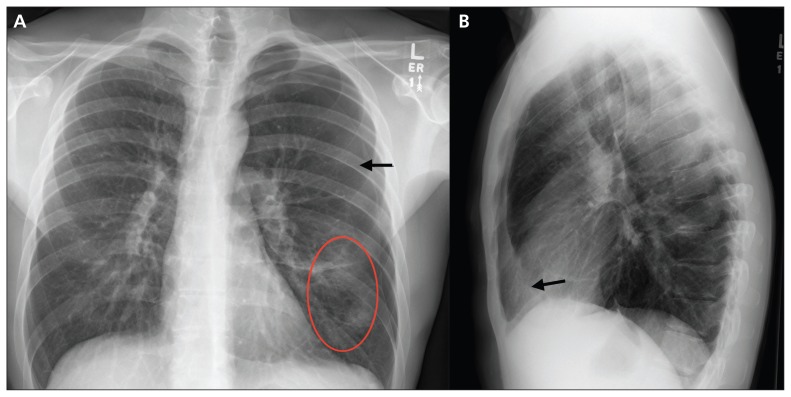
Figure 1:
Radiograph of the chest on admission of a 29-year-old man with type 1 diabetes who presented to the emergency department with shortness of breath and chest pain. (A) Posterior–anterior view showing left pneumothorax (arrow) and air space disease in the left lower lobe (circle). (B) Lateral view showing left pneumothorax (arrow). The left lower lobe consolidation seen on the posterior–anterior view is poorly identified on the lateral view.

