



Abstract
Heat shock proteins (HSPs) are constitutively expressed in murine skin. HSP27 is present in the epidermis and HSP70 can be found in both the epidermis and dermis. The purpose of this study was to investigate the role of these proteins in cutaneous chemical carcinogenesis and to determine if their effects on cell-mediated immune function were a contributing factor. In vivo inhibition of HSP27 and HSP70 produced a reduction in the T-cell mediated immune response to 7,12-dimethylbenz(a)anthracene (DMBA) and benzo(a)pyrene B(a)P in C3H/HeN mice and resulted in a state of antigen specific tolerance. When mice were pre-treated with anti-HSP27 and anti-HSP70 antibodies in vivo prior to subjecting them to a standard two-stage DMBA/12-O-tetradecanoylphorbol-13-acetate (TPA) cutaneous carcinogenesis protocol, the percentage of mice with tumors was much greater (p<0.05) in anti-HSP27 and HSP70 pre-treated animals compared to mice pre-treated with control antibody. Similar results were obtained when the data were evaluated as the cumulative number of tumors per group. Mice pre-treated with HSP27 and HSP70 antibodies developed more H-ras mutations and fewer DMBA specific cytotoxic T-lymphocytes. These findings indicate that in mice HSP27 and HSP70 play a key role in the induction of cell-mediated immunity to carcinogenic polyaromatic hydrocarbons. Bolstering the immune response to carcinogenic polyaromatic hydrocarbons may be an effective method for prevention of the tumors that they produce.
Keywords: Heat shock protein (HSP) 27; HSP70; Skin Carcinogenesis; 7,12-dimethylbenz(a)anthracene (DMBA); Contact Hypersensitivity
INTRODUCTION
Heat shock proteins (HSPs) are highly conserved intracellular proteins that are present in large quantities in all cells (1). Their expression is increased following exposure to heat, oxidative stress, toxins and glucose deprivation. These agents play an important role in the folding and unfolding of other proteins, serve as peptide chaperones, and help to transport proteins across membranes and within cells. Certain HSPs, including HSP27 and HSP70 are cytoprotective, impeding apoptotic pathways, thereby permitting cells to survive during conditions of stress, when it would be expected that there are increased amounts of unfolded proteins.
HSPs also play important roles in eliciting innate and adaptive immunity (2). HSPs, including HSP70, exert cytokine-like effects on maturation of antigen presenting cells (APC), enhancing tumor necrosis factor-α, interleukin (IL)-1-β, IL-6 and IL-12 secretion from monocytes, macrophages (3–5) and DCs (6). They induce surface expression of B7 and major histocompatibility complex (MHC) class II molecules on DCs (3, 7, 8), and stimulate the production of chemokines by macrophages and DC (9). Such findings support the concept that HSPs are endogenous signals for APC maturation, and may have a role in the regulation of immunity versus tolerance.
The effects of HSPs on the immune system have relevance for host defenses against tumors. For example, tumor cell lysates containing HSP70, as well as other HSPs, including gp96 and HSP90, have been used to protect animals against subsequent tumor challenge with autologous tumors. As a result, HSPs are being investigated as immunotherapeutic agents (10, 11). The role of HSPs in carcinogenesis, i.e. at earlier stages in cancer development prior to the time that tumors have formed, has not been investigated. This would have relevance for immunoprevention of tumors, in addition to their immunotherapeutic potential. In previous studies, we have shown that a cell mediated immune response occurs following topical application of carcinogenic polyaromatic hydrocarbons (PAHs) in selected strains of mice (12, 13), and have postulated that immunization against the chemical that causes the tumors or to proteins mutated by the chemical, rather than to the tumor itself, may be an effective method of preventing, as opposed to treating, chemically-induced tumors and may have more global activities.
Using proteomic mapping, we have shown that six molecular chaperones - HSP27, HSP60, HSP70, HSP84, ER60, and GRP78 - are constitutively expressed in the skin of C3H/HeN and BALB/c mice (12). Higher levels of HSP27 are found in the epidermis. HSP70 family members are expressed constitutively within keratinocytes and are elevated in both epidermis and dermis after skin samples are heat shocked in vivo and in vitro (14–16).
We have previously found that HSP27 and HSP70 are important in the development of allergic contact hypersensitivity to DNFB (17). We therefore postulated that HSPs may have relevance to the development of chemically induced skin tumors. To investigate this issue, we pretreated the skin in vivo with neutralizing antibodies to HSP27 and HSP70 and found that such a procedure not only reduced induction of the cell mediated immune response to the carcinogenic polychromatic hydrocarbon 7,12-dimethylbenz(a)anthracene (DMBA), but, in addition, rendered them immunologically tolerant to that molecule. Animals rendered immunologically tolerant to DMBA developed increased numbers of tumors when subjected to a DMBA initiation, 12-O-tetradecanoylphorbol-13-acetate (TPA) promotion skin carcinogenesis protocol. Thus, it may be possible to exploit the immunological properties of HSPs for the prevention of chemically induced tumors in addition to using them as an immunotherapeutic modality.
MATERIALS AND METHODS
Animals and Reagents
Adult female, 6–8 week old C3H/HeN mice were obtained from Charles River Laboratories (Wilmington, MA). Animals were maintained in accordance with institutional guidelines. Normal goat IgG and goat polyclonal anti-HSP27 and anti-HSP70 IgG were purchased from Santa Cruz Biotechnology CA, USA. Sheep anti-rat IgG dynabeads were from Life Technologies (Carlsbad,CA). Hybridoma lines GK1.5 (anti-CD4), Lyt-2 (anti-CD8) and HB-32 (anti-Iak) were acquired from ATCC (Manassas, VA). CD45R/B220 and recombinant GM-CSF were obtained from Pharmingen (San Diego, CA). DMBA, TPA, benzo(a)pyrene (B(a)P), and recombinant IL-4 and were purchased from Sigma Chemical Co. (St. Louis, MO).
In Vivo Treatment with anti-HSP Antibodies
In order to assess the contribution of HSP27 and 70 in contact hypersensitivity, the abdominal skin of mice was prepared by removing hair with an electric trimmer in conjunction with gentle brushing of the skin as described earlier (18). This was followed by administration of 2μg of control antibody, anti-HSP27, anti-HSP70 or a combination of anti-HSP27 and anti-HSP70 antibodies in PBS for 2 h under occlusion with a bio-occlusive dressing (Tegaderm®, 3M, Maplewood, MN) employing previously established techniques (17). The site was examined for erythema and edema using a modified Draize scoring system of 0 to 3 (17, 19). All animals employed for experiments had a Draize score of <1. Studies have shown that application of antibody to the shaved abdominal skin allowed the antibody to penetrate into the epidermis and dermis (17), and were confirmed for these experiments.
Contact hypersensitivity to DMBA and B(a)P
Contact hypersensitivity to DMBA was performed as described previously (20). Briefly, the shaved abdominal skin of C3H/HeN mice was sensitized on day 0 with 100 μl of a 0.1% solution of DMBA (w/v in acetone). Five days later, a challenge dose of 20 μl of 0.1% DMBA was painted on the ear after measuring baseline ear thickness. The increase in ear swelling was measured at 24h intervals to quantitate the magnitude of the contact hypersensitivity response. Contact hypersensitivity to B(a)P was conducted in the same manner with the exception that on day 0, 100 μl of a 0.1% solution of B(a)P was applied to the skin for sensitization and 20 μl of 0.1% B(a)P was applied to the ear for elicitation of the response.
Generation of bone marrow-derived dendritic cells (BMDC)
Bone marrow-derived dendritic cells were prepared from mice as described earlier with some modifications (21). Briefly, bone marrow cells were prepared from femurs and tibias of mice and were incubated in RPMI 1640 medium with a cocktail of antibodies against Iak, CD45R/B220, Lyt-2 and GK1.5 (2 μg/106 cells) on ice for 1 hour. The cells were subsequently washed once with HBSS after lysis of RBCs. Sheep anti-rat IgG dynabeads were used to remove different cellular populations from the cell suspension according to the manufacturer’s instructions. Cells were washed once with HBSS and cultured in 10% FCS RPMI 1640 media supplemented with recombinant mouse GM-CSF (10 ng/ml) and IL-4 (10 ng/ml) in 6-well plates (5 × 10⁵ cells/well). On day 5, half of the medium was replaced with fresh medium and cells were stimulated on the following day for the experiments.
Stimulation of DMBA primed lymph node cells with hapten-conjugated BMDC for cytokine production
In order to assess antigen specific cytokine production, BMDC were used for in vitro stimulation of primed lymph node cells as described (18, 22). Mice were pre-treated with anti-HSP27 and anti-HSP70 or isotype control antibody as described earlier in this section and were then sensitized with 100 μl of a 0.1% solution of DMBA (w/v in acetone) on day 0. On day +5, mice were sacrificed and lymph node cell suspensions were prepared by gentle pressure through a wire mesh screen. Ten million BMDC were suspended in 1 ml DMBA solution (10μM in DMSO) for 15 min. The cells were then washed three times with RPMI containing 5% FCS and re-suspended in the culture medium. DMBA primed lymph node cells (2 × 10⁶/ml) were stimulated with DMBA labeled BMDC (2 × 10⁵/ml). Cytokine concentrations in culture supernatants were measured 48 hours after culture using cytokine-specific (IFN-γ, IL-17, IL-4 and IL-10) enzyme linked immunosorbent assay (ELISA) kits from Life Technologies (Carlsbad, CA) according to the manufacturer’s instructions.
Assessment of immunological tolerance to DMBA
Tolerance to DMBA was assessed by a modification of the protocol of Schwarz et al. (23). On day -14, the abdominal skin of mice was treated with 2μg of isotype control antibody or a combination of anti-HSP27 and anti-HSP70 antibodies under occlusion with a bio-occlusive dressing. Immediately thereafter, 100μl of 0.1% DMBA was applied topically to the antibody treated site. On day 0, mice were re-sensitized on the dorsal skin, a site that had not been treated with antibody, with 100μl of 0.1% DMBA. Five days later mice were challenged with 20μl of 0.1% DMBA on the ear. The increase in ear swelling was measured at 24h intervals as described above. In order to evaluate the antigen specificity of tolerance, a separate panel of mice was treated in the same manner as above, but on day 0 that panel was sensitized with 100μl of 0.1% B(a)P and on day +5 was ear challenged with 20μl of 0.1% B(a)P.
In vivo cytotoxic T lymphocyte (CTL) activity
CTL activity was assessed using an in vivo antigen-specific cytotoxicity assay using methods by Inguilli (24) and Hermans et al. (25). Mice were topically treated with HSP27 and HSP70 antibodies as described above and were then sensitized at the same site with DMBA. Eight days later, mice received an intravenous (i.v.) injection of 5 × 10⁶ carboxyfluorescein diacetate succinimidyl ester (CFSE, Life Technologies)-labeled target spleen cells composed of two populations: CFSE low (6μM) labeled cells which were pre-pulsed with 50 μM DMBA for 25 min, washed and mixed 1:1 with un-pulsed CFSE high (12μM) labeled cells. After 16h spleens were harvested and processed into single cell suspensions for by flow cytometric analysis to quantify CFSE high and low cells. Flow cytometric acquisition of 2,000 CFSE high cells were detected per sample. The percent DMBA-specific cytotoxicity was calculated as follows: 100 * (1-(% CFSE low Exp/ Naive)). % Inhibition, which is calculated as follows: 100*(1-(Exp % cytotoxicity) Avg % cytotoxicity IgG).
Skin tumorigenesis
A two stage skin carcinogenesis protocol was employed to study the effect of HSP27 and HSP70 on DMBA tumorigenesis, in which DMBA was the initiating agent and TPA was the promoting agent, using methods that have been described previously (18). Anti-HSP (HSP27 and HSP70) antibodies were applied topically one time on the shaved dorsal skin of mice (10 mice/panel). Two hours later, the antibody treated site was painted with 100 μl of DMBA (0.1% w/v in acetone). One week after that, TPA (40 nmol in acetone) was applied twice weekly to the site that had been treated previously with DMBA. Mice were evaluated two times per week for tumors. Only tumors that had attained a size of one mm or greater and were present for two weeks or longer were counted.
Detection of H-ras mutation at codon 61
The H-ras mutation in DMBA induced skin tumors was performed as described with some modifications (26). Total RNA was isolated from the tumors using TRIzol Reagent (Life Technologies, Carlsbad CA) according to manufacturer’s instructions. 1μg of RNA from each sample was used as the starting material for RT-PCR (Promega, Madison WI). For PCR amplification of cDNA the following primers were used: Forward primer for both wild-type and mutant H-ras: 5’-CTAAGCCTGTTGTTTTGCAGGAC-3’, Reverse primer for wild-type H-ras: 5’-CATGGCACTATACTCTCTT-3’, and Reverse primer for mutant H-ras: 5’-CATGGCACTATACTCTCTA-3’. PCR was performed at 94°C for 5 min followed by 30 cycles of 94°C 60s, 55°C 60s and 72°C 60s. Codon 61 C→A point mutation creates an XbaI restriction enzyme site, which is utilized to distinguish mutant from wild-type H-ras. The PCR product was digested with XbaI and the products were separated by PAGE and visualized by ethidium bromide staining.
Statistical analysis
Data were analyzed by one-tailed Student’s t test, and the p values are indicated in the text and figure legends. The group difference was compared using ANOVA test followed by Tukey’s post hoc test for multiple comparison adjustment in one experiment. Differences were considered significant at p<0.05.
RESULTS
Heat shock proteins HSP27 and HSP70 play a role in the development of contact hypersensitivity to DMBA
In order to evaluate the role of HSP27 and HSP70 in the cell mediated response to DMBA, C3H/HeN mice were contact sensitized to DMBA after local treatment of the skin with anti-HSP27 or anti-HSP70 antibodies. Inhibition of the induction of DMBA contact hypersensitivity could be achieved by pretreatment with neutralizing antibodies to either HSP27 or HSP70 antibodies (Fig. 1A). Administration of both antibodies together gave an additive effect. There was no inhibition of the contact hypersensitivity response with isotype control antibodies (Fig. 1B). The effect of HSP27 and HSP70 neutralization on contact hypersensitivity to the polyaromatic hydrocarbons B(a)P was also assessed. There was inhibition of the induction of B(a)P contact hypersensitivity. Pre-treatment with neutralizing antibodies to either HSP27 or HSP70 antibodies inhibited B(a)P contact hypersensitivity also (Fig. 1C).
Figure 1.

Contact hypersensitivity to DMBA is inhibited by pretreatment of skin with antibodies to heat shock proteins 27 (HSP27) and 70 (HSP70). (A) The application of neutralizing antibodies to HSP27 or HSP70 to C3H/HeN mice (n=5 per group) significantly inhibited the CHS response and treatment with antibodies to HSP27 and HSP70 gave an additive effect (*,p<0.05). (B) Treatment with isotype control antibodies did not diminish the CHS response to DMBA (*, p<0.05). Results are expressed as the change in auricular thickness ± SEM.
Studies on the murine contact hypersensitivity response to DNFB have demonstrated that this response is mediated by IFN-γ and IL-17 (17, 22). Experiments were therefore also performed in which cells were isolated from the draining lymph nodes of mice sensitized to DMBA. The cells were then placed in culture with DMBA labeled BMDC. This served as an alternative method of assessing the immune response to the topically applied carcinogen. DMBA-labeled BMDC stimulated the production of IL-17 and IFN-γ by primed lymph node cells whereas they were not able to stimulate naïve lymph node cells (Fig. 2). Pretreatment of mice in vivo with anti-HSP27 and anti-HSP70 antibodies before DMBA application, significantly inhibited production of IL-17 and IFN-γ compared to untreated BMDC (Fig. 2, p<0.05). Moreover, pretreatment with anti-HSP antibodies resulted in a corresponding increase in IL-4 and IL-10.
Figure 2.

Effect of HSP27 and -70 on cytokine production by draining lymph node T-cells after DMBA sensitization in C3H/HeN mice. Sensitized T-cells were stimulated with bone marrow dendritic cells (BMDC). Lymph node cells from mice pre-treated with anti-HSP antibodies secreted significantly lower levels (*,p<0.05) of IL-17(A) and IFN-γ (B); and higher levels of IL-4 (C) and IL-10 (D) than control antibody treated mice.
Induction of DMBA-specific tolerance by HSP27 and HSP70 antibodies
Experiments were then conducted to determine whether treatment with anti-HSP antibodies (HSP27 and HSP70) followed by DMBA application had an effect on subsequent attempts to sensitize mice to that carcinogen. Animals that had been sensitized with DMBA on anti-HSP antibody treated skin were re-sensitized with 0.1% DMBA at a non-antibody treated site after a resting period of 14 days; they were then ear challenged with DMBA 5 days after that. The ear swelling response of mice pretreated with anti-HSP antibodies was suppressed compared to positive controls despite the fact that the second attempt to sensitize mice was through normal skin (Fig. 3A). This indicated that mice treated with anti-HSP antibody followed by carcinogen application had become tolerant to DMBA.
Figure 3.

Tolerance to DMBA follows HSP antibody treatment and is antigen specific. C3H/HeN mice (n=5 per group) were treated with anti-HSP antibodies on the abdomen after which 0.1% DMBA was applied to the same site. After 14 days, the mice were re-sensitized on the shaved back with either (A) 0.1% DMBA or (B) 0.1% B(a)P. Mice were ear challenged 5 days later with the same hapten that had been applied to the back. Ear swelling responses indicate that inhibition of CHS produced by anti-HSP antibodies was present in the DMBA sensitized mice (*,p<0.05), but not in the B(a)P sensitized mice (*, p>0.05). In another set of experiments, C3H/HeN mice (n=5 per group) were treated with anti-HSP antibodies on the abdomen after which 0.1% B(a)P was applied to the same site. After 14 days, the mice were re-sensitized on the shaved back with either (C) 0.1% B(a)P or (D) 0.1% DMBA. Mice were ear challenged 5 days later with the same hapten that had been applied to the back. Ear swelling responses indicate that inhibition of CHS produced by anti-HSP antibodies was present in the B(a)P sensitized mice (*,p<0.05), but not in the DMBA sensitized mice (*, p>0.05).Results are expressed as change in auricular thickness ± SEM.
To determine whether suppression of the induction of contact hypersensitivity was hapten specific and to exclude the possibility that the mice treated with a combination of anti-HSP27 and anti-HSP70 antibodies and DMBA were also non-responsive to other polyaromatic hydrocarbons given subsequently, the following experiment was performed. Panels of mice were treated with a combination of anti-HSP27 and anti-HSP70 antibodies followed immediately thereafter by application of DMBA to the antibody treated skin site. Two weeks later, animals were treated with either a sensitizing dose of B(a)P. Animals were ear challenged five days later. Although mice treated with DMBA plus anti-HSP antibodies and then re-sensitized with DMBA had a significantly suppressed response to DMBA, mice treated with anti-HSP antibodies plus DMBA, and then with B(a)P behaved like normal mice in their ear swelling response to B(a)P (Fig. 3B). In a reciprocal manner, mice that were treated with B(a)P plus anti-HSP antibodies and then re-sensitized with B(a)P had a significantly suppressed response to B(a)P (Fig. 3C), but mice that were treated with anti-HSP antibodies plus B(a)P and then re-sensitized with DMBA had a normal ear swelling response to DMBA (Fig. 3D). The results indicate that animals rendered unresponsive to DMBA are capable of developing a normal cell-mediated immune response to other polyaromatic hydrocarbons and that the combination of anti-HSP antibodies followed by hapten sensitization induces a state of specific immunologic unresponsiveness only to the carcinogen that was applied at the HSP treated skin site.
DMBA tolerant mice are more susceptible to cutaneous DMBA carcinogenesis
When mice were treated with anti-HSP (HSP27 and HSP70) antibodies and were then subjected to a DMBA initiation, TPA promotion skin carcinogenesis protocol, the number of tumors was significantly greater (p<0.05) in anti-HSP antibody treated mice compared to control antibody treated animals (Fig. 4A). Similar results were obtained when the data were evaluated as the cumulative number of tumors and the number of tumors per tumor bearing mouse (data not shown). Although animals in both treatment groups eventually developed tumors with this protocol, they arose more rapidly in anti-HSP antibody treated mice. For example, by week 16, 100% of anti-HSP antibody treated mice had developed tumors whereas only 40% of mice treated with control antibody had tumors, and these differences were significant (p<0.05) (Fig. 4B). The tumors from the immunologically unresponsive mice grew progressively and showed a significant (p<0.05) increase in tumor volume compared to tumors from control antibody pre-treated mice (Fig. 4 C&D).
Figure 4.
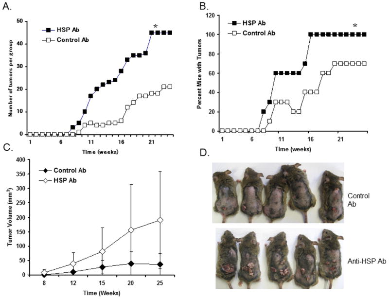
Effect of HSP27 and HSP70 antibodies on DMBA/TPA induced cutaneous carcinogenesis in C3H/HeN mice (n=10 per group). There was a significantly higher (*, p<0.05) number (A), percentage of tumors (B), and tumor volume (C) in the group of mice which was treated with anti-HSP antibodies. (D) Groups of mice with tumors after 25 weeks of DMBA/TPA treatment.
HSP27 and HSP70 contribute to reduced H-ras mutations in DMBA-induced tumors
DMBA forms adducts with DNA, the most important of which are activating mutations in the H-ras oncogene. One frequently occurring mutation is an A→T point mutation in codon 61, resulting in a change from glutamine (Q) to leucine (L) (26–30). The frequency of the H-ras mutation was significantly greater (p<0.05) in tumors from the anti-HSP antibody treated mice than in mice treated with the control antibody (Fig. 5).
Figure 5.
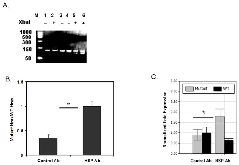
Increased expression of mutant H-ras in DMBA/TPA induced tumors from anti-HSP antibody treated mice after 25 weeks of DMBA/TPA treatment by RT-PCR. (A) Mutant H-ras is indicated by cleaved products (lanes 5&6) after digestion with XbaI. Lanes 1 and 2, naïve skin; lane 3, control antibody; lane 4, anti-HSP antibody; lane 5, control antibody; lane 6, anti-HSP antibody (after digestion with XbaI). (B) Densitometric analysis of mutant H-ras expression from gel, (C) qPCR for mutant H-ras expression in tumors. The frequency of the H-ras mutation was significantly greater (*, p<0.05) in tumors from the anti-HSP antibody treated mice than in mice treated with the control antibody.
HSP27 and HSP70 contribute to the generation of DMBA specific cytotoxic T lymphocytes
Experiments were performed to determine whether HSP27 and HSP70 were involved in the development of DMBA-specific cytotoxic T-lymphocytes. The skin of mice was pre-treated with anti-HSP27 and anti-HSP70 antibodies after which DMBA was applied. Eight days later, the treated mice received an intravenous mixture of DMBA-pulsed CFSE low and unpulsed CFSE high stained splenocytes. Sixteen hours later, splenocytes were harvested and analyzed by flow cytometry. There was a 52% inhibition of DMBA specific cytotoxicity in animals treated with anti-HSP antibodies compared to mice treated with control antibodies (Fig. 6). The results indicate that animals treated with HSP antibodies have a diminished ability to generate DMBA specific cytotoxic T cells.
Figure 6.

HSP antibody pretreatment inhibits development of DMBA specific CTLs. A. In vivo CTL assay of DMBA-pulsed CFSE low target spleen cells. Mice were pretreated with antibodies then sensitized, as indicated. After 8 days following treatment, mice (n=3 per group) received a 1:1 mixture of DMBA-pulsed CFSE low and unpulsed CFSE hi stained splenocytes, by i.v. After 16 hours, splenocytes were harvested and analyzed by flow cytometry. The % CFSE low population of total CFSE labeled cells is shown in each histogram. The % CFSE low population in naïve mice provided an input baseline control of 53% (confirming the 50:50 input mix). B. Bar graph of the mean ± SEM % DMBA specific cytotoxicity is shown for each group. Arrow indicates % Inhibition. *, p<0.05 by Student’s T test.
DISCUSSION
Cancer is a multistep process in which biochemical and molecular abnormalities accumulate in target cells in a sequential manner over long periods of time. Many of these steps have been identified using experimental animal models in which carcinogenic polyaromatic hydrocarbons are applied to the skin of animals (31, 32). Evaluation of the immune system in skin cancer development has focused primarily on immune responses to premalignant papillomas or invasive carcinomas. A vigorous host T-lymphocyte mediated immune response exists to PAH-induced tumors that have already progressed through the carcinogenesis pathway. These malignancies express tumor antigens on their cell surfaces that are recognized by immunocompetent lymphocytes (33). Immunization techniques against tumor antigens have been successful in protecting animals against the subsequent growth of tumors which express the same antigens, findings which support the immunosurveillance theory that was originally proposed by Thomas (34) and Burnet (35). However, in relation to the total time that it takes for tumors to develop, this is a late stage in the process. Previously, we have shown that administration of carcinogenic PAHs, such as DMBA, B(a)P, and 3-methylcholanthrene (3-MC), to the skin of mice results in an antigen specific response to the topically applied carcinogen, that is mediated by CD8+ T-cells; CD4+ T-cells have a regulatory role (12, 13, 18, 20). This raises the possibility that the development of a T-cell response to the chemical that causes the tumor, rather than to the tumor itself, may be an effective method of eradicating mutant cells and preventing these tumors from developing in the first place. Previous studies have supported that concept (13, 18). Specifically, the immune response to PAHs is genetically determined in part by polymorphisms in the class I genes within the MHC (13). Mice which develop an immune response to PAHs developed significantly fewer tumors when subjected to a DMBA cutaneous carcinogenesis protocol compared to MHC congenic mice that did not develop immunity to DMBA (13). Strains of mice that did develop an immune response to PAH also had a reduced number of DMBA-DNA adducts compared to those that did not (13). In addition, in mice subjected to a DMBA/TPA skin tumorigenesis protocol, greater numbers of tumors developed in CD8 deficient mice than wild-type mice, and fewer tumors occurred in CD4 deficient mice, indicating that CD8+ T-cells are effector cells and CD4+ T-cells have a regulatory role (18).
While suggestive that strains of mice that develop a T-cell response to PAHs are resistant to the carcinogenic effects of these agents, a more direct method of evaluating whether antigen specific T-cell mediated immunity to PAHs confers resistance to the tumors that they produce would be to demonstrate that mice rendered immunologically tolerant to a carcinogenic PAH are more susceptible to PAH-induced skin tumors compared to mice that develop a cell-mediated immune response that agent.
In this study, we observed that inhibition of the cell-mediated immune response to the carcinogen DMBA could be achieved by pretreating the skin with neutralizing antibodies to HSP27 and HSP70. Loss of the immune response to DMBA was antigen specific and led to long-term immunological unresponsiveness to that molecule. This enabled us to test, in a very direct manner, the hypothesis that the presence of a cell-mediated immune response to polyaromatic hydrocarbons confers resistance to their carcinogenic effects. We observed that this, in fact, was the case. Mice unresponsive to DMBA developed substantially greater numbers of tumors than mice in which a cell-mediated immune response was present. This was associated with fewer DMBA cytotoxic T-cells and more H-ras mutations, which are known to initiate DMBA-induced skin tumors.
The method we employed for the induction of immunological tolerance was to first pretreat the area of skin used for immunization with antibodies to HSP27 and HSP70. There are other methods of producing immunological tolerance to topically applied chemicals. These include oral or intravenous administration of hapten prior to sensitization, topical application of hapten to ultraviolet radiation exposed skin or intravenous injection of UV-irradiated hapten modified epidermal cells. However, most of those procedures require co-administration of the same or different carcinogenic agents (i.e. DMBA, ultraviolet radiation), which would obfuscate efforts to determine whether it was actually alterations in the immune response or differences in the amount of carcinogen that was administered to the animal. In vivo treatment with anti-HSP27 and anti-HSP70 antibodies does not have those complicating effects and thus had a number of advantages for these studies.
The generation of immune tolerance by HSPs has been seen in other model systems. In an autoimmune disease model, it has been shown that HSP70 can promote immunogenic APC function and prevent the induction of tolerance. HSP70 increased inflammatory responses and elevated CTL function after enhanced antigen presentation by dendritic cells (DCs), similar to what is observed after CD40 ligation. Thus, HSP70 promotes DC function and, together with antigen, triggered autoimmune disease in vivo. Initiation of T-cell activation and immune function are crucially dependent on immunostimulatory antigen presentation by professional antigen-presenting cells (APCs), most notably DCs (36). Immunological tolerance to self-antigens also depends on the interaction of T cells with APCs, and can result in T-cell deletion or induction of unresponsiveness (37, 38). In our model, pretreatment of mice with anti-HSP27 and anti-HSP70 antibodies significantly inhibited the production of IL-17 and IFN-γ compared to untreated carcinogen labeled BMDC, and there was a corresponding increase in IL-4 and IL-10 production under the same conditions.
HSPs are known to bind TLR4 (5). In other studies, we have found that TLR4 results in the production of IL-12 by dendritic cells, which in turn facilitates production of Tc1, IFN-γ producing cells that mediate a vigorous cell mediated immune response (15). We have further shown that IFN-γ plays an essential role in preventing the development of carcinogenic polyaromatic hydrocarbon-induced tumors (16). Thus, it seems reasonable to postulate that following treatment with a carcinogenic polyaromatic hydrocarbon, such as DMBA, HSPs are released in the extracellular space. In that location, they bind to TLR4 resulting in IL-12 production, and biasing dendritic cells towards activation of IFN-γ producing T-cells and cytotoxic T-cells. Those T-cells then proceed during the early stages of the chemical carcinogenesis pathway to eradicate mutant epidermal cells with the potential to eventually become DMBA-induced skin tumors. Thus, in the absence of HSPs, polyaromatic hydrocarbon-induced tumors are more likely to arise.
HSPs, including HSP27 and HSP70 have been associated with the development of cancer (39, 40). In some situations, their expression has been linked to a more aggressive behavior. HSP70 transgenic mice have an increased rate of T-cell lymphoma (41), and when HSP70 was transfected into fibrosarcoma cell lines they became more tumorigenic and resistant to the cytotoxic effects of TNF-α (42). In humans, increased HSP70 has been found in early liver carcinoma and its expression has been associated with increased tumor grade and poorer prognosis (43). Similarly, significantly higher levels of HSP27 have been found in leukemia, breast, ovarian, and endometrial cancer cell lines compared to non-transformed cells; and there is a direct correlation between HSP27 expression and the grade of ovarian carcinoma (44). HSP27 overexpression has been observed in glial tumors as well (45). On the other hand, high levels of HSPs have been associated with improved outcomes. This is the case for osteosarcoma (46) and renal cell carcinoma (47), in which high levels of HSP70 are found. Moreover, reduced tumor levels of HSP27 have been associated with more aggressive tumors in oral cancers (48). In addition, head and neck cancers in which there is diminished expression of HSP27 have the poorest survival rates (49). Thus, differences in HSP effects may depend on the type of tumor being investigated.
Polyaromatic hydrocarbons are a major causative agent for various types of epithelial cancer. They present in tobacco smoke, charcoal broiled food and automobile emissions, and are causative agents for cancers of the lung, head and neck, bladder and breast. Although the majority of non-melanoma skin cancers are caused by overexposure to ultraviolet light, there is clear evidence that carcinogenic polyaromatic hydrocarbons are an etiologic factor or cofactor in a significant proportion of squamous cell carcinomas of the skin as well (50). Vaccination against PAHs is analogous to prevention against cervical cancer by administration of HPV vaccines (51) or against liver cancer by hepatitis B vaccines. This strategy has not been employed before against chemical carcinogens. Efforts to bolster the immune response to carcinogenic PAHs or to proteins expressed by tumor-initiated cells could be an effective means of preventing their development.
Acknowledgments
Grant Support: NIH Grants P30 (CAE) AR050948, P30 AR050948, Veterans Administration Merit Review Award (CAE) 18-103-02 and a grant from the Department of Defense.
References
- 1.Young JC, V, Agashe R, Siegers K, Hartl FU. Pathways of chaperone-mediated protein folding in the cytosol. Nature reviews Molecular cell biology. 2004;5:781–791. doi: 10.1038/nrm1492. [DOI] [PubMed] [Google Scholar]
- 2.Srivastava P. Interaction of heat shock proteins with peptides and antigen presenting cells: chaperoning of the innate and adaptive immune responses. Annu Rev Immunol. 2002;20:395–425. doi: 10.1146/annurev.immunol.20.100301.064801. [DOI] [PubMed] [Google Scholar]
- 3.Basu S, Binder RJ, Suto R, Anderson KM, Srivastava PK. Necrotic but not apoptotic cell death releases heat shock proteins, which deliver a partial maturation signal to dendritic cells and activate the NF-kappa B pathway. Int Immunol. 2000;12:1539–1546. doi: 10.1093/intimm/12.11.1539. [DOI] [PubMed] [Google Scholar]
- 4.Sauter B, Albert ML, Francisco L, Larsson M, Somersan S, Bhardwaj N. Consequences of cell death: exposure to necrotic tumor cells, but not primary tissue cells or apoptotic cells, induces the maturation of immunostimulatory dendritic cells. J Exp Med. 2000;191:423–434. doi: 10.1084/jem.191.3.423. [DOI] [PMC free article] [PubMed] [Google Scholar]
- 5.Asea A, Kraeft SK, Kurt-Jones EA, Stevenson MA, Chen LB, Finberg RW, Koo GC, Calderwood SK. HSP70 stimulates cytokine production through a CD14-dependant pathway, demonstrating its dual role as a chaperone and cytokine. Nat Med. 2000;6:435–442. doi: 10.1038/74697. [DOI] [PubMed] [Google Scholar]
- 6.Moroi Y, Mayhew M, Trcka J, Hoe MH, Takechi Y, Hartl FU, Rothman JE, Houghton AN. Induction of cellular immunity by immunization with novel hybrid peptides complexed to heat shock protein 70. Proc Natl Acad Sci U S A. 2000;97:3485–3490. doi: 10.1073/pnas.070550797. [DOI] [PMC free article] [PubMed] [Google Scholar]
- 7.Cho BK, Palliser D, Guillen E, Wisniewski J, Young RA, Chen J, Eisen HN. A proposed mechanism for the induction of cytotoxic T lymphocyte production by heat shock fusion proteins. Immunity. 2000;12:263–272. doi: 10.1016/s1074-7613(00)80179-x. [DOI] [PubMed] [Google Scholar]
- 8.Kuppner MC, Gastpar R, Gelwer S, Nossner E, Ochmann O, Scharner A, Issels RD. The role of heat shock protein (hsp70) in dendritic cell maturation: hsp70 induces the maturation of immature dendritic cells but reduces DC differentiation from monocyte precursors. Eur J Immunol. 2001;31:1602–1609. doi: 10.1002/1521-4141(200105)31:5<1602::AID-IMMU1602>3.0.CO;2-W. [DOI] [PubMed] [Google Scholar]
- 9.Wang Y, Kelly CG, Karttunen JT, Whittall T, Lehner PJ, Duncan L, MacAry P, Younson JS, Singh M, Oehlmann W, Cheng G, Bergmeier L, Lehner T. CD40 is a cellular receptor mediating mycobacterial heat shock protein 70 stimulation of CC-chemokines. Immunity. 2001;15:971–983. doi: 10.1016/s1074-7613(01)00242-4. [DOI] [PubMed] [Google Scholar]
- 10.Li Z. Priming of T cells by heat shock protein-peptide complexes as the basis of tumor vaccines. Seminars in immunology. 1997;9:315–322. doi: 10.1006/smim.1997.0087. [DOI] [PubMed] [Google Scholar]
- 11.Srivastava PK, Callahan MK, Mauri MM. Treating human cancers with heat shock protein-peptide complexes: the road ahead. Expert Opin Biol Ther. 2009;9:179–186. doi: 10.1517/14712590802633918. [DOI] [PubMed] [Google Scholar]
- 12.Anderson C, Hehr A, Robbins R, Hasan R, Athar M, Mukhtar H, Elmets CA. Metabolic requirements for induction of contact hypersensitivity to immunotoxic polyaromatic hydrocarbons. J Immunol. 1995;155:3530–3537. [PubMed] [Google Scholar]
- 13.Elmets C, Athar M, Tubesing K, Rothaupt D, Xu H, Mukhtar H. Susceptibility to the Biological Effects of Polyaromatic Hydrocarbons Is Influenced by Genes of the Major Histocompatibility Complex. Proc Natl Acad Sci USA. 1998;95:14915–14919. doi: 10.1073/pnas.95.25.14915. [DOI] [PMC free article] [PubMed] [Google Scholar]
- 14.Huang CM, Foster KW, DeSilva T, Zhang J, Shi Z, Yusuf N, Van Kampen KR, Elmets CA, Tang DC. Comparative proteomic profiling of murine skin. J Invest Dermatol. 2003;121:51–64. doi: 10.1046/j.1523-1747.2003.12327.x. [DOI] [PubMed] [Google Scholar]
- 15.Muramatsu T, Tada H, Kobayashi N, Yamji M, Shirai T, Ohnishi T. Induction of the 72-kD heat shock protein in organ-cultured normal human skin. J Invest Dermatol. 1992;98:786–790. doi: 10.1111/1523-1747.ep12499953. [DOI] [PubMed] [Google Scholar]
- 16.Trautinger F, Trautinger I, Kindas-Mugge I, Metze D, Luger TA. Human keratinocytes in vivo and in vitro constitutively express the 72-kD heat shock protein. J Invest Dermatol. 1993;101:334–338. doi: 10.1111/1523-1747.ep12365491. [DOI] [PubMed] [Google Scholar]
- 17.Yusuf N, Nasti TH, Huang CM, Huber BS, Jaleel T, Lin HY, Xu H, Elmets CA. Heat shock proteins HSP27 and HSP70 are present in the skin and are important mediators of allergic contact hypersensitivity. J Immunol. 2009;182:675–683. doi: 10.4049/jimmunol.182.1.675. [DOI] [PMC free article] [PubMed] [Google Scholar]
- 18.Yusuf N, Nasti TH, Katiyar SK, Jacobs MK, Seibert MD, Ginsburg AC, Timares L, Xu H, Elmets CA. Antagonistic roles of CD4+ and CD8+ T-cells in 7,12-dimethylbenz(a)anthracene cutaneous carcinogenesis. Cancer Res. 2008;68:3924–3930. doi: 10.1158/0008-5472.CAN-07-3059. [DOI] [PMC free article] [PubMed] [Google Scholar]
- 19.Van Kampen KR, Shi Z, Gao P, Zhang J, Foster KW, Chen DT, Marks D, Elmets CA, Tang DC. Safety and immunogenicity of adenovirus-vectored nasal and epicutaneous influenza vaccines in humans. Vaccine. 2005;23:1029–1036. doi: 10.1016/j.vaccine.2004.07.043. [DOI] [PubMed] [Google Scholar]
- 20.Klemme JC, Mukhtar H, Elmets CA. Induction of contact hypersensitivity to dimethylbenz(a)anthracene and benzo(a)pyrene in C3H/HeN mice. Cancer Res. 1987;47:6074–6078. [PubMed] [Google Scholar]
- 21.Inaba K, Inaba M, Romani N, Aya H, Deguchi M, Ikehara S, Muramatsu S, Steinman RM. Generation of large numbers of dendritic cells from mouse bone marrow cultures supplemented with granulocyte/macrophage colony-stimulating factor. J Exp Med. 1992;176:1693–1702. doi: 10.1084/jem.176.6.1693. [DOI] [PMC free article] [PubMed] [Google Scholar]
- 22.He D, Wu L, Kim HK, Li H, Elmets CA, Xu H. IL-17 and IFN-gamma mediate the elicitation of contact hypersensitivity responses by different mechanisms and both are required for optimal responses. J Immunol. 2009;183:1463–1470. doi: 10.4049/jimmunol.0804108. [DOI] [PMC free article] [PubMed] [Google Scholar]
- 23.Schwarz A, Maeda A, Kernebeck K, van Steeg H, Beissert S, Schwarz T. Prevention of UV radiation-induced immunosuppression by IL-12 is dependent on DNA repair. J Exp Med. 2005;201:173–179. doi: 10.1084/jem.20041212. [DOI] [PMC free article] [PubMed] [Google Scholar]
- 24.Ingulli E. Tracing tolerance and immunity in vivo by CFSE-labeling of administered cells. Methods Mol Biol. 2007;380:365–376. doi: 10.1007/978-1-59745-395-0_23. [DOI] [PubMed] [Google Scholar]
- 25.Hermans IF, Silk JD, Yang J, Palmowski MJ, Gileadi U, McCarthy C, Salio M, Ronchese F, Cerundolo V. The VITAL assay: a versatile fluorometric technique for assessing CTL- and NKT-mediated cytotoxicity against multiple targets in vitro and in vivo. J Immunol Methods. 2004;285:25–40. doi: 10.1016/j.jim.2003.10.017. [DOI] [PubMed] [Google Scholar]
- 26.Pazzaglia S, Mancuso M, Primerano B, Rebessi S, Biozzi G, Covelli V, Saran A. Analysis of c-Ha-ras gene mutations in skin tumors induced in carcinogenesis-susceptible and carcinogenesis-resistant mice by different two-stage protocols or tumor promoter alone. Molecular Carcinogenesis. 2001;30:111–118. doi: 10.1002/1098-2744(200102)30:2<111::aid-mc1019>3.0.co;2-l. [DOI] [PubMed] [Google Scholar]
- 27.Quintanilla M, Brown K, Ramsden M, Balmain A. Carcinogen-specific mutation and amplification of Ha-ras during mouse skin carcinogenesis. Nature. 1986;322:78–80. doi: 10.1038/322078a0. [DOI] [PubMed] [Google Scholar]
- 28.Balmain A, Ramsden M, Bowden GT, Smith J. Activation of the mouse cellular Harvey-ras gene in chemically induced benign skin papillomas. Nature. 1984;307:658–660. doi: 10.1038/307658a0. [DOI] [PubMed] [Google Scholar]
- 29.Bizub D, Wood AW, Skalka AM. Mutagenesis of the Ha-ras oncogene in mouse skin tumors induced by polycyclic aromatic hydrocarbons. Proc Natl Acad Sci U S A. 1986;83:6048–6052. doi: 10.1073/pnas.83.16.6048. [DOI] [PMC free article] [PubMed] [Google Scholar]
- 30.Pelling JC, Fischer SM, Neades R, Strawhecker J, Schweickert L. Elevated expression and point mutation of the Ha-ras proto-oncogene in mouse skin tumors promoted by benzoyl peroxide and other promoting agents. Carcinogenesis. 1987;8:1481–1484. doi: 10.1093/carcin/8.10.1481. [DOI] [PubMed] [Google Scholar]
- 31.DiGiovanni J. Multistage carcinogenesis in mouse skin. Pharmacol Ther. 1992;54:63–128. doi: 10.1016/0163-7258(92)90051-z. [DOI] [PubMed] [Google Scholar]
- 32.Mukhtar H, Mercurio M, Agarwal R. Murine skin carcinogenesis: Relevance to humans. In: Mukhtar H, editor. Skin Cancer: Mechanisms and Human Relevance. CRC Press; Boca Raton, FL: 1995. pp. 3–8. [Google Scholar]
- 33.Vesely MD, Schreiber RD. Cancer immunoediting: antigens, mechanisms, and implications to cancer immunotherapy. Ann N Y Acad Sci. 2013;1284:1–5. doi: 10.1111/nyas.12105. [DOI] [PMC free article] [PubMed] [Google Scholar]
- 34.Thomas L. Reactions to homologous tissue antigens in relation to hypersensitivity. In: Lawrence HS, editor. Cellular and Humoral Aspects of the Hypersensitive States. Hoeber-Harper; New York: 1959. p. 529. [Google Scholar]
- 35.Burnet FM. Immunological Surveillance. Pergamon Press; Oxford: 1970. [Google Scholar]
- 36.Banchereau J, Palucka AK. Dendritic cells as therapeutic vaccines against cancer. Nature Reviews Immunology. 2005;5:296. doi: 10.1038/nri1592. [DOI] [PubMed] [Google Scholar]
- 37.Heath WR, Carbone FR. Cross-presentation, dendritic cells, tolerance and immunity. Annu Rev Immunol. 2001;19:47–64. doi: 10.1146/annurev.immunol.19.1.47. [DOI] [PubMed] [Google Scholar]
- 38.Steinman RM, Hawiger D, Nussenzweig MC. Tolerogenic dendritic cells. Annu Rev Immunol. 2003;21:685–711. doi: 10.1146/annurev.immunol.21.120601.141040. [DOI] [PubMed] [Google Scholar]
- 39.Murphy ME. The HSP70 family and cancer. Carcinogenesis. 2013;34:1181–1188. doi: 10.1093/carcin/bgt111. [DOI] [PMC free article] [PubMed] [Google Scholar]
- 40.Garrido C, Brunet M, Didelot C, Zermati Y, Schmitt E, Kroemer G. Heat shock proteins 27 and 70: anti-apoptotic proteins with tumorigenic properties. Cell Cycle. 2006;5:2592–2601. doi: 10.4161/cc.5.22.3448. [DOI] [PubMed] [Google Scholar]
- 41.Seo JS, Park YM, Kim JI, Shim EH, Kim CW, Jang JJ, Kim SH, Lee WH. T cell lymphoma in transgenic mice expressing the human Hsp70 gene. Biochem Biophys Res Commun. 1996;218:582–587. doi: 10.1006/bbrc.1996.0103. [DOI] [PubMed] [Google Scholar]
- 42.Jaattela M. Over-expression of hsp70 confers tumorigenicity to mouse fibrosarcoma cells. Int J Cancer. 1995;60:689–693. doi: 10.1002/ijc.2910600520. [DOI] [PubMed] [Google Scholar]
- 43.Chuma M, Sakamoto M, Yamazaki K, Ohta T, Ohki M, Asaka M, Hirohashi S. Expression profiling in multistage hepatocarcinogenesis: identification of HSP70 as a molecular marker of early hepatocellular carcinoma. Hepatology. 2003;37:198–207. doi: 10.1053/jhep.2003.50022. [DOI] [PubMed] [Google Scholar]
- 44.Geisler JP, Geisler HE, Tammela J, Wiemann MC, Zhou Z, Miller GA, Crabtree W. Heat shock protein 27: an independent prognostic indicator of survival in patients with epithelial ovarian carcinoma. Gynecol Oncol. 1998;69:14–16. doi: 10.1006/gyno.1998.4961. [DOI] [PubMed] [Google Scholar]
- 45.Aoyama A, Steiger RH, Frohli E, Schafer R, von Deimling A, Wiestler OD, Klemenz R. Expression of alpha B-crystallin in human brain tumors. Int J Cancer. 1993;55:760–764. doi: 10.1002/ijc.2910550511. [DOI] [PubMed] [Google Scholar]
- 46.Trieb K, Lechleitner T, Lang S, Windhager R, Kotz R, Dirnhofer S. Heat shock protein 72 expression in osteosarcomas correlates with good response to neoadjuvant chemotherapy. Hum Pathol. 1998;29:1050–1055. doi: 10.1016/s0046-8177(98)90412-9. [DOI] [PubMed] [Google Scholar]
- 47.Santarosa M, Favaro D, Quaia M, Galligioni E. Expression of heat shock protein 72 in renal cell carcinoma: possible role and prognostic implications in cancer patients. Eur J Cancer. 1997;33:873–877. doi: 10.1016/s0959-8049(97)00002-6. [DOI] [PubMed] [Google Scholar]
- 48.Lo Muzio L, Leonardi R, Mariggio MA, Mignogna MD, Rubini C, Vinella A, Pannone G, Giannetti L, Serpico R, Testa NF, De Rosa G, Staibano S. HSP 27 as possible prognostic factor in patients with oral squamous cell carcinoma. Histology and histopathology. 2004;19:119–128. doi: 10.14670/HH-19.119. [DOI] [PubMed] [Google Scholar]
- 49.Lo Muzio L, Campisi G, Farina A, Rubini C, Ferrari F, Falaschini S, Leonardi R, Carinci F, Stalbano S, De Rosa G. Prognostic value of HSP27 in head and neck squamous cell carcinoma: a retrospective analysis of 57 tumours. Anticancer Res. 2006;26:1343–1349. [PubMed] [Google Scholar]
- 50.Dika E, Fanti PA, Vaccari S, Patrizi A, Maibach HI. Causal relationship between exposure to chemicals and malignant melanoma? A review and study proposal. Reviews on environmental health. 2010;25:255–259. doi: 10.1515/reveh.2010.25.3.255. [DOI] [PubMed] [Google Scholar]
- 51.Lowy D. Human papillomavirus, cervical cancer prevention, and more. Vaccine. 2008;26(Suppl 10):iii–iv. doi: 10.1016/j.vaccine.2008.06.032. [DOI] [PubMed] [Google Scholar]