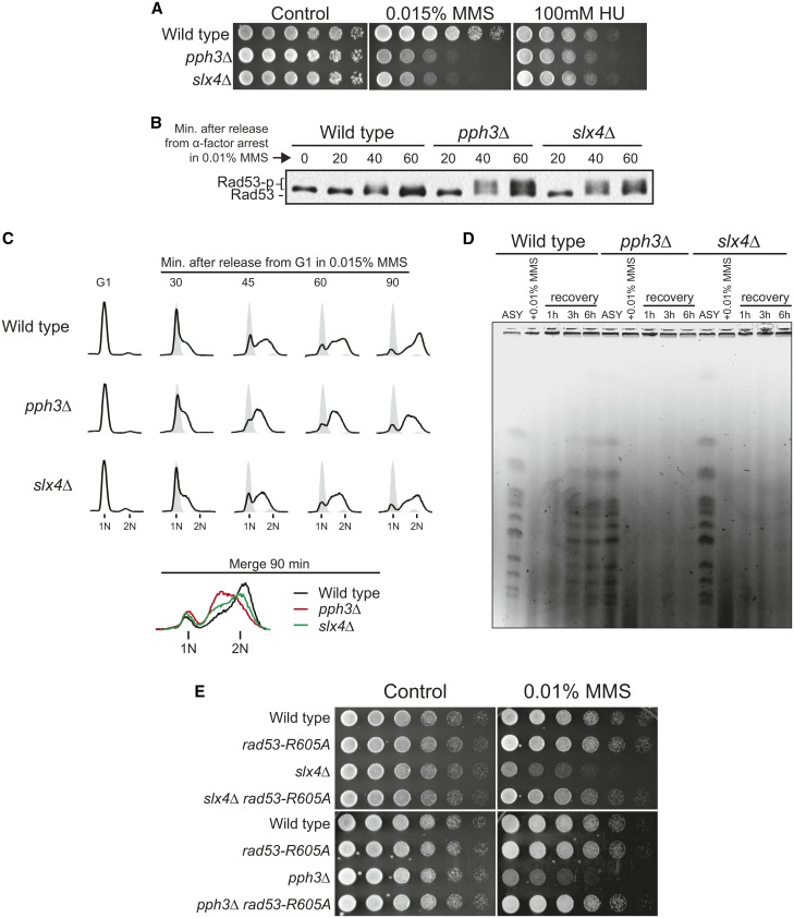
Figure 1.
Cells lacking either PPH3 or SLX4 show similar defects upon replication stress induced by MMS. (A) Serial dilution assays showing the effect of genotoxin treatment upon the sensitivity of wild-type, slx4Δ, and pph3Δ cells. Fourfold serial dilutions were spotted on YPD plates and grown for 2–3 days at 30°. (B) Anti-Rad53 immunoblots of wild-type, slx4Δ, and pph3Δ strains showing Rad53 phosphorylation status after MMS treatment. (C) S-phase progression analysis of wild-type, slx4Δ, and pph3Δ strains. For B and C, cells were arrested in G1 with α-factor and then released in medium containing MMS. Samples were collected in G1 and at different time points following release. (D) Analysis of fully replicated chromosomes measured by PFGE in wild-type, slx4Δ, and pph3Δ strains. Asynchronous (ASY) cells were treated with 0.01% MMS for 2 hr and then released in MMS-free medium for up to 6 hr. (E) Serial dilution assay showing the effect of a hypomorphic RAD53 allele (rad53-R605A) on MMS sensitivity of wild-type, slx4Δ, and pph3Δ strains.

