Abstract
A pencil core with an intact pencil tip was excised from the thigh of a 60-year-old male 53 years after a puncture wound. Histologic examination of the excised pencil core and the surrounding tissue revealed a foreign body reaction with abundant entrapped dark black pigment and chronic reparative changes, including dense sclerosis and focal granulation tissue formation.
Key Words: Pencil core granuloma, Graphite, Foreign body, Skin puncture
Case Report
A 60-year-old diabetic Caucasian male presented for his annual wellness visit. He indicated pain in an old injury site on the anterior left thigh. He narrated that when he was in second grade (age 7), he and a fellow student were irritating his neighbor in the classroom. His classmate retaliated by stabbing him in the left thigh with his pencil. The patient mentioned that the point broke off in his thigh and was never extracted. He subsequently developed a graphite tattoo at the site with a palpable subcutaneous papule. He complained that the site still hurt intermittently and after all this time, he wanted the lump and the reported pencil point to be removed. Examination of the anterior left thigh skin revealed a palpable 0.5-cm subdermal/subcutaneous elliptical lump in the skin. There was a tinge of black to blue to it as if it had been tattooed.
The patient was explained the indications, risks and benefits of the procedure and he provided consent. A time-out was performed. The area was prepped with ChloraPrep and draped in a sterile fashion. A 2-cm linear incision was made. The incision was partially opened by blunt and instrument dissection. A 0.6-cm diameter firm tan dermal-based soft tissue nodule with associated dark graphite precipitate was removed. In the midst of the removed tissue, a 4-mm cone-shaped pencil (graphite) tip was identified. The patient tolerated the procedure well. Two buried absorbable sutures were placed and three simple interrupted 4-0 vicryl sutures were used to close the wound. A triple antibiotic ointment was applied and the wound dressed in sterile fashion. The patient was put on cephalexin to prevent secondary infection. The surgical wound healed without complications or significant scarring.
Discussion
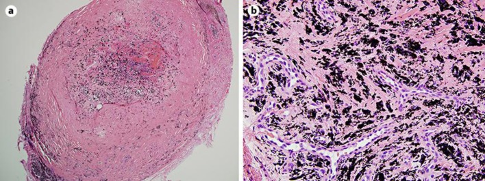
Graphite foreign body granulomas were reviewed in 1999 [1]. Pencil core granulomas continue to be infrequently reported in the literature [2,3,4,5,6]. The literature reports on hyperpigmented nodules in the skin secondary to a retained pencil core [5]. Hatano et al. [7] in 2000 reported on a pencil core granuloma from a pencil tip injury that had occurred about 30 years prior. Fukunaga et al. [8] in 2011 summarized 9 previously reported cases of pencil core granulomas, the oldest of which was 58 years old [9]. Our case involved a 53-year-old pencil core granuloma. Our case is unique in that we were able to recover the cone-shaped pencil tip and still write with it (fig. 1). In previous examinations of pencil core granuloma, giant cells and epithelioid cells had been observed [10]. Histologic examination in our case similarly revealed foreign body reaction with abundant entrapped dark black pigment and chronic reparative changes, including dense sclerosis and focal granulation tissue formation (fig. 2). Pencil core granulomas can display macroscopic and some radiologic features of melanoma [7]. Furthermore, one report suggested more rapid growth of lesions and greater degree of tissue injury by colored pencils when compared to non-colored graphite pencils [3]. A number of authors have commented on temporal changes in the granuloma, going from a relatively unchanged appearance to a rapid growth phase, ultimately leading to excision [3,4]. The decision to explore and excise a pencil core granuloma can be thus based on changes in the skin lesion and/or concern for possible malignancy.
Fig. 1.
a Tissue extracted from the wound site with recovered pencil core tip. b Pencil core tip with text written using it.
Fig. 2.
Histologic examination in our case similarly revealed foreign body reaction with abundant entrapped dark black pigment and chronic reparative changes, including dense sclerosis and focal granulation tissue formation. a H&E, ×20. b H&E, ×200.
Statement of Ethics
The patient gave written informed consent for this paper.
Disclosure Statement
The authors have no conflicts of interest to disclose.
References
- 1.Terasawa N, Kishimoto S, Kibe Y, Takenaka H, Yasuno H. Graphite foreign body granuloma. Br J Dermatol. 1999;141:774–776. doi: 10.1046/j.1365-2133.1999.3144c.x. [DOI] [PubMed] [Google Scholar]
- 2.Yoshitatsu S, Takagi T. A case of giant pencil-core granuloma. J Dermatol. 2000;27:329–332. doi: 10.1111/j.1346-8138.2000.tb02176.x. [DOI] [PubMed] [Google Scholar]
- 3.Shido H, Tamada I. Colored pencil-core granuloma on the forehead. Pediatr Dermatol. 2015;32:e58–e59. doi: 10.1111/pde.12524. [DOI] [PubMed] [Google Scholar]
- 4.Shibuya R, Endo Y, Fujisawa A, Tanioka M, Miyachi Y. Granuloma caused by carbon deposition in the dermis. Case Rep Dermatol Med. 2014;2014:686489. doi: 10.1155/2014/686489. [DOI] [PMC free article] [PubMed] [Google Scholar]
- 5.Lim GF, Lim SJ, Mahmoodi M, Radfar A. Enlarging hyperpigmented nodule on the right calf. Pencil-core granuloma. Int J Dermatol. 2013;52:933–934. doi: 10.1111/ijd.12027. [DOI] [PubMed] [Google Scholar]
- 6.Taguchi T, Terada Y. Images in clinical medicine. Pencil-core granuloma. N Engl J Med. 2012;366:2408. doi: 10.1056/NEJMicm1101530. [DOI] [PubMed] [Google Scholar]
- 7.Hatano Y, Komada S, Fujiwara S, Takayasu S. A case of pencil core granuloma with an unusual temporal profile. Dermatology. 2000;201:151–153. doi: 10.1159/000018460. [DOI] [PubMed] [Google Scholar]
- 8.Fukunaga Y, Hashimoto I, Nakanishi H, Seike T, Abe Y, Takaku M. Pencil-core granuloma of the face: report of two rare cases. J Plast Reconstr Aesthet Surg. 2011;64:1235–1237. doi: 10.1016/j.bjps.2011.01.017. [DOI] [PubMed] [Google Scholar]
- 9.Taylor B, Frumkin A, Pitha JV. Delayed reaction to ‘lead’ pencil simulating melanoma. Cutis. 1988;42:199–201. [PubMed] [Google Scholar]
- 10.Granick MS, Erickson ER, Solomon M. Pencil-core granuloma. Plast Reconstr Surg. 1992;89:136–138. [PubMed] [Google Scholar]


