Abstract
Objective:
The aims of this study are to propose a new set of Japanese diagnostic reference levels (DRLs) for 2014 and to study the impact of tube voltage and the type of reconstruction algorithm on patient doses. The volume CT dose index (CTDIvol) for adult and paediatric patients is assessed and compared with the results of a 2011 national survey and data from other countries.
Methods:
Scanning procedures for the head (non-helical and helical), chest and upper abdomen were examined for adults and 5-year-old children. A questionnaire concerning the following items was sent to 3000 facilities: tube voltage, use of reconstruction algorithms and displayed CTDIvol.
Results:
The mean CTDIvol values for paediatric examinations using voltages ranging from 80 to 100 kV were significantly lower than those for paediatric examinations using 120 kV. For adult examinations, the use of iterative reconstruction algorithms significantly reduced the mean CTDIvol values compared with the use of filtered back projection. Paediatric chest and abdominal scans showed slightly higher mean CTDIvol values in 2014 than in 2011. The proposed DRLs for adult head and abdominal scans were higher than those reported in other countries.
Conclusion:
The results imply that further optimization of CT examination protocols is required for adult head and abdominal scans as well as paediatric chest and abdominal scans.
Advances in knowledge:
Low-tube-voltage CT may be useful for reducing radiation doses in paediatric patients. The mean CTDIvol values for paediatric scans showed little difference that could be attributed to the choice of reconstruction algorithm.
Since the introduction of CT in the 1970s, it has been established worldwide as one of the most important imaging modalities in diagnostic radiology. In the past decade, various dose-reduction techniques, such as tube current modulation1 and low tube voltage,2 have been shown to reduce radiation exposure. In particular, the use of an iterative reconstruction (IR) algorithm, in contrast to a filtered back projection (FBP) algorithm, has provided diagnostically acceptable images using low-radiation-dose CT.3,4
Since estimates of the cancer risk attributable to the use of diagnostic X-rays have been reported,5,6 radiological technologists should aim to optimize scan parameters in order to avoid excessive radiation exposure. One powerful tool in this optimization applies the concept of diagnostic reference levels (DRLs). The DRLs of CT examinations are generally expressed in terms of the volume CT dose index (CTDIvol) or dose–length product. The DRL is used in medical imaging with ionizing radiation to indicate whether, in routine conditions, the patient dose from a specified procedure is unusually high or low; DRLs are usually reviewed at regular intervals and could be specific to a country or region.7 Surveys of DRLs for CT examination of adults8–11 and children12,13 have been reported in several countries.
The current DRLs in Japan were established as target values by the Japan Association of Radiological Technologists in 2006. The DRLs refer to a set of medical exposure guidelines, although there are several issues with these guidelines.14 First, no more than two examinations (head and abdomen) are listed in DRLs, and they contain no information about the CT examination of children. Second, the DRL for abdomen examination employs a 30-cm phantom, whereas a 32-cm phantom is more commonly used worldwide. Therefore, a new set of Japanese DRLs has become an urgent necessity. In 2011, Asada et al15 reported mean CTDIvol values for the head (non-helical and helical), chest and upper abdomen of both adults and children, which were obtained using a nationally distributed questionnaire. The aims of this study are to propose a new set of Japanese DRLs for 2014 and to study the impact of tube voltage and the type of reconstruction algorithm on patient doses. The CTDIvol for both adults and children have been assessed and compared with both the results of the 2011 survey and data from other countries.
METHODS AND MATERIALS
National questionnaire survey
A questionnaire was sent to 3000 facilities, which were taken from the list of Japanese Society of Radiological Technology members, with random two-stage sampling. These facilities comprised national hospitals, public medical organizations, social insurance agencies, public service corporations, medical corporations, educational corporations, social welfare corporations, companies and private medical health corporations in Japan.
The distributed questionnaire contained detailed questions on the CT scan parameters employed, including the manufacturer, specific CT scanner model, tube voltage (kV), tube current (mA), rotation time, number of channels, beam width, pitch factor and reconstruction algorithm (IR or FBP). Dose information was collected in terms of the displayed CTDIvol. The scanned anatomical regions were divided as follows: head (non-helical and helical), chest and upper abdomen (hereinafter called “abdomen”), for both adults and 5-year-old children. The questionnaire sought data for scanning performed on standard (average-sized) patients to represent usual practice, which excludes specialized examinations. The questionnaire also sought the displayed CTDIvol values with a 32-cm phantom for adult chest and abdomen examinations and with a 16-cm phantom for other examinations.
Analysis of collected data
The data were entered manually into an Excel® spreadsheet (Microsoft Corp., Redmond, WA). The first quartile (25th percentile), median (50th percentile) and third quartile (75th percentile) of CTDIvol values for each anatomical region were calculated directly from the total dose distribution. The national DRLs presented in this survey were determined using the 75th percentile of the CTDIvol, in accordance with the fact that the DRLs reported in other countries are usually based on the 75th percentile of the CTDIvol.8,9,16–18 The method of surveying CTDIvol values in Japan was the same as that used in the study by Asada et al15 in 2011, which serves as a basis for comparison with the present work.
A significant difference between the two groups was evaluated using Student's or Welch's t-test following the F-test, which was used in the analysis of variance. Statistical analyses were performed using Student's t-test when the two groups had equal variances, whereas Welch's t-test was used for unequal variances. A p-value of <0.05 was considered to be statistically significant. Data were occasionally missing at certain parts of the questionnaire. If the displayed CTDIvol was missing, the CTDIvol was estimated using the ImPACT CT Patient Dosimetry Calculator (CT Scanner Evaluation Centre, London, UK) based on the CT scan parameters, instead of the displayed CTDIvol, as discussed in a previous study.19
RESULTS
National questionnaire survey
The questionnaire was sent to 3000 facilities, and responses were received from 656 (21.9%). Sufficient details on the CTDIvol were provided by 584 (89.0%) of the 656 facilities. The 584 facilities that contributed to the survey represented 5.1% of all CT facilities in Japan.
Analysis of collected data
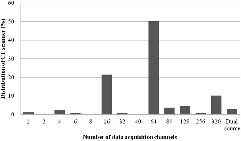
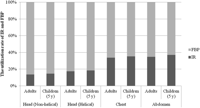
The collected data for 3004 scanner protocols from 584 facilities were analysed in this study. Multidetector row CT (MDCT) with 64 channels was found to be the most frequently used (50.2% of facilities) in these facilities, as shown in Figure 1, while an MDCT set-up with 16 channels was the second most frequently used (21.4%). There were 13 variations for 64-channel MDCT. The data for each anatomical region are listed in Table 1 according to the tube voltage (kV). A tube voltage of 120 kV was used in the majority of facilities for all anatomical regions, including CT examinations of children. However, voltages <120 kV were used in a higher percentage of CT examinations of children than for adult examinations. The mean CTDIvol values for paediatric examinations using voltages ranging from 80 to 100 kV were significantly lower than those for paediatric examinations using 120 kV (t-test, p < 0.05, Table 2). The utilization rate of the IR for the head region on adults and children was approximately 15% (Figure 2). On the other hand, the utilization rate of the IR algorithm for scans of the chest and abdomen on adults and children was approximately 35%. Table 3 lists the mean and median CTDIvol for each anatomical region, classified in terms of the image analysis algorithm used: IR or FBP. The mean CTDIvol values for adult examinations using IR were significantly lower than those of adult examinations using FBP (t-test, p < 0.05). However, there was no statistically significant difference in the mean CTDIvol values for paediatric scans between facilities employing IR and FBP algorithms, with the exception of abdominal CT scans.
Figure 1.
Distribution of CT scanners according to the number of acquisition channels. There were 13 variations for 64-channel multidetector row CT.
Table 1.
Distribution (%) of the tube voltage in each anatomical region
| Anatomical regions | Tube voltage (kV) |
|||||||
|---|---|---|---|---|---|---|---|---|
| n | 80 | 100 | 110 | 120 | 130 | 135 | 140 | |
| Head (non-helical) | ||||||||
| Adults | 536 | – | 0.2 | 0.4 | 93.8 | 4.3 | 0.4 | 0.9 |
| Children (5 years) | 318 | – | 6.0 | 1.9 | 89.6 | 2.2 | – | 0.3 |
| Head (helical) | ||||||||
| Adults | 482 | 0.2 | 0.8 | – | 95.4 | 2.7 | 0.2 | 0.4 |
| Children (5 years) | 271 | – | 5.5 | 1.1 | 93.0 | 0.4 | – | – |
| Chest | ||||||||
| Adults | 584 | – | 0.5 | 0.5 | 95.2 | 3.4 | 0.2 | 0.2 |
| Children (5 years) | 274 | 3.3 | 13.9 | 2.9 | 78.8 | 0.4 | – | – |
| Abdomen | ||||||||
| Adults | 570 | – | 0.5 | 0.4 | 95.3 | 3.7 | – | 0.2 |
| Children (5 years) | 269 | 2.2 | 14.5 | 1.9 | 79.6 | 1.1 | – | – |
Table 2.
Comparison of the distributions of CT dose index (CTDIvol) classified in terms of tube voltage used in paediatric examinations
| Anatomical regions | Tube voltage (kV) | CTDIvol |
p-value | ||
|---|---|---|---|---|---|
| n | Mean (mGy) | Coefficient of variation (%) | |||
| Head (non-helical) | 100 | 19 | 35.0 | 30 | <0.05 |
| 120 | 285 | 43.6 | 44 | ||
| Head (helical) | 100 | 15 | 31.5 | 35 | <0.05 |
| 120 | 252 | 42.4 | 43 | ||
| Chest | 80–100 | 47 | 5.0 | 63 | <0.05 |
| 120 | 216 | 10.5 | 87 | ||
| Abdomen | 80–100 | 45 | 6.4 | 84 | <0.05 |
| 120 | 214 | 12.6 | 76 | ||
Statistical analyses were performed using Welch's t-test.
Figure 2.
Utilization rate of the iterative reconstruction (IR). FBP, filtered back projection; y, years.
Table 3.
Comparison of the distributions of CT dose index (CTDIvol) using reconstruction algorithms
| Anatomical regions | Reconstruction algorithm | CTDIvol (mGy) |
Reduction rate (%) | p-value | |||
|---|---|---|---|---|---|---|---|
| n | Mean | Median | Coefficient of variation (%) | ||||
| Head (non-helical) | |||||||
| Adults | IR | 66 | 72.2 | 69 | 36 | 12 | <0.05a |
| FBP | 415 | 82.1 | 76 | 34 | |||
| Children (5 years) | IR | 44 | 41.6 | 38.3 | 42 | 6 | 0.4a |
| FBP | 254 | 44.2 | 41.1 | 43 | |||
| Head (helical) | |||||||
| Adults | IR | 77 | 66.7 | 64 | 40 | 11 | <0.05a |
| FBP | 362 | 75.2 | 72.2 | 32 | |||
| Children (5 years) | IR | 47 | 38.4 | 31.6 | 41 | 11 | 0.13a |
| FBP | 207 | 43.3 | 40 | 43 | |||
| Chest | |||||||
| Adults | IR | 183 | 10.4 | 10 | 42 | 28 | <0.05b |
| FBP | 359 | 14.4 | 12.9 | 69 | |||
| Children (5 years) | IR | 92 | 8.2 | 6 | 112 | 18 | 0.12a |
| FBP | 169 | 10 | 7.1 | 86 | |||
| Abdomen | |||||||
| Adults | IR | 184 | 15.8 | 14.8 | 49 | 16 | <0.05b |
| FBP | 344 | 18.7 | 17.2 | 46 | |||
| Children (5 years) | IR | 95 | 9.2 | 7.6 | 92 | 25 | <0.05a |
| FBP | 161 | 12.2 | 9.9 | 76 | |||
FBP, filtered back projection; IR, iterative reconstruction; n, number of CTDIvol values.
CTDIvol values in the adult chest and abdomen were calculated using a 32-cm phantom, while the others were calculated using a 16-cm phantom.
The CTDIvol in the head (non-helical) was from a scan of the posterior fossa.
Statistical analyses were performed using Student's t-test.
Statistical analyses were performed using Welch's t-test.
The distributions of CTDIvol for each anatomical region are summarized in Table 4. The CTDIvol data are presented in terms of the number of data points, mean, coefficient of variation (CV) and quartiles (25th percentile, median and 75th percentile). The mean CTDIvol values for all anatomical regions were higher than the medians.
Table 4.
Distributions of CT dose index (CTDIvol) for the CT examinations
| Anatomical regions | CTDIvol (mGy) |
|||||
|---|---|---|---|---|---|---|
| n | Mean | Coefficient of variation (%) | 25th percentile | Median | 75th percentile | |
| Head | ||||||
| Adults | 1018 | 77.4 | 34 | 60.0 | 73.0 | 92.3 |
| Children (5 years) | 589 | 42.7 | 43 | 30.0 | 40.0 | 50.0 |
| Non-helical | ||||||
| Adults | 536 | 81.0 | 34 | 62.4 | 75.3 | 93.9 |
| Children (5 years) | 318 | 43.3 | 43 | 30.7 | 40.0 | 50.0 |
| Helical | ||||||
| Adults | 482 | 73.4 | 34 | 58.6 | 70.9 | 90.4 |
| Children (5 years) | 271 | 42.0 | 43 | 29.8 | 39.0 | 50.8 |
| Chest | ||||||
| Adults | 584 | 13.0 | 66 | 8.7 | 11.5 | 16.2 |
| Children (5 years) | 274 | 9.5 | 93 | 4.0 | 6.6 | 12.0 |
| Abdomen | ||||||
| Adults | 570 | 17.7 | 48 | 12.0 | 16.0 | 21.5 |
| Children (5 years) | 269 | 11.3 | 81 | 5.0 | 8.4 | 14.1 |
All CT examinations (tube voltage values, reconstruction algorithms) have been included.
CTDIvol values in the adult chest and abdomen were calculated using a 32-cm phantom, while the others were calculated using a 16-cm phantom.
n indicates the number of CTDIvol values.
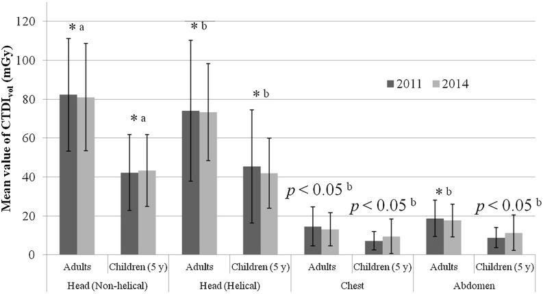
The changes in the mean CTDIvol values for each anatomical region from 2011 to 2014 are given in Figure 3. Although there was little difference in the mean CTDIvol values for CT head scans (non-helical and helical) on both adults and children between 2011 and 2014, the 2014 results for the chest and abdominal scans on children showed slightly higher mean CTDIvol values than in 2011 (t-test, p < 0.05). Furthermore, the adult chest scans showed a slightly lower mean CTDIvol in 2014 than in 2011 (t-test, p < 0.05). There was little difference in the mean CTDIvol values for CT abdominal scans on adults between 2011 and 2014.
Figure 3.
Change in the mean CT dose index (CTDIvol) values and standard deviations for each anatomical region from 2011 to 2014. *an insignificant change; astatistical analyses were performed using Student's t-test; bstatistical analyses were performed using Welch's t-test.
Table 5 compares the 75th percentile of the CTDIvol for each anatomical region in this study with the 75th percentiles of the CTDIvol values taken from surveys conducted in other countries. The 75th percentiles of the CTDIvol values in our study were mostly equal to those of other countries. However, the 75th percentiles of the CTDIvol for adult head and abdominal scans were slightly higher than those of other countries.
Table 5.
The 75th percentile of CT dose index (CTDIvol) obtained in this study for each anatomical region compared with the 75th percentile of CTDIvol values reported in other countries
| Country |
Switzerland12,18 |
Ireland9 |
Thailand13 |
Italy20 |
UK21 |
Portugal17 |
Korea16 |
This study |
|---|---|---|---|---|---|---|---|---|
| Year |
2008/2010 |
2010 |
2010 |
2011 |
2011 |
2012 |
2013 |
2014 |
| Number of facilities | 10/179a | 30 | 3 | 65 | 127 | 211 | 32 | 584 |
| Head | ||||||||
| Adults | 65 | 66 | – | 69 | 80 | 75 | 53 | 92 |
| Children (5 years) | 40 | – | 40 | – | 40 | 50 | – | 50 |
| Chest | ||||||||
| Adults | 10 | 9 | – | 15 | 12 | 14 | 13 | 16 |
| Children (5 years) | 10 | – | 10 | – | – | 5.6 | – | 12 |
| Abdomen | ||||||||
| Adults | 15 | 12 | – | 18 | 14 | 18 | 13 | 22 |
| Children (5 years) | 13 | – | 14 | – | – | – | – | 14 |
Values provided in milligrays.
CTDIvol values in the adult chest and abdomen were calculated using a 32-cm phantom, while the others were calculated using a 16-cm phantom.
Number of CT scanners.
DISCUSSION
Fukushima et al10 reported that the utilization rates of MDCT with 16 and 64 channels were 38.3% and 25.5%, respectively, in the Gunma prefecture of Japan in 2010. Asada et al15 reported that the use of MDCT with 64 channels (35.9%) was slightly more frequent than the use of MDCT with 16 channels (32.5%) in Japan in 2011. MDCT with 64 channels was used in half of the CT scanners included in the 2014 survey, implying that there is a general trend in Japan towards adopting MDCT with 64 channels. MDCT with 64 channels was also commonly used in Switzerland (approximately 30% of facilities),18 Ireland (approximately 40%)9 and Korea (approximately 36%).16
Many researchers have studied the use of low-tube-voltage CT for improving the image quality or reducing the radiation dose, particularly in paediatric patients.22,23 In this study, the utilization rate for tube voltages of 120 kV was equal for adult and paediatric head scans, although the utilization rate for this tube voltage for paediatric chest and abdominal scans was lower than that for adult scans. The tube voltage needed to penetrate the body of a child is lower than that of an adult, since a child is smaller.24 A tube voltage of 120 kV was most frequently used for paediatric chest and abdominal scans, similar to adult CT scans. However, tube voltages of 100 kV and, sometimes, as low as 80 kV were more frequently used to scan the chests and abdomens of children than of adults. Furthermore, the mean CTDIvol values for paediatric examinations using voltages ranging from 80 to 100 kV were significantly lower than those for paediatric examinations using 120 kV. Low-tube-voltage CT may therefore be useful for reducing radiation doses among paediatric patients.
Previous studies have reported that use of the IR algorithm could lead to a reduction in adult patient doses in the chest by approximately 40%25 and those in the abdomen by approximately 30%.3 In this study, the reduction of the CTDIvol in the chest (28%) was higher than in the abdomen (16%). Other works reported that the IR algorithms for head CT scanning had not been developed, in contrast to the CT scanning of the trunk.26,27 Similarly, the utilization rate of IR algorithms for adult head scans was approximately 50% lower than for adult chest and abdominal scans. The use of IR algorithms for adult head scans yielded CTDIvol values 10% lower than those obtained using FBP. In Japan, head CT scans constituted the largest share among all types of CT examinations.28 When IR algorithms are widely used in CT equipment, the DRL for head CT might be decreased further. However, the modification of any clinical protocol must consider patient dose and image quality to ensure sufficient diagnostic image quality. An important result was that none of the paediatric CT examinations exhibited a statistically significant difference between IR and FBP, except for those on the abdomen, as the CVs of the paediatric chest and abdominal scans were double those of adult scans. However, the CVs of the paediatric head scans were as high as those of the adult head scans. One reason for the higher CV in paediatric trunk CT is that a very high variation in weight and size exists in 5-year-old patients. Another reason is that paediatric trunk CT protocols vary widely between facilities. Therefore, paediatric trunk CT protocols may require further optimization.
Paediatric chest and abdominal scans showed slightly higher mean CTDIvol values in 2014 than in 2011. The reason for this increase is not completely clear. The use of MDCT with 64 channels increased from 2011 to 2014, although the diagnostic radiation dose could not be reduced. Therefore, only increasing the number of detector rows does not lead to a reduction in the CT radiation dose. Fukushima et al29 reported that the results of the first dose survey for each CT scanner in 2011 were provided to all hospitals/clinics, with the DRL set from all the data, and 1 year later, a second survey was performed in the same manner to reduce the CT radiation dose successfully. It is necessary to promote the optimal diagnostic radiation dose in Japan. Each facility can use the proposed DRL to optimize the diagnostic radiation dose, confirming that their typical dose for a standard size patient is not higher than the DRL dose because we are not aiming to achieve DRL values, but rather ones below DRL values. If the dose is higher or significantly lower than the DRL dose, a local review should be initiated to determine whether protection has been adequately optimized or whether corrective action is required.7 The CT radiation dose in Japan will be kept as low as reasonably achievable.
In this study, new DRLs for CT of adults and children in Japan are proposed on the basis of the analysis of data from 3004 scanner protocols. The 75th percentiles of each anatomical region for both adult and paediatric patients have been compared with those contained in data obtained from other countries9,12,13,16–18,20,21 (Table 5). The CTDIvol values for each anatomical region in this study were mostly very similar to those of the other countries, although the 75th percentile of the CTDIvol for the head and abdomen in adults was noticeably higher in Japan than in other countries. These CTDIvol values have not changed since the 2011 survey15 (Figure 3). This would ideally prompt an earnest attempt to reduce the diagnostic radiation dose of the adult head and abdomen.
The accuracy of the results of this questionnaire survey relies on the accuracy of the collected data. In this study, the analysed CTDIvol values were obtained using two different methods: the displayed CTDIvol and the estimated CTDIvol given by the ImPACT dose calculator. A previous study16 reported that there was no significant statistical difference between the CTDIvol values obtained from three different methods: reading from the CT display, ionization chamber measurement and a simulation method using the ImPACT dose calculator for head and body CT examinations. Furthermore, in this study, the percentage difference between the displayed CTDIvol and the CTDIvol estimated using the ImPACT dose calculator was 4.4% on average.
CONCLUSION
The DRLs for CT examinations of both adults and 5-year-old children in Japan were proposed based on the results of a national questionnaire survey. The proposed DRL for the adult head and abdomen was significantly higher than that reported in other countries, while the mean CTDIvol values of the chest and abdomen for children were slightly higher than those in the 2011 survey. This implies that further optimization of CT examination protocols is needed for adult head and abdominal scans and for paediatric chest and abdominal scans.
Low-tube-voltage CT may be useful for reducing radiation doses among paediatric patients. For adult examinations, the use of IR algorithms significantly reduced the mean CTDIvol values in comparison with the use of FBP. However, excluding abdominal scans, the mean CTDIvol values for paediatric scans showed little difference attributable to the choice of reconstruction algorithm.
FUNDING
This study was supported by a research grant from the Fujita Health University for the questionnaire investigation of patient exposure doses in diagnostic radiography in 2014 (group leader, Yasuki Asada).
Contributor Information
Y Matsunaga, Email: frankie217612@gmail.com.
A Kawaguchi, Email: hanamaru0918@gmail.com.
K Kobayashi, Email: k-koba@fujita-hu.ac.jp.
Y Kinomura, Email: i6euqj27@yahoo.co.jp.
M Kobayashi, Email: masa1121@fujita-hu.ac.jp.
Y Asada, Email: asada@fujita-hu.ac.jp.
K Minami, Email: kminami@fujita-hu.ac.jp.
S Suzuki, Email: ssuzuki@fujita-hu.ac.jp.
K Chida, Email: chida@med.tohoku.ac.jp.
REFERENCES
- 1.Kalra MK, Maher MM, Toth TL, Hamberg LM, Blake MA, Shepard JA, et al. Strategies for CT radiation dose optimization. Radiology 2004; 230: 619–28. doi: 10.1148/radiol.2303021726 [DOI] [PubMed] [Google Scholar]
- 2.Feuchtner GM, Jodocy D, Klauser A, Haberfellner B, Aglan I, Spoeck A, et al. Radiation dose reduction by using 100-kV tube voltage in cardiac 64-slice computed tomography: a comparative study. Eur J Radiol 2010; 75: e51–6. doi: 10.1016/j.ejrad.2009.07.012 [DOI] [PubMed] [Google Scholar]
- 3.Sagara Y, Hara AK, Pavlicek W, Silva AC, Paden RG, Wu Q. Abdominal CT: comparison of low-dose CT with adaptive statistical iterative reconstruction and routine-dose CT with filtered back projection in 53 patients. AJR Am J Roentgenol 2010; 195: 713–19. doi: 10.2214/AJR.09.2989 [DOI] [PubMed] [Google Scholar]
- 4.Didier RA, Vajtai PL, Hopkins KL. Iterative reconstruction technique with reduced volume CT dose index: diagnostic accuracy in pediatric acute appendicitis. Pediatr Radiol 2015; 45: 181–7. doi: 10.1007/s00247-014-3109-7 [DOI] [PMC free article] [PubMed] [Google Scholar]
- 5.Berrington de González A, Darby S. Risk of cancer from diagnostic X-rays: estimates for the UK and 14 other countries Lancet 2004; 363: 345–51. [DOI] [PubMed] [Google Scholar]
- 6.Pearce MS, Salotti JA, Little MP, McHugh K, Lee C, Kim KP, et al. Radiation exposure from CT scans in childhood and subsequent risk of leukaemia and brain tumours: a retrospective cohort study. Lancet 2012; 380: 499–505. doi: 10.1016/S0140-6736(12)60815-0 [DOI] [PMC free article] [PubMed] [Google Scholar]
- 7.International Commission of Radiological Protection. The 2007 recommendations of the International Commission on Radiological Protection. ICRP publication 103. Amsterdam, Netherlands: ICRP; 2007. [DOI] [PubMed]
- 8.Shrimpton PC, Hillier MC, Lewis MA, Dunn M. National survey of doses from CT in the UK: 2003. Br J Radiol 2006; 79: 968–88. [DOI] [PubMed] [Google Scholar]
- 9.Foley SJ, McEntee MF, Rainford LA. Establishment of CT diagnostic reference levels in Ireland. Br J Radiol 2012; 85: 1390–7. doi: 10.1259/bjr/15839549 [DOI] [PMC free article] [PubMed] [Google Scholar]
- 10.Fukushima Y, Tsushima Y, Takei H, Taketomi-Takahashi A, Otake H, Endo K. Diagnostic reference level of computed tomography (CT) in Japan. Radiat Prot Dosimetry 2012; 151: 51–7. doi: 10.1093/rpd/ncr441 [DOI] [PubMed] [Google Scholar]
- 11.Kharita MH, Khazzam S. Survey of patient dose in computed tomography in Syria 2009. Radiat Prot Dosimetry 2010; 141: 149–61. doi: 10.1093/rpd/ncq155 [DOI] [PubMed] [Google Scholar]
- 12.Verdun FR, Gutierrez D, Vader JP, Aroua A, Alamo-Maestre LT, Bochud F, et al. CT radiation dose in children: a survey to establish age-based diagnostic reference levels in Switzerland. Eur Radiol 2008; 18: 1980–6. doi: 10.1007/s00330-008-0963-4 [DOI] [PubMed] [Google Scholar]
- 13.Kritsaneepaiboon S, Trinavarat P, Visrutaratna P. Survey of pediatric MDCT radiation dose from university hospitals in Thailand: a preliminary for national dose survey. Acta Radiol 2012; 53: 820–6. doi: 10.1258/ar.2012.110641 [DOI] [PubMed] [Google Scholar]
- 14.Medical exposure guideline in Japan. [In Japanese.] Tokyo, Japan: the Japan Association of Radiological Technologists; 2006. Available from: http://www.jart.jp/activity/hibaku_guideline.html [Google Scholar]
- 15.Asada Y, Suzuki S, Kobayashi K, Kato H, Igarashi T, Tsukamoto A, et al. Investigation of patient exposure doses in diagnostic radiography in 2011 questionnaire. [In Japanese.] Jpn J Radiol Technol 2013; 69: 371–9. doi: 10.6009/jjrt.2013_JSRT_69.4.371 [DOI] [PubMed] [Google Scholar]
- 16.Kim MC, Han DK, Nam YC, Kim YM, Yoon J. Patient dose for computed tomography examination: dose reference levels and effective doses based on a national survey of 2013 in Korea. Radiat Prot Dosimetry 2015; 164: 383–91. doi: 10.1093/rpd/ncu293 [DOI] [PubMed] [Google Scholar]
- 17.Santos J, Foley S, Paulo G, McEntee MF, Rainford L. The establishment of computed tomography diagnostic reference levels in Portugal. Radiat Prot Dosimetry 2014; 158: 307–17. doi: 10.1093/rpd/nct226 [DOI] [PubMed] [Google Scholar]
- 18.Treier R, Aroua A, Verdun FR, Samara E, Stuessi A, Treub PR. Patient doses in CT examinations in Switzerland: implementation of national diagnostic reference levels. Radiat Prot Dosimetry 2010; 142: 244–54. doi: 10.1093/rpd/ncq279 [DOI] [PubMed] [Google Scholar]
- 19.Miyazaki O, Sawai H, Murotsuki J, Nishimura G, Horiuchi T. Nationwide radiation dose survey of computed tomography for fetal skeletal dysplasias. Pediatr Radiol 2014; 44: 971–9. doi: 10.1007/s00247-014-2916-1 [DOI] [PubMed] [Google Scholar]
- 20.Palorini F, Origgi D, Granata C, Matranga D, Salerno S. Adult exposures from MDCT including multiphase studies: first Italian nationwide survey. Eur Radiol 2014; 24: 469–83. doi: 10.1007/s00330-013-3031-7 [DOI] [PubMed] [Google Scholar]
- 21.Shrimpton P, Hillier M, Meeson S, Golding S. Doses from computed tomography (CT) examinations in the UK–2011. Review. London, UK: Public Health England; 2014. 1–129. [Google Scholar]
- 22.Yu L, Bruesewitz MR, Thomas KB, Fletcher JG, Kofler JM, McCollough CH. Optimal tube potential for radiation dose reduction in pediatric CT: principles, clinical implementations, and pitfalls. Radiographics 2011; 31: 835–49. doi: 10.1148/rg.313105079 [DOI] [PubMed] [Google Scholar]
- 23.Dougeni E, Faulkner K, Panayiotakis G. A review of patient dose and optimisation methods in adult and paediatric CT scanning. Eur J Radiol 2012; 81: e665–83. doi: 10.1016/j.ejrad.2011.05.025 [DOI] [PubMed] [Google Scholar]
- 24.International Commission of Radiological Protection. Radiological protection in paediatric diagnostic and interventional radiology. ICRP publication 121. Amsterdam, Netherlands: ICRP; 2013. [DOI] [PubMed] [Google Scholar]
- 25.Hu XH, Ding XF, Wu RZ, Zhang MM. Radiation dose of non-enhanced chest CT can be reduced 40% by using iterative reconstruction in image space. Clin Radiol 2011; 66: 1023–9. doi: 10.1016/j.crad.2011.04.008 [DOI] [PubMed] [Google Scholar]
- 26.Kilic K, Erbas G, Guryildirim M, Arac M, Ilgit E, Coskun B. Lowering the dose in head CT using adaptive statistical iterative reconstruction. AJNR Am J Neuroradiol 2011; 32: 1578–82. doi: 10.3174/ajnr.A2585 [DOI] [PMC free article] [PubMed] [Google Scholar]
- 27.Ren Q, Dewan SK, Li M, Li J, Mao D, Wang Z, et al. Comparison of adaptive statistical iterative and filtered back projection reconstruction techniques in brain CT. Eur J Radiol 2012; 81: 2597–601. doi: 10.1016/j.ejrad.2011.12.041 [DOI] [PubMed] [Google Scholar]
- 28.Nuclear Safety Research Association. Newly published living environment radiation (calculation of the population dose). Tokyo, Japan: NSRA; 2011. [Google Scholar]
- 29.Fukushima Y, Taketomi-takahashi A, Nakajima T, Tsushima Y. Prefecture-wide multi-centre radiation dose survey as a useful tool for CT dose optimisation: report of GUNMA radiation dose study. Radiat Prot Dosimetry 2014; 162: 1–6. doi: 10.1093/rpd/ncu323 [DOI] [PubMed] [Google Scholar]


