Abstract
Epithelial ovarian cancer (EOC) is the fourth leading cause of death due to cancer in women and comprises distinct histological subtypes, which vary widely in their genetic profiles and tissues of origin. It is therefore imperative to understand the etiology of these distinct diseases. Ovarian clear cell carcinoma (OCCC), a very aggressive subtype, comprises >10% of EOCs. In the present study we show that mitochondrial superoxide dismutase (Sod2) is highly expressed in OCCC compared to other EOC subtypes. Sod2 is an antioxidant enzyme that converts highly reactive superoxide (O2•−) to hydrogen peroxide (H2O2) and oxygen (O2), and our data demonstrate that Sod2 is pro-tumorigenic and -metastatic in OCCC. Inhibiting Sod2 expression reduces OCCC ES-2 cell tumor growth and metastasis in a chorioallantoic membrane (CAM) model. Similarly, cell proliferation, migration, spheroid attachment and outgrowth on collagen, and Akt-phosphorylation are significantly decreased with reduced expression of Sod2. Mechanistically, we show that Sod2 has a dual function in supporting OCCC tumorigenicity and metastatic spread. First, Sod2 maintains highly functional mitochondria, by scavenging O2•−, to support the high metabolic activity of OCCC. Secondly, Sod2 alters the steady-state ROS balance to drive H2O2-mediated migration. While this higher steady-state H2O2 drives pro-metastatic behavior it also presents a doubled-edged sword for OCCC, as it pushed the intracellular H2O2 threshold to enable more rapid killing by exogenous sources of H2O2. Understanding the complex interaction of antioxidants and ROS may provide novel therapeutic strategies to pursue for the treatment of this histological EOC subtype.
Keywords: Sod2, ovarian clear cell carcinoma, mitochondrial function, metastasis, metabolism
Introduction
Ovarian clear cell carcinomas (OCCC) represent approximately 10%–25 of all epithelial ovarian cancer (EOC), depending on ethnic background (1). It is now evident that OCCC differs widely from the more common high-grade serous adenocarcinoma. While the primary tumor mass of OCCC is found on the ovary, its origin is not thought to be the ovary or fallopian tube, but rather stem from endometrioid tissue and endometriosis. Due to the ROS stress associated with endometrioisis OCCC has been characterized as a stress-responsive cancer (2,3). Gene expression studies have shown increased expression of a number of stress-related and metabolic genes, in particular those related to hypoxic insult, glycolysis and antioxidant defense mechanisms (4). The Nrf2 stress response pathway has been implicated in driving some of these changes, including the enhanced expression of mitochondrial manganese-containing superoxide dismutase (Sod2) (5).
Sod2 is a nuclear-expressed mitochondria-targeted antioxidant enzyme, which catalyzes the conversion of two molecules of superoxide anion (O2•−) to hydrogen peroxide (H2O2) and oxygen (O2). Since a small amount of O2•− leakage occurs during normal oxidative phosphorylation from the mitochondrial electron transport chain, Sod2 is of importance in preventing redox-mediated damage of mitochondrial proteins and preserving mitochondrial function. Enhanced O2•− production can occur in response to stress, such as hypoxia, and has been linked to a number of cancer types. For this reason, Sod2 was initially characterized as a tumor suppressor gene (6). However, recent data now point to a dichotomous role of Sod2 during tumor progression. Evidence suggests that Sod2 expression is often increased during metastatic progression, potentially as an adaptation to enhanced levels of intra- and extracellular ROS (7). While Sod2 may initially prevent reactive oxygen species (ROS)-mediated DNA damage to facilitate tumor initiation, increased Sod2 expression appears to conversely contribute to the metastatic phenotype by altering redox-signaling pathways (8). We and others have observed that increased Sod2 expression correlates with a shift to higher intracellular H2O2 levels, contributing to pro-metastatic behavior (7,9,10).
In the present study we set out to interrogate the role of Sod2 in OCCC and found a dual function for this mitochondrial antioxidant in OCCC tumorigenicity. Sod2 not only provides a protective role in scavenging mitochondrial O2•−, thereby maintaining high mitochondrial function and proliferation, but also alters the steady-state ROS balance to drive H2O2-mediated migration and metastasis of OCCC cells.
Materials and Methods
Oncomine Data and Ovarian cancer cell line microarray data
Oncomine.org was used to screen Sod2 expression in ovarian cancer histological subtypes (Supplementary Fig. S1). Two representative data sets are shown in Fig. 1A & B (GEO Accession no. GSE2109 & GSE6008). Microarray data of the following ovarian cancer cell lines was obtained using the GeneChip Human Genome U133A 2.0 Array (affymetrix; GEO Accession no.: GSE25428) (4,11). Data represents expression of Sod2 probe 215223_s_at (log2 RMA normalized). OCCC: JHOC-5, JHOC-7, JHOC-8, JHOC-9, KOC-5C, KOC-7C, OVISE, OVTOKO, RMG-1, RMG-2, RMG-5, TAYA, TOV-21-G. Serous adenocarcinoma: CAOV3, Fuov1, HEY, Hey-A8, Hey-Ce, JHOS-2, JHOS-3, JHOS-4, M41, M41-cisR, OV90, OVARY1847, OVCA420, OVCA429, OVCA432, OVCAR3, PEO1, PEO4, SKOV3. Mucinous: JHOM-1, JHOM-2B, MCAS, OMC-3. Endometrioid: OVK-18, TOV-112D. Adenocarcinoma: A2780 (A2780-J), A2780J-cisR, DOV13, OVCAR2, OVCAR5, OVCAR8. Teratocarcinoma: CH1, PA1. Undifferentiated: TYK-nu, TYK-nu cisR. Prior to microarray analysis, cell lines were authenticated by STR analysis at the Fragment Analysis Facility, Johns Hopkins University (PowerPlex 1.2 System; Promega) or at the University of Colorado Cancer Center (AmpFℓSTR Identifier Plus PCR Kit, Applied Biosystems) (11).
Figure 1.
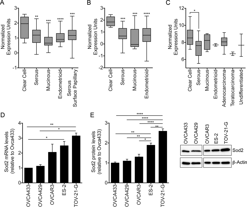
Increased Sod2 mRNA expression is observed in ovarian clear cell carcinomas (OCCC) compared to other ovarian carcinoma histological subtypes. Two representative data sets from ovarian cancer micro-array studies are displayed as box and whisker plots (data obtained through oncomine.org). A. Clear cell (n=16), Serous (n=24), Mucinous (n=8), Endometrioid (n=29), Serous Surface Papillary (n=107); One-way ANOVA (p<0.0002), Tukey’s post-test **** p<0.0001, ***p<0.001, **p<0.01 (GSE2109). B. Clear cell (n=8), Serous (n=41), Mucinous (n=13), Endometrioid (n=37),; One-way ANOVA (p<0.0001), Tukey’s post-test ***p<0.001 (GSE6008). C, Sod2 mRNA expression was significantly increased in a panel of OCCC lines, listed in Methods section, compared to serous adenocarcinoma cell lines (Affymetrix array; ANOVA Tukey’s post-test *p<0.05)
D, Semi-quantitative real-time RT-PCR was performed to assess Sod2 mRNA expression in serous ovarian cancer cell lines OVCA433, OVCA429 and OVCAR3 and OCCC cell lines ES-2 and TOV-21-G (data expressed relative to OVCA433, which displayed lowest Sod2 expression). E, Immunoblot analysis and densitometric quantification of Sod2 protein expression in ovarian cancer cell lines (For D & E: Mean ± SEM, n=3, ANOVA, Tukey’s post-test **** p<0.0001, ***p<0.001, **p<0.01, * p<0.05).
Cell lines and cell culture conditions
At commencement of this study ES-2 and TOV-21-G cells were newly obtained from American Type Culture Collection (ATCC, Manassas, VA). Authenticity was verified by ATCC using STR analysis. ES-2 cells were maintained in McCoy’s 5A media + 10% FBS and TOV-21-G cells in 40% Media199/40% MCBD supplemented with 20% FBS and sodium bicarbonate. Cells were maintained at 37°C with 5% CO2.
Sod2 knockdown using RNA interference
Scramble non-targeting control and Sod2-specific siRNA oligonuecliotides were synthesized by Life Technologies/Dharmacon. 5’- CAACAGGCCUUAUUCCACU-3’ and 5’- AAGUAAACCACGAUCGUUA-3’ sequences were used as siSod2_#1 and siSod2_#2 respectively (Supplementary Fig.S2) and 10pmol transfected into cells using lipofectamine RNAiMax (Invitrogen). Short hairpin RNA (shRNA) with non-targeting scramble sequence or targeting Sod2 (shSod2_#1: 5’-CTGACGGCTGCATCTGTTGGTGTCCAAGG-3’, and shSod2_#2: 5’-ACCTGAACGTCACCGAGGAGAAGTACCAG-3’) in pGFP-V-RS vector (Origene; TG309190) were used to stably transfect ES-2 cells (Fig. 2; Supplementary Fig.S3). The clone expressing shSod2_#1 was used in Figures 2–7.
Figure 2.
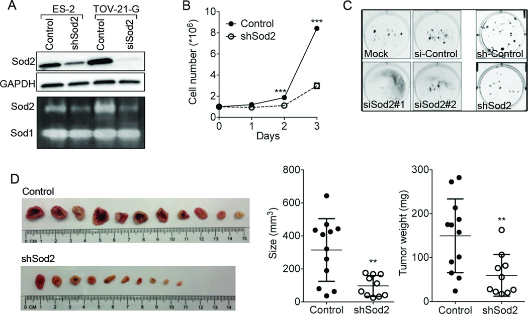
Inhibition of Sod2 expression abrogates OCCC tumorigenicity A, Sod2 expression and activity were reduced using RNA interference in OCCC cell lines ES-2 (shSod2_#1) and TOV-21-G (siRNASod2_#1). Sod2 expression was analyzed using western blotting (upper panels) and activity determined using in-gel zymography (lower panel). B, Cell proliferation rate was decreased with reduced Sod2 expression (shSod2_#1), analyzed by cell counting using trypan blue exclusion assay (mean ± SEM. ANOVA Tukey’s post-test; ****p<0.0001). C, Sod2 RNA interference decreases single cell survival of ES-2 cells in clonogenicity assays. D, Reduced Sod2 levels significantly inhibited the tumor size and weight of ES-2 tumors grown in the ex ovo CAM model. Fertilized chicken eggs were incubated at 37 °C for 7 days after inoculating the cells (shSod2_#1 or scramble transfected ES-2) onto the chorioallantoic membrane, after which tumors were collected and measured (control, n=12; shSod2, n=10; mean ± SD. Student t-test; **p < 0.01).
Figure 7.
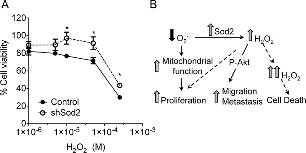
Exogenous H2O2 treatment significantly decreases cell viability of cells with higher steady state H2O2 levels A, ES-2 scramble control cells show significantly lower cell viability than shSod2 ES-2 cells using crystal violet assays, in response to increasing concentrations of H2O2 for 72 h. Each data point represents an average of 6 replicates ± SEM. Tukey’s post-test *p<0.05. B, Proposed mechanisms of Sod2-mediated OCCC tumorigenesis and metastasis. OCCC has high Sod2 expression, which provides efficient superoxide scavenging and maintenance of high mitochondrial function to drive increased cell proliferation. In addition, Sod2 shifts the intracellular ROS balance from O2·− to H2O2, which drives tumor cell migration and metastasis. This concomitantly enhances the intracellular H2O2 threshold enabling more rapid killing by exogenous H2O2.
Immunoblotting
Protein expression was analyzed by standard western blotting using antibodies Antibodies were from Cell Signaling Technology (Boston, MA; pAkt-s473, Akt, pFAK-Y397, FAK, p-p130cas-Y165, p130cas) or Abcam (Cambridge, MA; Sod2). Primary Antibodies were diluted in blocking solution (5% nonfat milk in TBS with 0.1% tween 20, 1:1000), and incubated overnight at 4 °C. Blots were visualized using Femto and Pico ECL chemiluminescence substrate (Thermo scientific, Rockford, IL) and imaged using a ChemiDoc MP system (BioRad). Densitometric analysis was performed using ImageJ software (NIH). Each protein band was normalized to the respective GAPDH or β-Actin loading control band.
Sod2 Zymography
Sod2 activity was analyzed using Sod2 in-gel zymography as previously described (12). Briefly, cell lysates were loaded on non-denaturing acrylamide gels, followed by electrophoresis. Sod2 activity is visualized by the inhibition of nitroblue tetrazolium reduction.
Clonogenicity & cell viability
Single cell survival clonogenicity assays were performed as previously described (13). Briefly, 100 cells were plated in each well of a 6 well plate colonies visualized after 10 days using crystal violet. Viability was assessed by cell counting using trypan blue (1%) staining or crystal violet uptake assays (13).
Chorioallantoic Membrane Assay (CAM)
Each CAM was inoculated with 5× 10⁵ ES-2 cells stably expressing either scramble-shRNA-GFP or Sod2-shRNA_#1-GFP that were suspended in 50 µl PBS (with 1 mM MgCl2, 0.5 mM CaCl2, 100 U/mL penicillin, and 100 µg/mL streptomycin), essentially as previously described (14). Tumors were allowed to form for 7 days prior to termination of the experiments by sacrificing the chick embryo. Tumors on the CAM were removed and measured. Chorioallantoic membrane and chick embryo organs (liver and lung) were collected for tumor metastasis analysis by surveying for GFP-labeled cells.
Seahorse XF24 extracellular flux analysis
Oxygen consumption rate (OCR), extracellular acidification rate (ECAR) and mitochondria stress tests were measured using the Seahorse XF243 Extracellular Flux Analyzer (Seahorse Bioscience; Billerica, MA), as described previously (13). Cells were plated at a density of 40,000 cells/well and media replaced with XF media the following day 1hr prior to the assay. Three measurements of OCR and ECAR were taken at baseline and after each injection of the following mitochondrial stress test compounds: oligomycin (1µM; complex V inhibitor); FCCP (0.75µM; proton gradient uncoupler); antimycin A (1µM; complex III inhibitor). Basal and maximal respiration were normalized by subtracting non-mitochondrial OCR (i.e. after Antimycin A addition). Respiratory reserve capacity was calculated as the difference between maximal and basal OCR. ATP-linked OCR was derived as the difference between basal and Oligomycin A inhibited OCR. Data was normalized to total protein content in each well.
Wound healing assay
Cell migration was assessed in serum free media by wound healing assays using Ibidi inserts (Martisried, Germany) and quantified after 72hrs. Ibdi inserts were removed from a monolayer of GFP-labeled cells to expose the cell-free wound area. Fluorescence images were taken after 72 hrs of migration and overlayed with corresponding images at time 0hr. Pixels representing GFP-labeled cells were quantified within the wound area using Image J and corrected by subtracting any GFP-detected cells in the same area at time 0.
Spheroid attachment assay
Cells were plated at a density of 1000 cells per well in ultra-low attachment 96 well plates (Corning) and incubated for 5 days. Spheroids were transferred to 24 well plates with or without Collagen I coating. Percentage outgrowth was calculated by subtracting the area covered by migrating cells onto the collagen matrix from the area of the spheroid at time 0h.
Live cell imaging of MitoSox oxidation
As an indicator of mitochondrial O2•− the oxidation and fluorescence of the redox-sensitive MitoSox Red dye (Life Technologies) was monitored by live cell imaging according to manufacture’s instructions. Cells were imaged to detect oxidation and fluorescence of MitoSox using a Leica SP5 II AOBS confocal microscope following incubation with dye in HBSS for 30 min at 37°C. The Manganese Porphyrin O2•− scavenger ortho tetrakis(N-n-butoxyethylpyridinium-2-yl) porphyrin (MnTnBuOE-2-PyP5+) was generously provided by Dr. Ines Batinic Haberle (Duke University).
Measurement of cellular H2O2 using catalase activity assay
Concentration of cellular H2O2 was determined by measuring the rate of inactivation of catalase with amino1,2,4-triazol (ATZ), according to Yusa et al. (15). ATZ irreversibly inactivates catalase by covalently binding with intermediate compound I, formed following oxidation of catalase by H2O2. Briefly, cells were treated with 20 mM ATZ for different time intervals (15 and 30 min). Cells were washed with PBS and protein lysates were prepared in 50 mM phosphate buffer (pH=7.4) with protease inhibitors. Decomposition of H2O2 by catalase in protein lysates was analyzed using ultraviolet spectroscopy at 240nm wavelength. H2O2 concentration was determined using the equation [H2O2] = k/k1, where k is the empirically-determined pseudo first order rate constant of catalase inactivation in the cells by ATZ (Figure 6A), while k1 is the rate of compound I formation (1.7 × 10⁷ M−1 s−1).
Figure 6.
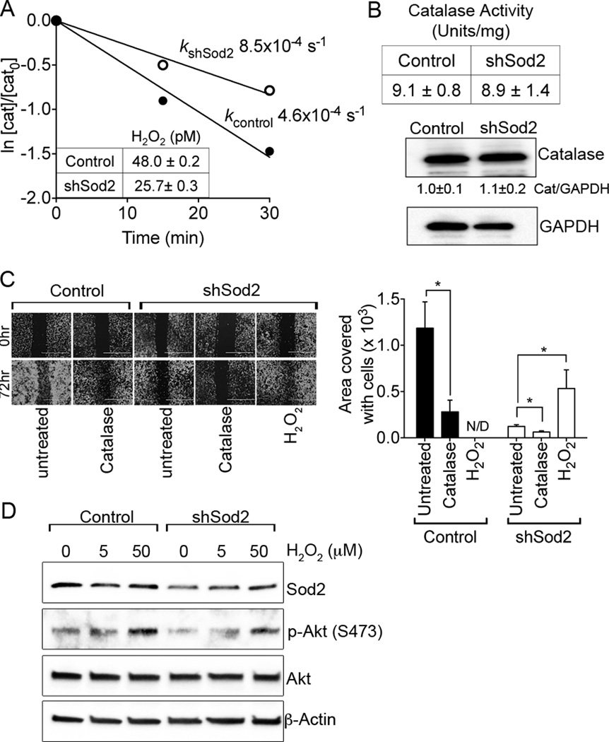
H2O2 contributes to OCCC migration. A, Steady-state H2O2 levels decrease with reduced Sod2 expression. Steady-state H2O2 levels were derived in ES-2 cells stably transfected with either scramble shRNA Control (closed circles) or shRNA targeting Sod2 (open circles), using aminotriazole inhibition of catalase kinetics assays, as described in methods. First order decay curves for catalase inhibition by aminotrizaole were used to determine rate constants of inactivation (k; one representative decay curve from 3 replicate experiments shown). H2O2 concentration were determined using the equation [H2O2] = k/k1, where k1 is the rate of catalase compound I formation (1.7 × 10⁷ M−1 s−1). H2O2 data represents an average of three experiments ± SD. B. There was no change in catalase activity and catalase protein expression between control and shRNA-Sod2 cells. C, H2O2 scavenging by catalase abrogates migration of ES-2 cells, while H2O2 can stimulate migration in cells with low Sod2 expression. Migration was analyzed by wound healing assays using ibidi inserts and the area covered by cells quantified following 72 hrs of migration (normalized to day 0). Scavenging of endogenous H2O2 by catalase significantly reduces the migration of ES-2 scramble-transfected control cells. Recombinant catalase protein (500 units/mL) was added in serum free media at time 0 and left on cells for 72hrs. Conversely, migration of shSod2 cells was significantly increased with treatment of 5 µM H2O2, while this level of H2O2 was toxic to control cells, and effects on migration could therefore not be determined (N/D). Data represents one of two independent cellular migration experiments (n=5, mean ± SEM. ANOVA Tukey’s post-test *p<0.05). D. H2O2 can stimulate Akt signaling in OCCC. Dose dependent increases in phospho-Akt S473 levels were observed by western blotting following 2hrs of H2O2 treatment with indicated doses in serum free media.
Statistical analysis
All data presented are representative of at least three independent experiments and expressed as mean ± SEM, unless otherwise stated. Statistical data analysis (ANOVA with Tukey’s post-test or student t-test) was performed using GraphPad Prism Software v6. p < 0.05 was considered to be significant.
Results
Sod2 expression is significantly increased in OCCC compared to other EOC histological subtypes
Mining publicly available expression data (Oncomine.org) revealed that Sod2 mRNA levels are elevated in OCCC compared to other ovarian cancer histological subtypes (Supplementary Fig. S1). Two representative data sets displayed in Figure 1A and B show that levels of this antioxidant enzyme are statistically higher than in any other ovarian cancer subtype. Similarly, significantly higher Sod2 mRNA expression was observed by microarray analysis in a panel of OCCC cell lines when compared to cell lines of high grade serous adenocaricnoma origin (Fig. 1C). This was further verified by assessing Sod2 expression in cultured ovarian cancer cell lines, using semi quantitative real-time RT-PCR and western blotting (Fig. 1D and E).
Reduced Sod2 levels significantly inhibit OCCC cell proliferation in vitro and tumor formation in the CAM model
To further investigate the role of Sod2 we utilized two OCCC cell lines, ES-2 and TOV-21-G. Sod2 expression was inhibited by shRNA and siRNA transfection, which was demonstrated to lead to a concomitant decrease in Sod2 enzyme activity (Fig. 2A, Supplementary Fig. S2 & S3A). Following Sod2 expression knockdown, ES-2 cell proliferation rate (Fig. 2B, Supplementary Fig. S3B) and clonogenicity (Fig. 2C, Supplemental Fig. 3SC) were significantly attenuated. This appeared to be Sod2 concentration dependent. A 30% reduction in Sod2 levels mediated by stable shRNA transfection reduced clonogenicity by approximately 50% (shSod2_#1), while a 70% Sod2 decrease almost completely abrogated the cells ability to survive in this assay (shSod2_#2; Supplemental Fig. S3C). Analysis of PARP cleavage and Annexin V staining suggested that the decrease in cell viability observed in cells with 30% Sod2 knock-down is not related to a significant increase in apoptosis (Supplemental Fig. S4). This cell line, referred to as shSod2 in subsequent Figures, was chosen for subsequent studies to achieve pathophysiologically relevant changes in Sod2 expression (rather than complete loss), which also closely reflects Sod2 levels observed in non-OCCC cell lines OVCA433 and OVCA429 (Fig. 1E). The CAM ex ovo model was used to further test the role of Sod2 on ES-2 tumorigenicity. A significant decrease in tumor size and weight was observed in tumors grown from ES-2 cells with reduced Sod2 expression (shSod2; Fig. 2D). In addition, the shRNA-Sod2 tumors exhibited less vascularization compared to the control groups (Fig. 2D), suggesting that Sod2 may contribute to both proliferation and the recruitment of blood vessels to the tumor.
Sod2 maintains OCCC mitochondrial respiration
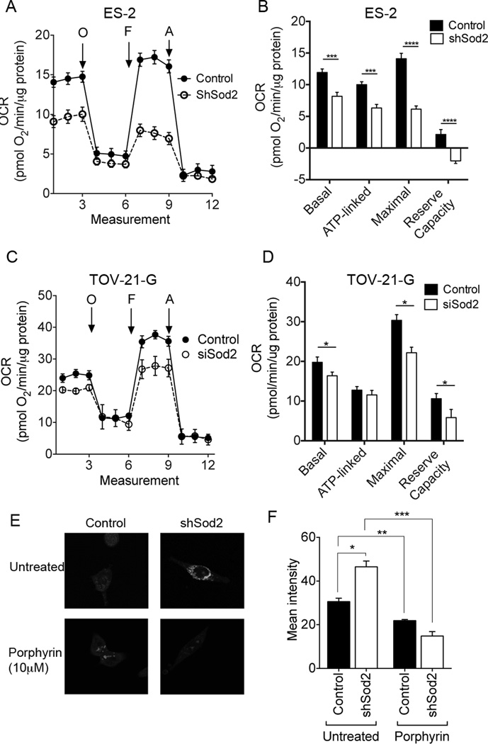
We previously demonstrated that OCCC cell lines are highly energetic, and depend on both oxidative phosphorylation and glycolysis for their energy needs (13). Given that Sod2 has a primary role in protecting mitochondria from excess O2•−, the effect of Sod2 knock-down on mitochondrial respiration was assessed. Oxygen consumption rate (OCR), representing mitochondrial oxidative phosphorylation, and extracellular acidification rate (ECAR), correlating with glycolytic activity, was measured using extracellular flux analysis in both ES-2 and TOV-21-G OCCC cell lines following Sod2 knock-down (13,16). Stable shRNA-Sod2 decreases in ES-2 and transient siRNA-mediated knock-down of Sod2 in TOV-21-G cells resulted in significant reduction in basal OCR compared to control scramble RNAi transfected cells (Fig. 3A–D, Supplementary Fig. S5A). Further, respiratory reserve capacity, a measure of the cells’ ability to enhance respiration in response to physiological cues and stress, was significantly inhibited with reduced Sod2 levels (Fig. 3B & D), suggesting that Sod2 plays a major role in maintaining mitochondrial health to support maximal respiration. Although slight, a consistent negative effect on OCR was observed in stable shRNA-Sod2 cells following FCCP treatment (Fig. 3B). A wide range of FCCP concentrations were tested on these cells, but none were able to enhance OCR with Sod2 loss. FCCP can be inhibitory at high concentrations and it has been speculated that this may be due to a loss in the ability of mitochondria to accumulate respiratory substrates (16). While not tested here, it is possible that sustained Sod2 expression decreases, and concomitant increases in mitochondrial O2•− levels, may exacerbate this FCCP-depended OCR inhibition, thereby influencing mitochondrial membrane integrity and substrate transport. No significant increases in ECAR were observed between control and sh- or si-RNA transfected cells, suggesting that decreases in Sod2 expression do not influence a compensatory shift towards glycolysis (Supplementary Fig. S5B–D). The above observations show that both si- and shRNA mediated decreases in Sod2 reduced basal OCR and respiratory reserve capacity, indicating that Sod2 is important in maintaining mitochondrial respiration in OCCC.
Figure 3.
Sod2 preserves OCCC mitochondrial respiration A, Reduced Sod2 levels significantly attenuated the mitochondrial oxidative phosphorylation in stably transfected ES-2 cells (shSod2_#1). Oxygen Consumption Rate (OCR) was measured using extracellular flux analysis and pharmacological manipulation of mitochondrial activity used to interrogate bioenergetics parameters. Oligomycin A (O) was used to derive ATP-linked OCR, FCCP (F) to stimulate maximal OCR, and Antimycin A (A) to inhibit all mitochondrial OCR. B, Basal oxygen consumption rate (OCR), ATP linked OCR, maximum OCR and respiratory reserve capacity (Max OCR- basal OCR), were significantly decreased with stable shRNA-mediated Sod2 knock down in ES-2 (one representative of three experiments is shown, mean ± SEM, control n=8, shSod2 n=9. ANOVA, Tukey’s post-test ***p<0.001, **** p<0.0001). C, Transiently transfected siRNA targeting Sod2 in TOV-21-G cells showed significantly reduced mitochondrial oxidative phosphorylation (siRNA construct #1). D, Basal OCR, maximum OCR and respiratory reserve capacity (Max OCR- basal OCR), were significantly decreased with Sod2 knock down in TOV-21-G cells. (one representative of three experiments is shown, mean ± SEM, control n=4, siSod2 n=5, ANOVA Tukey’s post-test *p<0.05). E, Superoxide-mediated oxidation of MitoSox redox-sensitive dye was measured in live cells. Increased MitoSox fluorescence was observed in ES-2 cells stably transfected with shRNA-Sod2_#1 compared to scramble-transected control cells, suggesting that the amount of mitochondrial superoxide accumulation is higher in cells with reduced Sod2 expression. MitoSox florescence was abrogated by addition of 10 µM of the porphyrin superoxide scavenger ortho tetrakis(N-n-butoxyethylpyridinium-2-yl) porphyrin (MnTnBuOE-2-PyP5+) (a representative of three experiments is shown, mean ± SEM, n=4, ANOVA, ANOVA Tukey’s post-test *p<0.05, **p < 0.01, ***p < 0.001).
To assess whether a decrease in Sod2 expression result in compromised O2•− scavenging, which may be one of the causes of compromised mitochondrial function, the presence of mitochondrial O2•− was evaluated using the mitochondria-targeted redox-sensitive dye MitoSox. As expected, increased oxidation and consequential enhanced fluorescence of MitoSox were observed in the Sod2 knockdown cells compared to controls (Fig. 3E & F). Addition of the Sod2-mimetic porphyrin (MnTnBuOE-2-PyP5+), which acts as a O2•− scavenger, reduced MitoSox oxidation and in both control and shRNA-Sod2 groups (Fig. 3E & F).
Sod2 knockdown attenuates metastasis of cancer cells in the CAM model
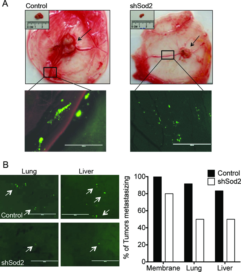
We have previously demonstrated that enhanced Sod2 expression is implicated with metastatic progression (7,8,17). To investigate the role of Sod2 during OCCC metastasis, the appearance of metastatic lesions of GFP-labeled ES-2 cells were investigated in the CAM tumor model. Single cells and micrometer-sized cellular clusters were highly abundant throughout the membrane in the control group, which could be observed 2–3 cm from the tumor (Fig. 4A). In contrast, metastatic spread from shRNA-Sod2 knockdown tumors was limited to the appearance of single cells in the membrane confined to an approximate 1–1.5 cm radius from the tumor (Fig. 4A). Further, lung metastases in the chick embryo were observed in 11 of the 12 controls compared to only 5 of 10 embryo’s in the shRNA-Sod2 group (Fig. 4B). Further, 10 of 12 control tumors metastasized into the liver, whereas only 4 of10 liver metastases were observed in the shRNA-Sod2 group (Fig. 4B). While clusters of five or more cells were found in the lungs and livers of control groups, only single cells were detected in the Sod2 knockdown group (Fig. 4B).
Figure 4.
Metastatic spread of ES-2 cells is attenuated with reduced Sod2 expression in the ex ovo CAM tumor model. A, Representative images show the location of the primary tumors and micro-metastasis of GFP-labeled cells in the membrane (scale bar = 1mm, CAM tumor study carried out as in Fig. 2). B, Reduced Sod2 expression inhibited tumor metastasis into the chick embryo and CAM (shSod2_#1). Quantification of percentage metastasis of tumor cells into CAM, liver and lung of chick embryo. Representative images show the percent of tumors with metastatic cancer cells present in the chicken embryo liver and lung (control, n=12; shSod2, n=10).
Sod2 levels modulate cell migration and tumor spheroid outgrowth
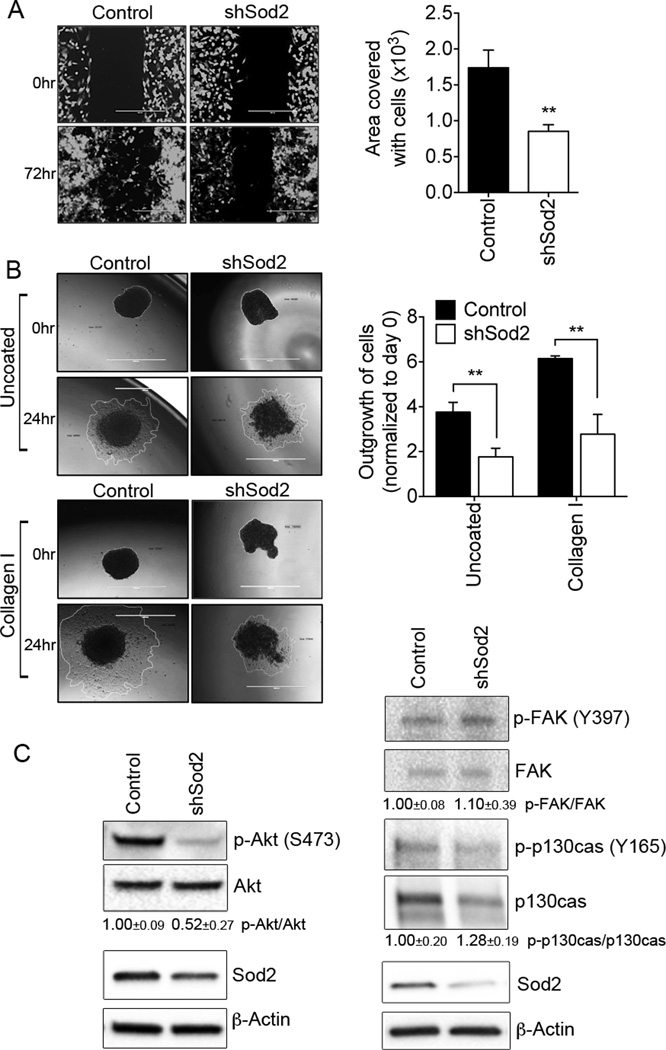
Due to the significant abrogation of metastatic spread in response to Sod2 expression decreases, the role of Sod2 on cell migration was further investigated. Cell migration, assessed by wound healing assays, was significantly inhibited with reduced Sod2 expression in ES-2 cells (Fig. 5A; Supplementary Fig. S6). Further, the ability of cellular spheroid clusters to attach and cells to migrate from the spheroid onto collagen-I and un-coated surfaces was also compromised with reduced Sod2 expression (Fig 5B). Anchorage independent spheroid formation is a commonly observed phenotype of ovarian cancer cells metastasizing via the transcoelomic route through the IP cavity and these have been shown the ability to attach on the peritoneum to form metastatic lesions. These data suggest that Sod2 plays an important role in tumor spheroid metastasis (Fig. 5B). While spheroids of equal size were chosen for this assay, it should be noted that Sod2 knock-down also decreased ES-2 spheroid growth in anchorage independence (data not shown).
Figure 5.
A, Cell migration is significantly inhibited with Sod2 knockdown. A. Migration was analyzed by wound healing assays using ibidi inserts and the area covered by cells quantified following 72 hrs of migration (normalized to day 0). Data from one representative experiment is shown (n=6, mean ± SEM, t-test; **p < 0.01, scale bar = 500µm). B, Representative images of attachment and outgrowth of ES-2 spheroids on uncoated and collagen-I coated surfaces (scale bar = 1mm). Reduced Sod2 levels significantly inhibited the outgrowth of spheroids in both uncoated and collagen-I coated surfaces. Data from one representative experiment is shown (n=4, mean ± SEM, ANOVA, Tukey’s post-test; **p < 0.01). C. Akt-phosphorylation is dependent on Sod2 expression. Protein expression of phosphorylated and total levels of Akt, p130cas, and FAK were analyzed by western blotting. Data from one representative experiment is shown. Levels of phosphorylated proteins were quantified by densitometry and normalized to total levels (n=3, mean ± SEM).
To gain mechanistic insights into the signaling pathways that may be altered by Sod2-mediated metastasis, phosphorylation profiles of Akt, p130cas and focal adhesion Kinase (FAK) were investigated. These were chosen based on previous observations of their redox regulation and involvement in tumor cell migration (8,9,18,19). shRNA-Sod2 cells exhibited a 50% decrease in phospho-Akt levels compared to scramble control cells, while there was no appreciable change observed in phosphorylation of FAK or the focal adhesion adapter protein p130cas (Fig. 5C). The effects on Akt phosphorylation were also Sod2 concentration dependent, where cells with lower Sod2 expression demonstrated a more striking decrease in Akt phosphorylation (Supplementary Fig S6). These data suggest that Akt signaling may be important in driving Sod2-mediated tumrigenicity and metastasis of OCCC.
OCCC cell migration is H2O2 dependent
Sod2 is the primary enzyme involved in converting O2•− to H2O2 within the mitochondria. While it serves as a protective mechanism to maintain mitochondrial function (Fig. 3) by removal of damaging O2•−, a shift towards increasing levels of H2O2 has also been observed in response to enhanced Sod2 expression (8,10,18,20,21). Due to its relative stability and ease in traversing cellular membranes, H2O2 can mediate redox signaling, including events that drive migration (9). To test if Sod2 changes the steady-state H2O2 levels in OCCC, we assessed intracellular H2O2 status in control and shRNA-Sod2 ES-2 cells using a biochemical assay based on the irreversible inhibition of catalase by aminotriazole (15,17). Steady-state levels of H2O2 were reduced approximately 2-fold in Sod2 knockdown cells compared to controls (Fig. 6A), while baseline catalase activity and protein expression were comparable (Fig. 6B).
In order to test whether OCCC cell migration is H2O2 depended, wound-healing assays were carried in the presence of catalase, which catalyzes the conversion of H2O2 to H2O and O2. As previously demonstrated, exogenous application of recombinant catalase resulted in accumulation of catalase within ES-2 cells (Supplementary Figure S7) (9). Catalase significantly reduced the migration of both control and shRNA-Sod2 cells, suggesting that H2O2 is a promoter of ES-2 cell migration (Fig. 6C). Conversely, treatment with low levels of H2O2 (5 µM) significantly reversed the slow migration of shRNA-Sod2 cells (Fig. 6C). Further, 5 and 50 µM H2O2 treatment was able to increase Phospho-Akt levels (Fig. 6D), while catalase expression abrogated Akt phosphorylation (Supplementary Fig. S8), suggesting that this may be an important redox-dependent signaling pathway in OCCC.
It was noted that ES-2 control cells were not able to tolerate long-term exposure to low dose H2O2 during migration assays (Fig. 6C). To examine this further, cell viability was assessed in response to H2O2. A significant reduction in cell survival in response to H2O2 was observed in the control group compared to cells with decreased Sod2 expression (Fig. 7A). This suggests that a higher intracellular steady-state H2O2 milieu in OCCC predisposes cells to enhanced killing by additional exposure to low level exogenous H2O2. The above data imply that high Sod2 expression provides several advantages to OCCC, by protecting mitochondrial function through scavenging of O2•− and driving H2O2 dependent migration. While these attributes are advantageous for OCCC survival and metastatic progression, an enhanced intracellular steady-state H2O2 level presents a double-edged sword, as these cells are consequentially more susceptible to H2O2 toxicity (Fig. 7B).
Discussion
Although the five different EOC histological subtypes share the same primary tumor location on the ovaries, it is now evident that these are distinct diseases with vastly different tissue origins, and genetic and epigenetic profiles (2,11). In the present study, we show that Sod2 is highly expressed in OCCC compared to other EOC histological subtypes, and that this mitochondrial antioxidant plays a significant role in OCCC tumorigenicity and metastasis.
Intracellular ROS are maintained within a narrow range, tightly regulated by the balance of the rate of ROS production and ROS scavenging/detoxifying by antioxidant enzymes. This balance is often disrupted in the context of cancer, due to high ROS production as a consequence of changes in metabolism or the tumor environment (e.g. hypoxia), and the resulting changes in antioxidant expression. Since the mitochondrial respiratory chain is the major site of O2•− generation within cells, Sod2 plays an important role in maintaining cellular ROS balance. Based on the above findings Sod2 appears to play a dual role in enhancing OCCC tumorigenicity; first by protecting cells from mitochondrial O2•− damage; and second, by shifting the steady-state ROS balance towards H2O2.
We recently demonstrated that a distinguishing feature of OCCC is their unique metabolic phenotype. Compared to serous adenocarcinoma cells, OCCC cell lines were significantly more energetic, displaying both very high levels of mitochondrial oxidative phosphorylation and glycolytic flux (13). Our data suggest that Sod2 is intricately involved in maintaining this high rate of oxygen consumption, potentially by preserving mitochondrial function as a consequence of O2•− scavenging (Fig. 3). By preventing damage mediated by O2•− or secondary products of mitochondrial electron transport chain complexes, Sod2 likely supports the high rate of OCCC proliferation, clonogenicity and tumor growth. The present study suggests that inhibiting mitochondrial antioxidant defenses may provide and alternate strategy to therapeutically target OCCC.
In addition to scavenging O2•− and maintaining mitochondrial health for optimal cell proliferation, we believe that Sod2 has an additional role in promoting the aggressiveness of OCCC, by shifting steady-state H2O2 levels and driving pro-metastatic behavior (Fig. 4–6). It has been previously shown that high expression of Sod2 is associated with metastatic progression (8,10,17,20–23), and that dependence of cancer cell migration is related to cellular H2O2 production (9,19,24,25). For example, we have shown that steady-state increases in H2O2 can lead to induction of the focal adhesion kinase pathway and migration of metastatic bladder cancer cells and cells with enforced Sod2 expression (9,19). This was mediated by oxidation-dependent inhibition of the phosphatase PTPN12, leading to enhanced phosphorylation of p130cas and Rac1 activation. In addition, work from the Melendez’ group has shown that Sod2 expression significantly contributes to the expression of the matrix-degrading enzyme MMP-1 in a H2O2-dependent manner (21), and that the Sod2/H2O2-dependent inhibition of the dual lipid protein tyrosine phosphatase PTEN enhances Akt/GSK3β/VEGF-dependent angiogenesis (18), both processes contributing significantly to metastasis. Our present data suggest that Sod2 may similarly contribute to metastatic progression of OCCC by activating Akt signaling (Fig 5 and 6). In addition to its pro-survival function, Akt has been shown to influence metastasis and cell migration by regulating cytoskeletal rearrangement, pro-metastatic cell signaling and gene transcription (26). These results are of specific importance to OCCC, which, unlike other ovarian cancer histological subtypes, has been characterized by high frequency Akt pathway activation. 70% of early and 68% of late stage OCCC cases have been shown to display phospho-Akt (S473) staining (27). About 38% of OCCC cases show PTEN loss (28) and 40% of cases PI3K activating mutations (29). Our data imply that Sod2-dependent Akt phosphorylation may also contribute to high activation of Akt signaling in OCCC. Since Akt-phosphorylation was highly susceptible to H2O2 treatment, it suggests that this signaling pathway is redox regulated in OCCC, with a plausible mechanism for this being the oxidation of PTEN (18).
Although an increase in Sod should theoretically not result in higher levels of H2O2 production based on the kinetic properties of Sod2 (30), a number of studies have demonstrated increases in H2O2 levels that correlate with Sod2 expression (18,31–33). While the reason for this observation in OCCC has not been investigated, there are plausible explanations for this increase in steady-state H2O2 as a consequence of Sod expression. These primarily relate to changes in the reaction rates within the mitochondrial electron transport chain (ETC). For example, it has been proposed that Sod2 in the mitochondria may alter the flux of O2·− from some quinone/semiquinone/hydroquinone triads, such as coenzyme Q, thereby driving the reaction into the direction of O2·− production, potentially leading to enhanced localized dismutation to H2O2 by Sod2 (31,34). Alternatively, inhibition of cytochrome c oxidase by nitric oxide, arising as a consequence of Sod2 expression may influence the reduction state of the ETC and drive O2·− and H2O2 production (35). Our observation that Sod2 knock-down also decreases H2O2 levels, suggests that Sod2 is involved in regulating H2O2 balance within cells and this may contribute to H2O2-mediated redox signaling.
While Sod2 appears to contribute to H2O2-mediated metastatic progression, an enhanced steady-state H2O2 milieu may also present a disadvantage to OCCC cells. Our data suggest that cells with high Sod2 levels and concomitant increases in intracellular H2O2 are more susceptible to exogenous sources of redox stress (Fig. 7A). This likely puts cells closer to the cytotoxic threshold of H2O2, which is reached once cells are further challenged by exogenous ROS. Interestingly, OCCC do not appear to have enhanced expression of Catalase to provide additional scavenging of excess H2O2 (Fig. 6A). While sub-lethal levels of H2O2 have been shown to contribute to redox signaling, high levels of H2O2 can elicit tumor cell death by a number of pathways, including apoptosis, protein/DNA damage and mitochondrial dysfunction (36–38). Further, it was recently reported that H2O2 exposure of tumor cells with enforced Sod2 expression can result in Sod2 peroxidase activity, leading to mitochondrial damage and dysfunction (39). An increased H2O2 steady-state has been observed in a number of cancer cells (17,40–42) and lends credence to the idea that this higher H2O2 threshold may be exploited therapeutically. In this regard, the use of high-dose ascorbic acid, which is oxidized within tumor cells to produce H2O2, has recently been revisited for use in cancer treatment (43–45) and has shown promise in early clinical trials in advanced stage cancers (46,47). Ascorbate and concomitant H2O2 – mediated DNA damage and apoptosis were shown to enhance ovarian cancer cell death and increased chemosensitivity (48). While that study was not focused on OCCC, this type of treatment may be of particular benefit to this histological subtype given the high expression of Sod2. It is important to highlight that cancer cells with enhanced Sod2 expression may respond differently to ROS-producing agents, depending on both the type and cellular location of the ROS/reactive nitrogen species generated. For instance, Sod2 may enhance scavenging of O2•−, and therefore provide chemoresistance benefits to the tumor cells in response to these ROS. Conversely, while an increase in steady-state H2O2 facilitates redox-signaling beneficial to the cancer cells, this higher threshold may facilitate H2O2-mediated OCCC cell death in response to further insult by exogenous sources of H2O2. Understanding the complex interaction of antioxidants and ROS in OCCC is therefore of importance and may provide novel therapeutic avenues to pursue for this histological subtype of ovarian cancer.
Supplementary Material
Acknowledgments
The ortho tetrakis(N-n-butoxyethylpyridinium-2-yl) porphyrin (MnTnBuOE-2-PyP5+) was generously provided by Dr. Ines Batinic-Haberle (Duke University). We thank Dr. J. Andres Melendez (SUNY Poly) for helpful discussions and Dr. Nate Cady for use of his microscope. We appreciate the help of Mr. Jeffrey Richards for assistance with spheroid assays.
Financial Support: This work was supported by NIH/NCI grant R00CA143229
Footnotes
The authors have no conflicts of interest to declare.
References
- 1.Karst AM, Drapkin R. Ovarian cancer pathogenesis: a model in evolution. Journal of oncology. 2010;2010:932371. doi: 10.1155/2010/932371. [DOI] [PMC free article] [PubMed] [Google Scholar]
- 2.Vaughan S, Coward JI, Bast RC, Jr, Berchuck A, Berek JS, Brenton JD, et al. Rethinking ovarian cancer: recommendations for improving outcomes. Nature reviews Cancer. 2011;11(10):719–725. doi: 10.1038/nrc3144. [DOI] [PMC free article] [PubMed] [Google Scholar]
- 3.Mandai M, Matsumura N, Baba T, Yamaguchi K, Hamanishi J, Konishi I. Ovarian clear cell carcinoma as a stress-responsive cancer: influence of the microenvironment on the carcinogenesis and cancer phenotype. Cancer letters. 2011;310(2):129–133. doi: 10.1016/j.canlet.2011.06.039. [DOI] [PubMed] [Google Scholar]
- 4.Yamaguchi K, Mandai M, Oura T, Matsumura N, Hamanishi J, Baba T, et al. Identification of an ovarian clear cell carcinoma gene signature that reflects inherent disease biology and the carcinogenic processes. Oncogene. 2010;29(12):1741–1752. doi: 10.1038/onc.2009.470. [DOI] [PubMed] [Google Scholar]
- 5.Konstantinopoulos PA, Spentzos D, Fountzilas E, Francoeur N, Sanisetty S, Grammatikos AP, et al. Keap1 mutations and Nrf2 pathway activation in epithelial ovarian cancer. Cancer Res. 2011;71(15):5081–5089. doi: 10.1158/0008-5472.CAN-10-4668. [DOI] [PubMed] [Google Scholar]
- 6.Bravard A, Sabatier L, Hoffschir F, Ricoul M, Luccioni C, Dutrillaux B. SOD2: a new type of tumor-suppressor gene? Int J Cancer. 1992;51(3):476–480. doi: 10.1002/ijc.2910510323. [DOI] [PubMed] [Google Scholar]
- 7.Hempel N, Carrico PM, Melendez JA. Manganese superoxide dismutase (Sod2) and redox-control of signaling events that drive metastasis. Anti-cancer agents in medicinal chemistry. 2011;11(2):191–201. doi: 10.2174/187152011795255911. [DOI] [PMC free article] [PubMed] [Google Scholar]
- 8.Connor KM, Hempel N, Nelson KK, Dabiri G, Gamarra A, Belarmino J, et al. Manganese superoxide dismutase enhances the invasive and migratory activity of tumor cells. Cancer Res. 2007;67(21):10260–10267. doi: 10.1158/0008-5472.CAN-07-1204. [DOI] [PubMed] [Google Scholar]
- 9.Hempel N, Bartling TR, Mian B, Melendez JA. Acquisition of the metastatic phenotype is accompanied by H2O2-dependent activation of the p130Cas signaling complex. Mol Cancer Res. 2013;11(3):303–312. doi: 10.1158/1541-7786.MCR-12-0478. [DOI] [PMC free article] [PubMed] [Google Scholar]
- 10.Liu Z, Li S, Cai Y, Wang A, He Q, Zheng C, et al. Manganese superoxide dismutase induces migration and invasion of tongue squamous cell carcinoma via H2O2-dependent Snail signaling. Free Radic Biol Med. 2012;53(1):44–50. doi: 10.1016/j.freeradbiomed.2012.04.031. [DOI] [PMC free article] [PubMed] [Google Scholar]
- 11.Yamaguchi K, Huang Z, Matsumura N, Mandai M, Okamoto T, Baba T, et al. Epigenetic determinants of ovarian clear cell carcinoma biology. Int J Cancer. 2014;135(3):585–597. doi: 10.1002/ijc.28701. [DOI] [PMC free article] [PubMed] [Google Scholar]
- 12.Beauchamp C, Fridovich I. Superoxide dismutase: improved assays and an assay applicable to acrylamide gels. Anal Biochem. 1971;44(1):276–287. doi: 10.1016/0003-2697(71)90370-8. [DOI] [PubMed] [Google Scholar]
- 13.Dier U, Shin DH, Hemachandra LP, Uusitalo LM, Hempel N. Bioenergetic analysis of ovarian cancer cell lines: profiling of histological subtypes and identification of a mitochondria-defective cell line. PLoS One. 2014;9(5):e98479. doi: 10.1371/journal.pone.0098479. [DOI] [PMC free article] [PubMed] [Google Scholar]
- 14.Deryugina EI, Quigley JP. Chick embryo chorioallantoic membrane model systems to study and visualize human tumor cell metastasis. Histochemistry and cell biology. 2008;130(6):1119–1130. doi: 10.1007/s00418-008-0536-2. [DOI] [PMC free article] [PubMed] [Google Scholar]
- 15.Yusa T, Beckman JS, Crapo JD, Freeman BA. Hyperoxia increases H2O2 production by brain in vivo. J Appl Physiol (1985) 1987;63(1):353–358. doi: 10.1152/jappl.1987.63.1.353. [DOI] [PubMed] [Google Scholar]
- 16.Dranka BP, Hill BG, Darley-Usmar VM. Mitochondrial reserve capacity in endothelial cells: The impact of nitric oxide and reactive oxygen species. Free Radic Biol Med. 2010 Jan 23;482010:905–914. doi: 10.1016/j.freeradbiomed.2010.01.015. [DOI] [PMC free article] [PubMed] [Google Scholar]
- 17.Hempel N, Ye H, Abessi B, Mian B, Melendez JA. Altered redox status accompanies progression to metastatic human bladder cancer. Free Radic Biol Med. 2009;46(1):42–50. doi: 10.1016/j.freeradbiomed.2008.09.020. [DOI] [PMC free article] [PubMed] [Google Scholar]
- 18.Connor KM, Subbaram S, Regan KJ, Nelson KK, Mazurkiewicz JE, Bartholomew PJ, et al. Mitochondrial H2O2 regulates the angiogenic phenotype via PTEN oxidation. J Biol Chem. 2005;280(17):16916–16924. doi: 10.1074/jbc.M410690200. [DOI] [PubMed] [Google Scholar]
- 19.Hempel N, Melendez JA. Intracellular redox status controls membrane localization of pro- and anti-migratory signaling molecules. Redox Biol. 2014;2:245–250. doi: 10.1016/j.redox.2014.01.005. [DOI] [PMC free article] [PubMed] [Google Scholar]
- 20.Ranganathan AC, Nelson KK, Rodriguez AM, Kim KH, Tower GB, Rutter JL, et al. Manganese superoxide dismutase signals matrix metalloproteinase expression via H2O2-dependent ERK1/2 activation. J Biol Chem. 2001;276(17):14264–14270. doi: 10.1074/jbc.M100199200. [DOI] [PubMed] [Google Scholar]
- 21.Nelson KK, Ranganathan AC, Mansouri J, Rodriguez AM, Providence KM, Rutter JL, et al. Elevated sod2 activity augments matrix metalloproteinase expression: evidence for the involvement of endogenous hydrogen peroxide in regulating metastasis. Clin Cancer Res. 2003;9(1):424–432. [PubMed] [Google Scholar]
- 22.Nonaka Y, Iwagaki H, Kimura T, Fuchimoto S, Orita K. Effect of reactive oxygen intermediates on the in vitro invasive capacity of tumor cells and liver metastasis in mice. Int J Cancer. 1993;54(6):983–986. doi: 10.1002/ijc.2910540620. [DOI] [PubMed] [Google Scholar]
- 23.Bur H, Haapasaari KM, Turpeenniemi-Hujanen T, Kuittinen O, Auvinen P, Marin K, et al. Oxidative stress markers and mitochondrial antioxidant enzyme expression are increased in aggressive Hodgkin lymphomas. Histopathology. 2014;65(3):319–327. doi: 10.1111/his.12389. [DOI] [PubMed] [Google Scholar]
- 24.Polytarchou C, Hatziapostolou M, Papadimitriou E. Hydrogen peroxide stimulates proliferation and migration of human prostate cancer cells through activation of activator protein-1 and up-regulation of the heparin affin regulatory peptide gene. J Biol Chem. 2005;280(49):40428–40435. doi: 10.1074/jbc.M505120200. [DOI] [PubMed] [Google Scholar]
- 25.Hurd TR, DeGennaro M, Lehmann R. Redox regulation of cell migration and adhesion. Trends in cell biology. 2012;22(2):107–115. doi: 10.1016/j.tcb.2011.11.002. [DOI] [PMC free article] [PubMed] [Google Scholar]
- 26.Xue G, Hemmings BA. PKB/Akt-dependent regulation of cell motility. Journal of the National Cancer Institute. 2013;105(6):393–404. doi: 10.1093/jnci/djs648. [DOI] [PubMed] [Google Scholar]
- 27.Sasano T, Mabuchi S, Kuroda H, Kawano M, Matsumoto Y, Takahashi R, et al. Preclinical Efficacy for AKT Targeting in Clear Cell Carcinoma of the Ovary. Mol Cancer Res. 2015;13(4):795–806. doi: 10.1158/1541-7786.MCR-14-0314. [DOI] [PMC free article] [PubMed] [Google Scholar]
- 28.Hashiguchi Y, Tsuda H, Inoue T, Berkowitz RS, Mok SC. PTEN expression in clear cell adenocarcinoma of the ovary. Gynecologic oncology. 2006;101(1):71–75. doi: 10.1016/j.ygyno.2005.09.047. [DOI] [PubMed] [Google Scholar]
- 29.Kuo KT, Mao TL, Jones S, Veras E, Ayhan A, Wang TL, et al. Frequent activating mutations of PIK3CA in ovarian clear cell carcinoma. Am J Pathol. 2009;174(5):1597–1601. doi: 10.2353/ajpath.2009.081000. [DOI] [PMC free article] [PubMed] [Google Scholar]
- 30.Liochev SI, Fridovich I. The effects of superoxide dismutase on H2O2 formation. Free Radic Biol Med. 2007;42(10):1465–1469. doi: 10.1016/j.freeradbiomed.2007.02.015. [DOI] [PubMed] [Google Scholar]
- 31.Buettner GR, Ng CF, Wang M, Rodgers VG, Schafer FQ. A new paradigm: manganese superoxide dismutase influences the production of H2O2 in cells and thereby their biological state. Free Radic Biol Med. 2006;41(8):1338–1350. doi: 10.1016/j.freeradbiomed.2006.07.015. [DOI] [PMC free article] [PubMed] [Google Scholar]
- 32.Dasgupta J, Subbaram S, Connor KM, Rodriguez AM, Tirosh O, Beckman JS, et al. Manganese superoxide dismutase protects from TNF-alpha-induced apoptosis by increasing the steady-state production of H2O2. Antioxid Redox Signal. 2006;8(7–8):1295–1305. doi: 10.1089/ars.2006.8.1295. [DOI] [PubMed] [Google Scholar]
- 33.Zhang HJ, Zhao W, Venkataraman S, Robbins ME, Buettner GR, Kregel KC, et al. Activation of matrix metalloproteinase-2 by overexpression of manganese superoxide dismutase in human breast cancer MCF-7 cells involves reactive oxygen species. J Biol Chem. 2002;277(23):20919–20926. doi: 10.1074/jbc.M109801200. [DOI] [PubMed] [Google Scholar]
- 34.Song Y, Buettner GR. Thermodynamic and kinetic considerations for the reaction of semiquinone radicals to form superoxide and hydrogen peroxide. Free Radic Biol Med. 2010;49(6):919–962. doi: 10.1016/j.freeradbiomed.2010.05.009. [DOI] [PMC free article] [PubMed] [Google Scholar]
- 35.Buerk DG, Lamkin-Kennard K, Jaron D. Modeling the influence of superoxide dismutase on superoxide and nitric oxide interactions, including reversible inhibition of oxygen consumption. Free Radic Biol Med. 2003;34(11):1488–1503. doi: 10.1016/s0891-5849(03)00178-3. [DOI] [PubMed] [Google Scholar]
- 36.Hyslop PA, Hinshaw DB, Halsey WA, Jr, Schraufstatter IU, Sauerheber RD, Spragg RG, et al. Mechanisms of oxidant-mediated cell injury. The glycolytic and mitochondrial pathways of ADP phosphorylation are major intracellular targets inactivated by hydrogen peroxide. J Biol Chem. 1988;263(4):1665–1675. [PubMed] [Google Scholar]
- 37.Ahmad KA, Iskandar KB, Hirpara JL, Clement MV, Pervaiz S. Hydrogen peroxide-mediated cytosolic acidification is a signal for mitochondrial translocation of Bax during drug-induced apoptosis of tumor cells. Cancer Res. 2004;64(21):7867–7878. doi: 10.1158/0008-5472.CAN-04-0648. [DOI] [PubMed] [Google Scholar]
- 38.Imlay JA, Chin SM, Linn S. Toxic DNA damage by hydrogen peroxide through the Fenton reaction in vivo and in vitro. Science. 1988;240(4852):640–642. doi: 10.1126/science.2834821. [DOI] [PubMed] [Google Scholar]
- 39.Ansenberger-Fricano K, Ganini D, Mao M, Chatterjee S, Dallas S, Mason RP, et al. The peroxidase activity of mitochondrial superoxide dismutase. Free Radic Biol Med. 2013;54:116–124. doi: 10.1016/j.freeradbiomed.2012.08.573. [DOI] [PMC free article] [PubMed] [Google Scholar]
- 40.Szatrowski TP, Nathan CF. Production of large amounts of hydrogen peroxide by human tumor cells. Cancer Res. 1991;51(3):794–798. [PubMed] [Google Scholar]
- 41.Zieba M, Suwalski M, Kwiatkowska S, Piasecka G, Grzelewska-Rzymowska I, Stolarek R, et al. Comparison of hydrogen peroxide generation and the content of lipid peroxidation products in lung cancer tissue and pulmonary parenchyma. Respiratory medicine. 2000;94(8):800–805. doi: 10.1053/rmed.2000.0825. [DOI] [PubMed] [Google Scholar]
- 42.Lim SD, Sun C, Lambeth JD, Marshall F, Amin M, Chung L, et al. Increased Nox1 and hydrogen peroxide in prostate cancer. Prostate. 2005;62(2):200–207. doi: 10.1002/pros.20137. [DOI] [PubMed] [Google Scholar]
- 43.Chen Q, Espey MG, Krishna MC, Mitchell JB, Corpe CP, Buettner GR, et al. Pharmacologic ascorbic acid concentrations selectively kill cancer cells: action as a pro-drug to deliver hydrogen peroxide to tissues. Proc Natl Acad Sci U S A. 2005;102(38):13604–13609. doi: 10.1073/pnas.0506390102. [DOI] [PMC free article] [PubMed] [Google Scholar]
- 44.Cameron E, Campbell A. The orthomolecular treatment of cancer. II. Clinical trial of high-dose ascorbic acid supplements in advanced human cancer. Chem Biol Interact. 1974;9(4):285–315. doi: 10.1016/0009-2797(74)90019-2. [DOI] [PubMed] [Google Scholar]
- 45.Levine M, Padayatty SJ, Espey MG. Vitamin C: a concentration-function approach yields pharmacology and therapeutic discoveries. Advances in nutrition (Bethesda, Md) 2011;2(2):78–88. doi: 10.3945/an.110.000109. [DOI] [PMC free article] [PubMed] [Google Scholar]
- 46.Hoffer LJ, Levine M, Assouline S, Melnychuk D, Padayatty SJ, Rosadiuk K, et al. Phase I clinical trial of i.v. ascorbic acid in advanced malignancy. Annals of oncology : official journal of the European Society for Medical Oncology / ESMO. 2008;19(11):1969–1974. doi: 10.1093/annonc/mdn377. [DOI] [PubMed] [Google Scholar]
- 47.Monti DA, Mitchell E, Bazzan AJ, Littman S, Zabrecky G, Yeo CJ, et al. Phase I evaluation of intravenous ascorbic acid in combination with gemcitabine and erlotinib in patients with metastatic pancreatic cancer. PLoS One. 2012;7(1):e29794. doi: 10.1371/journal.pone.0029794. [DOI] [PMC free article] [PubMed] [Google Scholar]
- 48.Ma Y, Chapman J, Levine M, Polireddy K, Drisko J, Chen Q. High-dose parenteral ascorbate enhanced chemosensitivity of ovarian cancer and reduced toxicity of chemotherapy. Science translational medicine. 2014;6(222):222ra18. doi: 10.1126/scitranslmed.3007154. [DOI] [PubMed] [Google Scholar]
Associated Data
This section collects any data citations, data availability statements, or supplementary materials included in this article.


