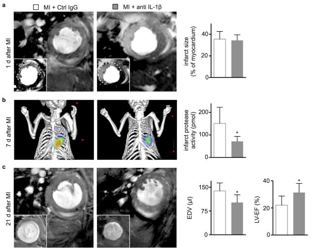
Figure 6.
In vivo imaging of anti-IL-1β treatment effects on infarct protease activity and post-MI remodeling. (a) Evaluation of post-MI remodeling in ApoE−/− mice by cardiac MRI. Each panel shows the short axis of the mid left-ventricular segment at the end of diastole. Infarct size was measured as gadolinium enhanced hypokinetic area on day 1 after MI. (b) Imaging infarct protease activity 7d after MI in ApoE−/− mice by Fluorescence Molecular Tomography-Computed Tomography (FMT/CT) (n = 8 per group). (c) End-diastolic volumes (EDV) and left-ventricular ejection fraction (LV-EF) were examined serially on day 1 and 21 and are displayed as absolute values on day 21 (n = 6 per group, mean ± SD, *p<0.05, Mann-Whitney test).

