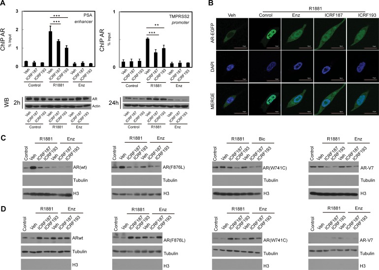
Figure 3. ICRF187 and ICRF193 inhibit AR recruitment to target promoters and AR nuclear localization.
(A) LNCaP cells were cultured in RPMI1640 medium containing 5% CSS and treated with vehicle, 1uM of ICRF187 or 1uM of ICRF193 in addition to vehicle, 10nM of R1881 or 10uM of ENZ treatment for 2 hours. Three independent ChIP experiments were performed using the AR antibody. Precipitated DNA fragment were used as templates to amplify the PSA enhancer and the TMPRSS2 promoter by real-time PCR. Data represented mean ± SEM (n = 3) and plotted as percentage of input. P < 0.01 ** and P < 0.001 as *** (student's t-test). AR protein levels under 2 and 24 hour treatment were detected by Western blotting. (B) LNCaP cells expressing EGFP-AR were cultured in RPMI1640 medium containing 5% CSS. Cells were treated with vehicle, 10nM of R1881, 10nM of R1881 plus 10 μM of ENZ, 10nM of R1881 plus 1uM of ICRF187, or 10nM of R1881 plus 1uM of ICRF193 for 6 hours. Cells were then fixed with 4% paraformaldehyde and mounted with DAPI. Representative confocal microscopic images showed AR localization (Green) and nucleus (Blue). (C–D) 293T cells were transfected with plasmids encoding wild type AR, AR(F876L), AR(W741C) and AR-V7. Cells were treated with vehicle, 1uM of ICRF187 or 1uM of ICRF193 in addition to 10nM of R1881, 10uM of ENZ or 10uM of bicalutamide for 24 hours. Nuclear (C) and cytosol (D) protein extract were immunoblotted with AR, tubulin and Histone H3 antibodies. Three independent experiments were performed and one set of Western blotting images are presented.

