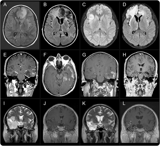
Figure 3. MRI findings in patients with relapsing symptoms post–herpes simplex encephalitis.
Axial fluid-attenuated inversion recovery sequences of patients 1 (A, B) and 2 (C, D) during herpes simplex virus encephalitis (HSE) (A, C) and during relapsing symptoms due to autoimmune encephalitis (B, D). In both cases, there is an interval change due to areas of encephalomalacia, brain atrophy, and white matter changes. Panels E–H correspond to T1 sequences with contrast from patient 3 obtained during HSE (E), a few weeks later during relapsing symptoms due to autoimmune encephalitis (F, G), and after symptom improvement (H). Note that the areas of contrast enhancement during autoimmune encephalitis resolved after symptom improvement. Panels I–L correspond to patient 4 during HSE (I, T2; J, T1 with contrast) and 1 year later (K, T2; L, T1 with contrast). In this patient, the relapsing symptoms post-HSE were not recognized as autoimmune encephalitis for 1 year; during this year, he did not receive immunotherapy and had persistent symptoms and contrast enhancement in MRI.

