SUMMARY
When assessing vitamin D status, measure serum 25-hydroxyvitamin D concentration as this reflects total body vitamin D reserves.
Recent Australasian guidelines outline who should be tested for vitamin D deficiency, who should be treated and when repeat testing should be performed.
A 25-hydroxyvitamin D threshold of at least 50 nanomol/L at the end of winter is a suitable treatment target. Measurement can be repeated after three months of repletion, and thereafter less frequently unless new risk factors for vitamin D deficiency arise.
When interpreting vitamin D pathology reports, practitioners should be aware that some laboratories quote reference limits which are based on overseas rather than Australian guidelines.
Key words: vitamin D tests, vitamin D deficiency, vitamin D supplements, 25-hydroxyvitamin D
Introduction
Vitamin D is an important hormone required for bone and muscle development as well as preservation of musculoskeletal function.1,2 Vitamin D deficiency can be detected by measuring 25-hydroxyvitamin D in serum.
Vitamin D physiology
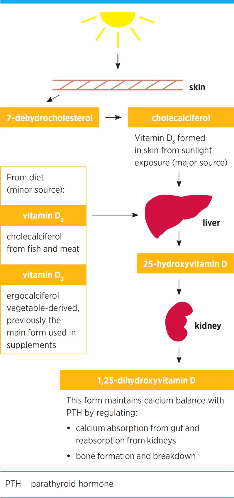
Multiple metabolites of vitamin D are present in the circulation (see Fig.). Vitamin D is synthesised in the skin following ultraviolet B radiation exposure. It can also be obtained from the diet. There are two major circulating forms of vitamin D: 25-hydroxyvitamin D and 1,25-dihydroxyvitamin D. Two steps are involved in the metabolic activation of vitamin D in the body. The second step produces 1,25-dihydroxyvitamin D and occurs in the kidney plus many other body tissues.
Fig.

Vitamin D metabolism
Vitamin D has three main functions:
enhancing intestinal calcium and phosphate absorption
inhibiting parathyroid hormone production
formation and mineralisation of bone.
While 1,25-dihydroxyvitamin D is the functionally active vitamin D metabolite, deficiency is defined according to the measured concentration of circulating 25-hydroxyvitamin D. The serum concentration of 25-hydroxyvitamin D and not 1,25-dihydroxyvitamin D is associated with fracture risk.1 25-hydroxyvitamin D is a good reflection of substrate available for local synthesis of 1,25-dihydroxyvitamin D. Due to diminishing ultraviolet B light exposure, 25-hydroxyvitamin D concentrations decline in winter.
Vitamin D deficiency
Moderate to severe vitamin D deficiency (25-hydroxyvitamin D <30 nanomol/L) is causally associated with osteomalacia and rickets in children. Mild vitamin D deficiency (25-hydroxyvitamin D <50 nanomol/L) was first associated with hip fracture and subsequently other osteoporotic fractures. Correction of vitamin D deficiency and adequate calcium intake have been cornerstones of osteoporosis management. Most evidence for fracture reduction with current antiresorptive therapies has been from trials where participants were vitamin D and calcium replete, or if not, they were receiving adequate supplementation.
Vitamin D receptor expression has been found in tissues other than bone. Conversion to the active metabolite can be achieved through local enzymatic action. Consequently, vitamin D may exert paracrine or autocrine extra-skeletal effects.3 These effects have generated much research but most studies are observational. Outcomes from these studies have several inherent biases. The major bias is that illness can result in reduced outside activities, diminished sunlight exposure and low 25-hydroxyvitamin D. Low concentrations of 25-hydroxyvitamin D could be a consequence, rather than a cause, of disease. Two recent systematic reviews have concluded there is insufficient evidence to establish a role for vitamin D replacement in extra-skeletal disease. Several large randomised clinical trials in Australia and overseas are planned or underway and may help resolve this issue definitively.4,5
When should 25-hydroxyvitamin D be measured?
The Royal College of Pathologists of Australasia published a position statement to clarify the role of vitamin D testing in vitamin D deficiency, with guidelines for who should be tested, and when repeat testing should be performed.6 The recommendations, broadly consistent with current evidence, advocate testing in individuals at increased risk of vitamin D deficiency and provide clinical indications for vitamin D measurement (see Box).
Box. Major risk factors for vitamin D deficiency 6 .
Adults
Signs, symptoms and/or planned treatment of osteoporosis or osteomalacia
Increased alkaline phosphatase with otherwise normal liver function tests
Hyperparathyroidism, hypo- or hypercalcaemia, hypophosphataemia
Malabsorption (e.g. cystic fibrosis, short bowel syndrome, inflammatory bowel disease, untreated coeliac disease, bariatric surgery)
Deeply pigmented skin, or chronic and severe lack of sun exposure for cultural, medical, occupational or residential reasons
Drugs known to decrease 25-hydroxyvitamin D (mainly anticonvulsants)
Chronic renal failure and renal transplant recipients
Children
Signs, symptoms and/or planned treatment of rickets
Infants of mothers with established vitamin D deficiency
Exclusively breastfed babies in combination with at least one other risk factor
Siblings of infants or children with vitamin D deficiency
Re-testing
Repeat testing is commonly advised, because the nadir of parathyroid hormone suppression following supplementation with cholecalciferol (25-hydroxyvitamin D3) can take at least three months and the response can vary between individuals. Consequently, repeat testing after three months is recommended in most guidelines. In patients already taking long-term replacement (including when combined with other treatments such as a bisphosphonate) or those who have a higher fracture risk, repeat testing annually at the end of winter may be helpful, especially if risk factors for vitamin D deficiency have changed.
Methods of measuring 25-hydroxyvitamin D
Initial methods using liquid chromatography or competitive protein binding were cumbersome and not suited to routine laboratory analysis. Subsequent assays used a simpler extraction method which separated 25-hydroxyvitamin D from its binding protein and allowed quantification of total 25-hydroxyvitamin D using a radio-labelled antibody. However, as test requests increased, this manually intensive method became impractical.
Automated assays use a variety of proprietary reagents to release 25-hydroxyvitamin D from its binding protein, and different antibody detection methods. These methods have been problematic and subject to interference from other antibodies present in the sample. These can cause falsely high results, or suboptimal release of 25-hydroxyvitamin D from its binding protein resulting in falsely low results. Initial automated immunoassays were also not optimally standardised.7
To resolve these limitations, newer assays using liquid chromatography and more specific detectors containing two mass spectrometers were developed. These methods have not been widely adopted as they require expensive hardware and technical expertise. The lack of a reference standard also meant that disagreement between these methods was still a problem.
The US National Institute for Standards and Technology developed separate serum-based standard reference materials to help minimise inter-method disagreement and reduce bias. A reference method using liquid chromatography tandem mass spectrometry measurement from the University of Ghent has been adopted by the US Centers for Disease Control and Prevention. The first vitamin D standardisation certification program administered by the US Centers for Disease Control and Prevention is now in place. More than eight methods have achieved certification in this program including several automated, commercially available immunoassays. To achieve annual certification, tests must have a bias of ±5% (closeness to the true result) and an imprecision (reproducibility) of 10% or less. Consequently, routine immunoassay methods are improving and inter-method disagreement is diminishing as testified in external quality assurance programs, such as the one administered by the Royal College of Pathologists of Australasia and the Australasian Association of Clinical Biochemists. All Australian laboratories providing routine laboratory testing are required to be enrolled in appropriate external quality assurance programs.
What is the target concentration of 25-hydroxyvitamin D?
Surrogate measures indicate that a 25-hydroxyvitamin D threshold of 50 nanomol/L is a suitable target for treatment. Supplementation of patients at highest risk for fracture should aim to achieve above this target.
No clinical studies investigating the effectiveness of calcium and vitamin D treatment on fracture reduction have recruited people based on their 25-hydroxyvitamin D concentrations. Also, no intervention studies with calcium and vitamin D targeted the 25-hydroxyvitamin D concentration required for fracture prevention. Consequently, the threshold of 50 nanomol/L is determined by surrogate measures which relate fracture risk factors to vitamin D concentrations.
Fractures
An observational study of American women found hip fractures were more common in women with 25-hydroxyvitamin D concentrations below 47.5 nanomol/L.8
Parathyroid hormone
Parathyroid hormone was the first biomarker to indicate that a 25-hydroxyvitamin D threshold of 50 nanomol/L was adequate based on the change in parathyroid hormone with cholecalciferol and calcium therapy.9 This threshold has been verified in a larger study.10
Bone turnover markers and bone density
Data using biochemical bone turnover markers show that the 25-hydroxyvitamin D threshold for higher bone resorption and hence higher fracture risk is closer to 50 nanomol/L than to 75 nanomol/L.11
Data from over 1200 community-dwelling men over the age of 65 years found a 25-hydroxyvitamin D below 49 nanomol/L was associated with higher rates of loss of hip bone density.12
Calcium absorption
The change in serum calcium following oral calcium loading has been used as a surrogate measure of fractional calcium absorption.13 This estimate is less accurate than dual stable isotopic calcium studies which use two calcium isotopes − one isotope is ingested and one is infused to correct for renal and gastrointestinal recycling. A study assessing fractional calcium absorption (using dual stable isotopic calcium) in individuals before and after cholecalciferol supplementation found that absorption was 3% higher when 25-hydroxyvitamin D was above 100 nanomol/L compared to when it was 55 nanomol/L, a negligible difference.14
Interpreting test results
Practitioners should pay attention to the measured amount of 25-hydroxyvitamin D but be cautious of quoted reference limits reported by some laboratories. The different threshold limits quoted by laboratories are not due to methodological differences, but to differences in the interpretation of data from surrogate measures and to the use of overseas, rather than Australian, guidelines.
Vitamin D supplementation
Most supplements in Australia provide cholecalciferol 500−1000 IU (vitamin D3) either as a single supplement or combined with calcium. Some clinicians advise a higher dose in patients with severe vitamin D deficiency (25-hydroxyvitamin D <12.5 nanomol/L) compared with less severe forms. A higher dose may also be required in patients taking anticonvulsant drugs, those with obesity or nephrotic syndrome, or following biliopancreatic bypass surgery.
Daily calcium with 800 IU of cholecalciferol was effective at preventing non-vertebral and hip fractures in elderly French women.15 In a West Australian study of hip fractures in patients with vitamin D deficiency, a daily dose of cholecalciferol 1000 IU was sufficient to attain 25-hydroxyvitamin D concentrations greater than 50 nanomol/L in patients adherent to treatment.16
Evidence from an Australian randomised controlled study in 2200 women at high risk of hip fracture has questioned the use of annual high-dose cholecalciferol therapy.17 Risk was based on maternal history of hip fracture, past personal fracture history or self-reported falls. Women receiving oral cholecalciferol 500 000 IU annually experienced 26% more fractures than those receiving placebo. This was attributed to a 31% higher rate of falls in the first three months after dosing. In view of these results, daily, weekly or even monthly vitamin D replacement therapy can probably be safely used, but annual high-dose replacement should be avoided.
Conclusion and recommendations
Vitamin D is one of the most commonly requested tests. Replacement of vitamin D should be started when circulating levels of 25-hydroxyvitamin D are low (<50 nanomol/L at the end of winter) and when patients are at increased risk of falls or fractures. Annual testing of 25-hydroxyvitamin D at the same laboratory, at the end of winter in patients who are concerned about fracture risk or falls is appropriate management in 2014.
Footnotes
Conflict of interest: The author has received financial support from MSD, Novartis, Sanofi-Aventis, Servier, Eli Lilly and Amgen for conference attendance.
References
- 1.Glendenning P, Prince RL. What is the therapeutic target level of 25-hydroxyvitamin D in osteoporosis and how accurately can we measure it? Intern Med J 2012;42:1069-72. [DOI] [PubMed] [Google Scholar]
- 2.Joshi D, Center JR, Eisman JA. Vitamin D deficiency in adults. Aust Prescr 2010;33:103-6. [Google Scholar]
- 3.Bouillon R, Van Schoor NM, Gielen E, Boonen S, Mathieu C, Vanderschueren D, et al. Optimal vitamin D status: a critical analysis on the basis of evidence-based medicine. J Clin Endocrinol Metab 2013;98:E1283-304. [DOI] [PubMed] [Google Scholar]
- 4.Autier P, Boniol M, Pizot C, Mullie P. Vitamin D status and ill health: a systematic review. Lancet Diabetes Endocrinol 2014;2:76-89. [DOI] [PubMed] [Google Scholar]
- 5.Chowdhury R, Kunutsor S, Vitezova A, Oliver-Williams C, Chowdhury S, Kiefte-de-Jong JC, et al. Vitamin D and risk of cause specific death: systematic review and meta-analysis of observational cohort and randomised intervention studies. BMJ 2014;348:g1903. [DOI] [PMC free article] [PubMed] [Google Scholar]
- 6.The Royal College of Pathologists of Australasia. Position statement. Use and interpretation of vitamin D testing. 2013. www.rcpa.edu.au/Library/College-Policies/Position-Statements/Use-and-Interpretation-of-Vitamin-D-Testing [cited 2015 Jan 7]
- 7.Glendenning P, Inderjeeth CA. Vitamin D: methods of 25 hydroxyvitamin D analysis, targeting at risk populations and selecting thresholds of treatment. Clin Biochem 2012;45:901-6. [DOI] [PubMed] [Google Scholar]
- 8.Cauley JA, Lacroix AZ, Wu L, Horwitz M, Danielson ME, Bauer DC, et al. Serum 25-hydroxyvitamin D concentrations and risk for hip fractures. Ann Intern Med 2008;149:242-50. [DOI] [PMC free article] [PubMed] [Google Scholar]
- 9.Malabanan A, Veronikis IE, Holick MF. Redefining vitamin D insufficiency. Lancet 1998;351:805-6. [DOI] [PubMed] [Google Scholar]
- 10.Lips P. Vitamin D deficiency and secondary hyperparathyroidism in the elderly: consequences for bone loss and fractures and therapeutic implications. Endocr Rev 2001;22:477-501. [DOI] [PubMed] [Google Scholar]
- 11.Jesudason D, Need AG, Horowitz M, O’Loughlin PD, Morris HA, Nordin BE. Relationship between serum 25-hydroxyvitamin D and bone resorption markers in vitamin D insufficiency. Bone 2002;31:626-30. [DOI] [PubMed] [Google Scholar]
- 12.Ensrud KE, Taylor BC, Paudel ML, Cauley JA, Cawthon PM, Cummings SR, et al. Osteoporotic Fractures in Men Study Group . Serum 25-hydroxyvitamin D levels and rate of hip bone loss in older men. J Clin Endocrinol Metab 2009;94:2773-80. [DOI] [PMC free article] [PubMed] [Google Scholar]
- 13.Heaney RP, Dowell MS, Hale CA, Bendich A. Calcium absorption varies within the reference range for serum 25-hydroxyvitamin D. J Am Coll Nutr 2003;22:142-6. [DOI] [PubMed] [Google Scholar]
- 14.Hansen KE, Jones AN, Lindstrom MJ, Davis LA, Engelke JA, Shafer MM. Vitamin D insufficiency: disease or no disease? J Bone Miner Res 2008;23:1052-60. [DOI] [PMC free article] [PubMed] [Google Scholar]
- 15.Chapuy MC, Arlot ME, Duboeuf F, Brun J, Crouzet B, Arnaud S, et al. Vitamin D3 and calcium to prevent hip fractures in the elderly women. N Engl J Med 1992;327:1637-42. [DOI] [PubMed] [Google Scholar]
- 16.Glendenning P, Chew GT, Seymour HM, Gillett MJ, Goldswain PR, Inderjeeth CA, et al. Serum 25-hydroxyvitamin D levels in vitamin D-insufficient hip fracture patients after supplementation with ergocalciferol and cholecalciferol. Bone 2009;45:870-5. [DOI] [PubMed] [Google Scholar]
- 17.Sanders KM, Stuart AL, Williamson EJ, Simpson JA, Kotowicz MA, Young D, et al. Annual high-dose oral vitamin D and falls and fractures in older women: a randomized controlled trial. JAMA 2010;303:1815-22. [DOI] [PubMed] [Google Scholar]


