Abstract
Hypothesis
Visual and vestibular cues provide different information about spatial orientation.
Background
A previous study we performed showed that visual and vestibular cues are fused when the brain judges the roll-tilt direction. However it was unclear if the motion perception threshold measured in visual-vestibular conditions will be better than visual or vestibular thresholds at high frequencies.
Methods
An innovative method of vestibular evaluation - the measurement of vestibular thresholds, was used. This sensitive test may represent a promising clinical tool in the evaluation and the treatment of vestibular and other equilibrium disorders. We used a Moog mobile platform with dedicated software. Four subjects were tested at 1, 2, 3, 4 and 5 Hz with adaptively decreasing amplitude. Subjects were asked to indicate the direction of motion in 3 conditions: vestibular only – subjects roll tilted in the dark, visual only – a visual scene was tilted in front of subjects, and combined visual - vestibular – subjects rotated while watching a stationary visual scene.
Results
Visual and vestibular thresholds significantly differed as a function of frequency. They were significantly different at high and low frequencies, while not statistically different at intermediate frequencies. Across all frequencies, the visual-vestibular thresholds are different from vestibular thresholds, although they do not differ significantly from visual thresholds.
Conclusions
Vestibular and visual thresholds at low and high frequencies are significantly different which support the fact that they use different perception pathways. The brain may determine the body motion in space during roll tilt motion by integrating vestibular and visual inputs. The combined estimate of motion is better than the vestibular input and is not significantly better than the visual cues alone. This research may be useful in work-up of vertiginous disorders with impaired integration of vestibular and visual cues (motion-sickness and migraine dizziness).
Introduction
Perception of body motion relies on sensory cues from many organs, including the visual and vestibular systems. Although the relative role of visual and vestibular cues has been well studied in reflexive pathways, much less is known about the contributions of visual and vestibular cues to self-motion perception. This is especially concerning because previous studies have demonstrated that vestibulo-ocular reflexes (VOR) use different central nervous system pathways from those of vestibular perception [1;2].
One technique that has recently experienced a resurgence in the self-motion perception literature is the use of thresholds to quantify how precisely the brain recognizes motion using different sensory cues. Some of these studies also measure thresholds with different stimulus frequencies to determine the underlying dynamics of the sensory pathways. For example, Grabherr et al 2008 compared vestibular perception threshold at different frequencies in healthy subjects who underwent yaw rotation [3] The study concluded that the vestibular thresholds plateau for frequencies above 0.5 Hz and above. consistent with the previous findings by Benson in 1989 regarding roll-tilt motion [4]. However Grabherr`s study was limited to yaw rotation while Benson`s study tested only frequencies in the range 0.05–1.11 Hz due to technical limitations [3,4]. Another recent study of vestibular thresholds showed that visual and vestibular cues are fused when the brain judges roll-tilt motion [5].
In this study, we ask whether the dynamics of visual and vestibular perception differ. We do so by measuring roll-tilt perception thresholds as a function of frequency, to yield a more objective measurement than those made previously using magnitude estimation tasks. We tested in three conditions: “vestibular” in which subjects were rotated in the dark, “visual” in which subjects watched a moving visual scene, and “visual-vestibular” in which subjects were rotated while viewing the same visual scene. We find that there is a statistically significant difference between the visual and vestibular thresholds across the frequencies tested (1, 2, 3, 4, 5 Hz).
This study of basic physiology may eventually lead to important clinical implications. Given that many vestibular patients report perceptual symptoms that are undiagnosed [6], and given that clinical tests focus almost entirely on reflexive responses, a better understanding of self-motion perceptual processing may lead to improved patient care. This work is part of a longer-term goal of measuring thresholds of human perception of motion at various frequencies and velocity of motion to create a “vestibulogram” in a fashion similar to its audiological equivalent, the audiogram [3].
Materials and methods
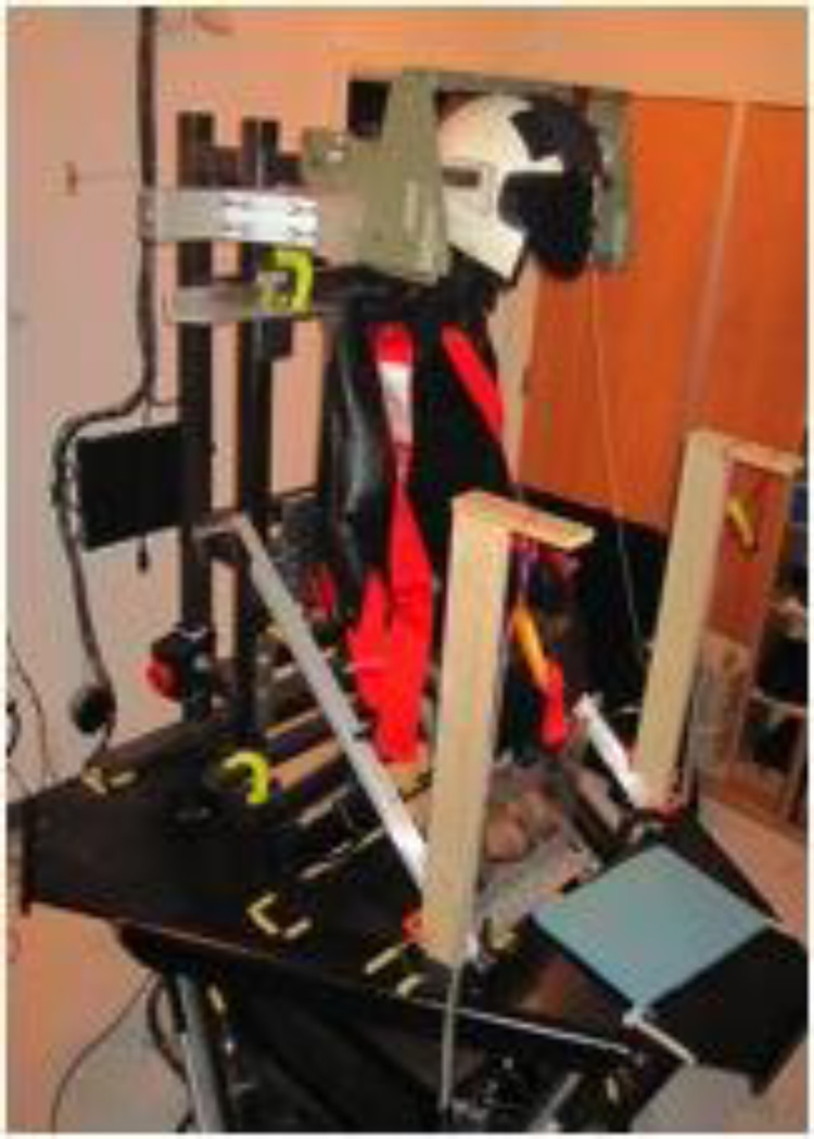
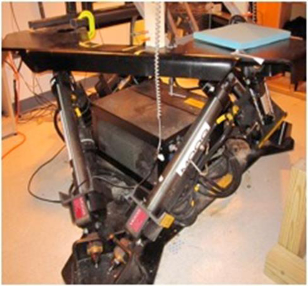
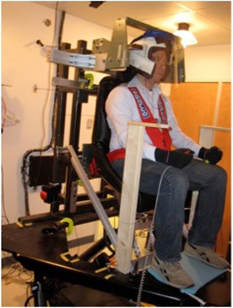
An innovative method of vestibular evaluation - the measurement of vestibular thresholds, was used. We used a Moog mobile platform (Moog Industrial Group- East Aurora, NY, USA) with dedicated software (Fig 1 and 2). The subjects were seated in a chair with a 5-point harness. For all roll tilt stimuli the chair was oriented and positioned in a way that the resultant rotation axis fell midway between the ears at a level as near the vestibular apparatus as possible. The head was held in place via an adjustable helmet. Tactile cues were distributed as evenly as possible using pads/cushions. To minimize the influence of non-vestibular cues regarding motion direction, all skin surfaces including the face were be covered (e.g., light gloves were provided and subjects were be asked to wear clothing that covered all skin surface); a clear visor that moved with the helmet surrounded the face. Earplugs with built-in speakers were inserted in each ear (Fig. 3). The earplugs acted to reduce external noise by about 20 dB; to mask any remaining auditory motion cues, “white noise” was applied at a level of about 60 dB SPL. All trials were performed in a light-tight room.
Fig 1.
Chair on the mobile MOOG platform, adapted to immobilize the patient during testing
Fig 2.
The system of hydraulic pistons guarantees wide range of motion of the MOOG platform.
Fig. 3.
Position of the subject during the testing sessions
In our study we chose roll-tilt motion; with the axis of roll rotation at the center of the head, we measured the roll tilt thresholds for 4 normal humans (2 males, 2 females). All the subjects had been previously screened to exclude the presence of vestibular disorders. The screening consisted of a questionnaire as well as caloric electro-nystagmography, Dix-Hallpike testing, angular VOR evoked via rotation and posture control measures. Motion stimuli consisted of single cycles of sinusoidal acceleration, because they reproduce closely the natural movements of the head [citation].
The subjects were tested at increasing frequencies (1, 2, 3, 4 and 5 Hz) of sinusoidal acceleration motion with adaptively decreasing amplitude of motion. They were asked to indicate the direction of motion in 3 conditions: vestibular only – subjects roll tilted in the dark, visual only – a visual scene was tilted in front of subjects, and combined visual - vestibular – subjects rotated while watching a stationary visual scene. The subjects indicated the direction (left or right) of perceived roll motion by pressing a button in their hand upon completion of the movement. In our experiments the motion was roll tilt with the axis of roll at the level of the ears.
Thresholds levels were selected using a hybrid method. First, an adaptive “3-down, 1-up” staircase paradigm: after three consecutive correct answers the amplitude of the motion would decrease, while after one wrong answer the amplitude of the motion would increase. Second, levels were presented that were a fixed ratio of the thresholds computed in the staircase. The direction-recognition threshold was determined by fitting a cumulative Gaussian to the data acquired. We defined the direction-recognition threshold as the value at which the subject correctly detects motion 84.1% of the times. The values of thresholds obtained for each subject defined a curve plotting angular velocity (deg/s) against frequency (1, 2, 3, 4 and 5 Hz) in a way equivalent to that of an audiogram. When data were combined across subjects, a geometric mean and standard deviation were used because threshold data are distributed logarithmically [3].
An institutional review board approval was obtained at Massachusetts Eye and Ear Infirmary (# 98-09-027).
Results
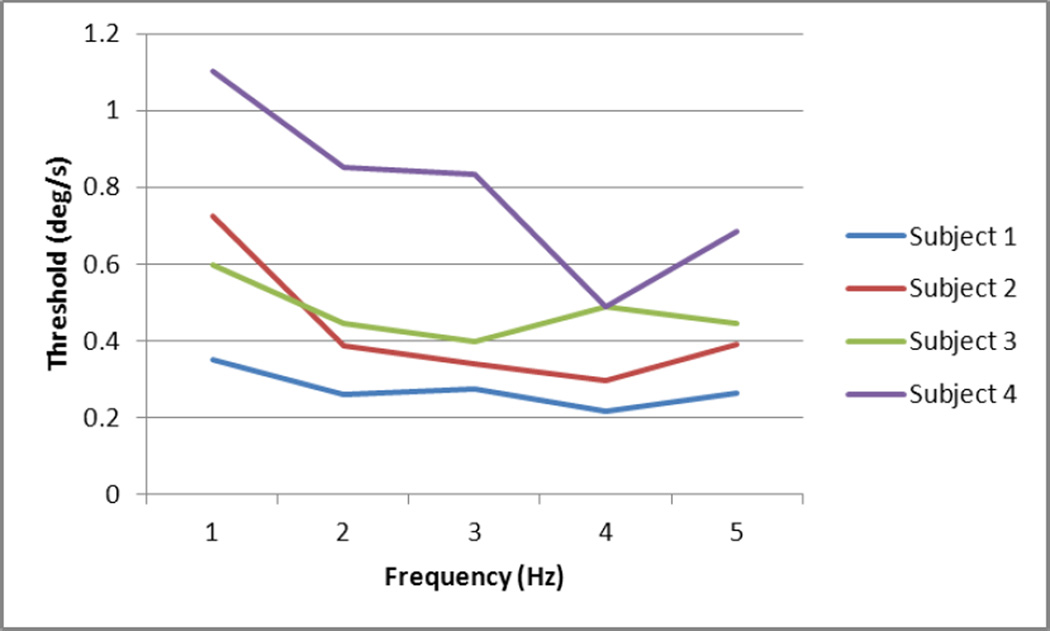
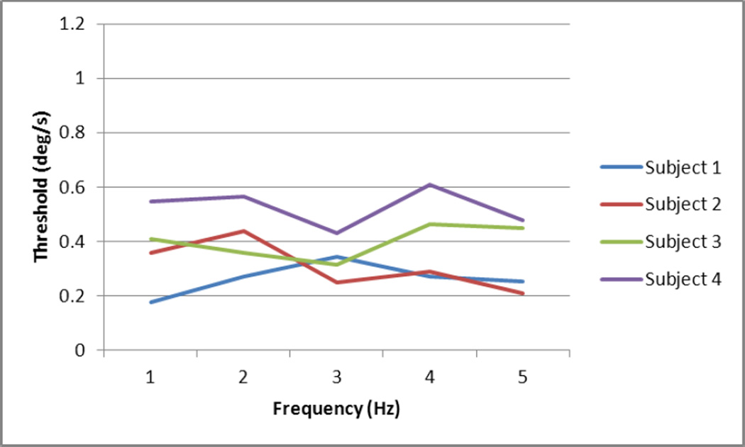
In the “visual only” condition all subjects showed a trend towards velocity at threshold as the frequency of the motion increased (Fig 4). In the “vestibular only” condition the velocity at threshold for all subjects tended to decrease with the increasing frequency (Fig 5). In the “visual-vestibular” condition the thresholds remain grossly steady across the different frequencies (Fig 6).
Fig 4.
Visual conditions – Motion perception thresholds in angular velocity (deg/s) plotted with frequency (Hz). The motion perception threshold increases in all subjects for higher frequencies of roll tilt motion, reflecting increasing difficulty recognizing a visual motion with decreasing duration.
Fig 5.
Vestibular conditions – Motion perception thresholds in angular velocity (deg/s) plotted with frequency (Hz). The thresholds of motion perception decrease as the frequency of motion increases and the duration of the motion decreases.
Fig 6.
Visual-vestibular conditions – Motion perception thresholds in angular velocity (deg/s) plotted with frequency (Hz).
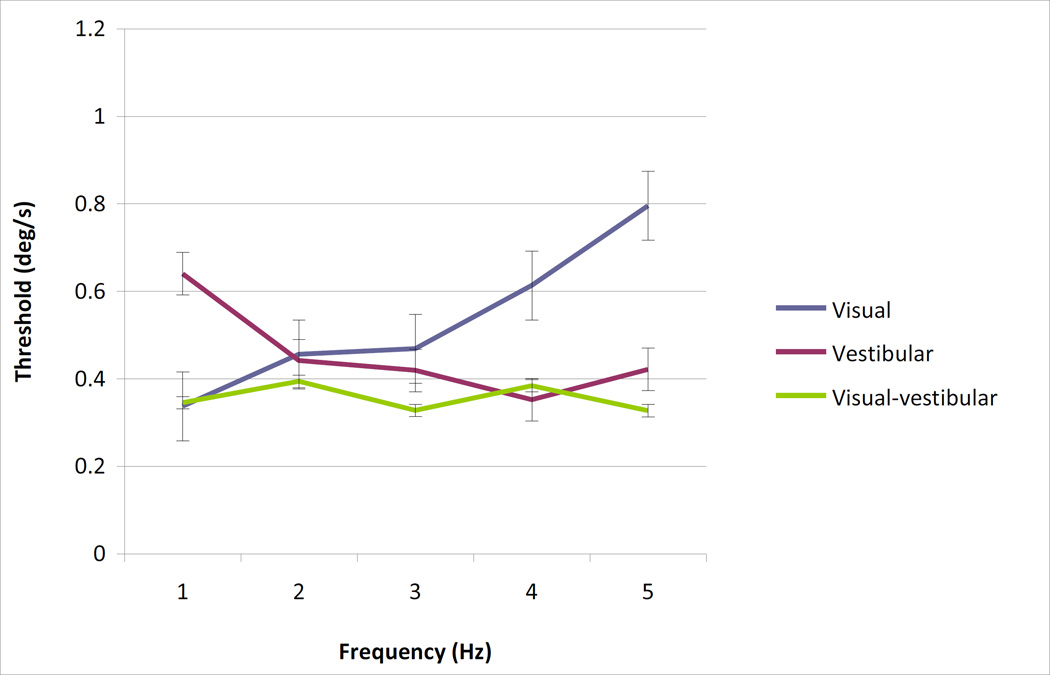
Figure 7 shows the mean thresholds across the four subjects for each frequency in visual-only, vestibular-only and visual-vestibular conditions. The error bars represent standard error. Over the frequencies measured in the study, the visual-vestibular thresholds are essentially flat (ANOCOVA; p>0.5), as opposed to the individual responses which are gradually rising for vision and falling for vestibular (ANOCOVA; p<0.01). The slopes of visual and vestibular are significantly different (ANOVA interaction term between frequency and condition; p<0.01). At 1 Hz, vestibular thresholds are significantly higher than visual thresholds (one-way unpaired t-test; p=0.028). At 5 Hz, visual thresholds are significantly higher than both vestibular thresholds (one-way unpaired t-test; p=0.025) and visual-vestibular thresholds (one-way unpaired t-test; p=0.016). At intermediate frequencies most differences do not reach the level of statistical significance.
Fig 7.
Thresholds for visual, vestibular and combined conditions for all 4 subjects
Pooled across frequencies, the visual-vestibular thresholds are significantly different from vestibular thresholds (2-way rmANOVA; p=0.054), although they do not differ significantly from visual thresholds (2-way rmANOVA; p=0.115).
Discussion
Previous studies have demonstrated that vestibulo-ocular reflexes (VOR) use different central nervous system pathways from those of vestibular perception [1;2]; despite some significant work done recently, our understanding of vestibular perception is limited compared to our understanding of the VOR and compared to our understanding of perception for other sensory systems (e.g., vision). As mentioned above, the perception of motion however uses central pathways, which have been shown to be anatomically and physiologically distinct. The measurement of direction-recognition thresholds to evaluate the function of the vestibular system has the potential to improve clinical testing. In our study we chose single cycle accelerations because we believe that they closely reproduce the natural movements of the head.
In our experiments, the values of thresholds obtained for each subject defined a curve plotting angular velocity (deg/s) against frequency (1, 2, 3, 4 and 5 Hz) in a way equivalent to that of an audiogram. We present angular velocity at threshold and not displacement at threshold for two reasons: first, for each frequency, these parameters are linearly related to one another [3]. Second, based on the recent studies, it is the velocity of motion that best accounts for vestibular thresholds at high frequencies. This is also consistent with literature showing that the semicircular canals are sensitive primarily to angular velocity.
A recent study by Karmali et al [5] showed that the predominance of vestibular and visual cues on motion perception likely varies with frequency. They reported that the vestibular and visual cues contribute equally at 0.05 Hz, the vestibular system predominates over the visual at 5 Hz and the visual cues have preponderance over the vestibular ones at 0.1 – 2 Hz. They also found that vestibular-visual thresholds are consistent with optimal fusion of vestibular and visual cues.
Our investigation showed that the motion perception thresholds measured in visual-vestibular (or combined) conditions are significantly lower than the thresholds obtained in vestibular conditions and not significantly lower than visual conditions for the frequencies from 1 to 5 Hz. In addition, vestibular and visual thresholds are statistically different at high and low frequencies (1 Hz; 4 and 5 Hz), but not at the intermediate frequencies (2 and 3 Hz) tested. This finding is a further step towards a better understanding of the physiology of human perception of motion and is one of the first studies focused on determining the thresholds of roll-tilt motion perception and the contribution of visual and vestibular input in spatial orientation.
There may be a functional importance of the cross-over between 2 and 3 Hz in which visual and vestibular cues are of similar reliability. This is close to the natural frequency of 2 Hz that dominates many human behaviors and their accompanying head movements, including walking, cycling and finger tapping [7]. We suggest as phenomenological speculation that the two senses may have evolved to provide the best complementary perception at this frequency; if one sense is lost, a higher level of precision is maintained at 2 Hz than at any other frequency.
Our study was performed in healthy subjects and serves as a baseline for but given the sensitivity of the measurement of vestibular threshold we find it as a potentially promising modality for the diagnosis and the follow-up of certain central and peripheral vestibular disorders with important impact of dizziness on patient’s quality of life. Further studies are needed to assess the vestibular thresholds in diseases for which no accurate diagnostic tests exist such as vestibular migraine [8], motion sickness, “mal de debarquement” and whiplash injury. Those studies will help determine the role of this promising test in the clinical environment of the Otolaryngology office.
Conclusions
The brain determines body motion in space during roll tilt motion using vestibular, visual and mixed visual-vestibular cues. Vestibular and visual thresholds at high and low frequencies are significantly different which support the fact that they use different perception pathways. The combined estimate of motion is significantly better than the vestibular input and is not significantly better with visual cues alone. The methods and results of this research may be useful in the work-up of patients with symptoms caused by impaired integration of vestibular and visual cues (motion-sickness and migraine dizziness).
Acknowledgments
We would like to thank the subjects for volunteering to participate. This research was supported by the NIH/NIDCD grant DC04158. IRB MEEI # 98-09-027
Contributor Information
Vartan Mardirossian, Department of Otolaryngology - Head and Neck Surgery, Boston University Medical Center, Boston, MA
Faisal Karmali, Massachusetts Eye and Ear Infirmary, Boston, MA; Department of Otology and Laryngology, Harvard Medical School, Boston MA, USA.
Daniel M. Merfeld, Massachusetts Eye and Ear Infirmary, Boston, MA; Department of Otology and Laryngology, Harvard Medical School, Boston, MA, USA.
References
- 1.Merfeld DM, et al. Vestibular Perception and Action Employ Qualitatively Different Mechanisms. II. VOR and Perceptual Responses During Combined Tilt&Translation. J Neurophysiol. 2005;94(1):199–205. doi: 10.1152/jn.00905.2004. [DOI] [PubMed] [Google Scholar]
- 2.Merfeld DM, et al. Vestibular Perception and Action Employ Qualitatively Different Mechanisms. I. Frequency Response of VOR and Perceptual Responses During Translation and Tilt. J Neurophysiol. 2005;94(1):186–198. doi: 10.1152/jn.00904.2004. [DOI] [PubMed] [Google Scholar]
- 3.Grabherr L, et al. Vestibular thresholds for yaw rotation about an earth-vertical axis as a function of frequency. Exp Brain Res. 2008;186(4):677–681. doi: 10.1007/s00221-008-1350-8. [DOI] [PubMed] [Google Scholar]
- 4.Benson AJ, Hutt EC, Brown SF. Thresholds for the perception of whole body angular movement about a vertical axis. Aviat Space Environ Med. 1989;60:205–213. [PubMed] [Google Scholar]
- 5.Karmali Faisal, Lim Koeun, Adatia Adil, Claret Romain, Nicoucar Keyvan, Merfeld Daniel M. Perceptual roll-tilt thresholds demonstrate visual-vestibular fusion. presented at the 40th Annual meeting of Neuroscience; Nov. 13–17, 2010; San Diego, CA. [Google Scholar]
- 6.Perez N, Garmendia I, Garcia-Granero M, Martin E, Garcia-Tapia R. Factor analysis and correlation between Dizziness Handicap Inventory and Dizziness Characteristics and Impact on Quality of Life scales. Acta Otolaryngol Suppl. 2001;545:145–154. doi: 10.1080/000164801750388333. [DOI] [PubMed] [Google Scholar]
- 7.MacDougall HG, Moore ST. Marching to the beat of the same drummer: the spontaneous tempo of human locomotion. J Appl Physiol. 2005;99:1164–1173. doi: 10.1152/japplphysiol.00138.2005. [DOI] [PubMed] [Google Scholar]
- 8.Lewis RF, Priesol AJ, Nicoucar K, Lim K, Merfeld DM. Dynamic tilt thresholds are reduced in vestibular migraine. J Vestib Res. 2011 Jan 1;21(6):323–330. doi: 10.3233/VES-2011-0422. [DOI] [PMC free article] [PubMed] [Google Scholar]


