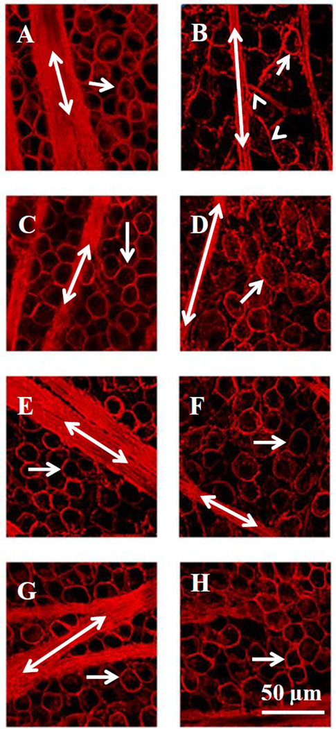
Figure 3.
Confocal images illustrating the neuroprotective effect of PNU-282987. The left column represents control untreated images obtained from different rats (A, C, E and G). RGCs (arrow) and axon fascicles (double labeled arrows) were labeled with an antibody against Thy 1.1. The corresponding confocal images in the right column were obtained from the same animal and from the same retinal geographic location as shown in the right column, but the episcleral veins were injected with 2 M NaCl to induce glaucoma-like conditions (B) or treated with 100 µM (D), 500 µM (F) or 2 mM PNU-282987 (H) eye drops before and after the procedure to induce glaucoma-like conditions. The arrowhead points to defasciculating axon strands.

