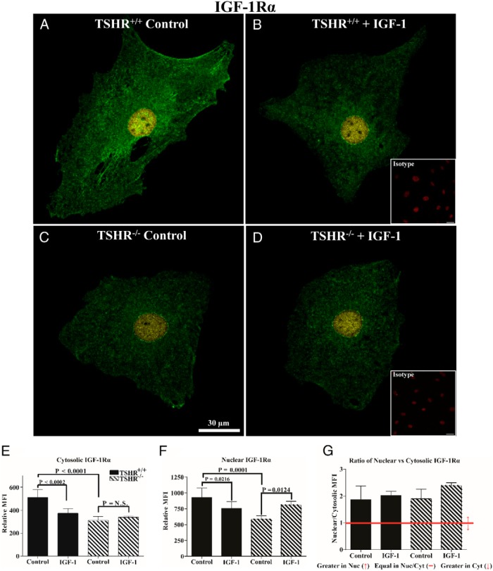
Figure 2.
Analysis of IGF-1Rα subcellular distribution by immunofluorescence in TSHR+/+ and TSHR−/− mouse lung fibroblasts. A–D, Immunofluorescence staining for IGF-1Rα (green and yellow) and PI (red, insets) demonstrates cellular localization of the receptor protein by confocal microscopy in lung fibroblasts isolated from TSHR+/+ (panels A and B) and TSHR−/− mice (panels C and D). Fibroblasts were inoculated on coverslips as described in Materials and Methods. Cells were either left untreated (panels A and C, control) or were treated with 100 ng/mL IGF-1 for 16 hours (panels B and D, IGF-1). Yellow signal represents nuclear IGF-1Rα colocalizing with PI and green represents cytosolic localization. Isotype control Abs (panels B and D, insets) demonstrate an absence of background labeling. Fluorescence intensities of the cytosolic (E) and nuclear (F) compartments and the ratios (G) of the signal intensities in the two cellular compartments were quantified. Intensity measurements from five different cells from each group were used in the analysis. Data are expressed as mean ± SD. Bar, 30 μm.

