ABSTRACT
Tooth wear or tooth surface loss is a normal physiological process and occurs throughout life but is considered pathological when the degree of destruction is excessive or the rate of loss is rapid, causing functional, aesthetic or sensitivity problems. The importance of tooth wear as a dental problem has been increasingly recognized. The findings of a study in Trinidad indicate that the prevalence of tooth wear in a Trinidadian population is comparable to the United Kingdom (UK) and, indeed, that the level of moderate and severe wear is nearly twice as high. The aetiology of tooth wear is attributed to four causes: erosion, attrition, abrasion and abfraction. Erosion is generally considered to be the most prevalent cause of tooth wear in the UK and Europe. Acids that cause dental erosion originate mainly from the diet or the stomach and, to a lesser extent, the environment. Underlying medical problems can contribute to influence the progress of tooth wear due to erosion and the patient may not be aware of these conditions. Moderate to severe tooth wear poses a significant clinical challenge to dental practitioners and may result in treatment that is more complex and costly to the patient, both in terms of finances and time spent in the dental chair. This paper provides an overview of aetiology and diagnosis of tooth wear, in particular tooth wear due to erosion, so that medical and dental practitioners may recognize tooth wear early, institute preventive measures and manage patients appropriately.
Keywords: Dental erosion, medical conditions, tooth wear
RESUMEN
El desgaste dental o la pérdida de la superficie del diente es un proceso fisiológico normal y ocurre durante toda la vida, pero se considera patológico cuando el grado de destrucción es excesivo o el ín-dice de pérdida es rápido, causando así problemas funcionales, estéticos o de sensibilidad. El desgaste dental como problema odontológico ha ganado cada vez más importancia. Los resultados de un estudio realizado en Trinidad indican que la prevalencia del desgaste dental en una población de Trinidad y Tobago, es comparable a la del Reino Unido (UK), y que – en efecto – los niveles de desgaste moderados y severos son casi dos veces más altos. La etiología del desgaste dental se atribuye a cuatro causas: erosión, atrición, abrasión y abfracción. La erosión se considera generalmente la causa más frecuente del desgaste dental en el Reino Unido y Europa. Los ácidos que causan la erosión dental se originan principalmente de la dieta o el estómago y – en menor medida – del medio ambiente. Los problemas médicos subyacentes pueden contribuir al progreso del desgaste dental por erosión, y el paciente puede no tener conciencia de estas condiciones. El desgaste dental moderado a severo plantea un desafío clínico significativo para los profesionales dentales, y puede resultar en tratamientos muy complejos y costosos para el paciente, tanto en términos de finanzas como de tiempo en el sillón dental. Este documento proporciona una visión general de la etiología y diagnóstico del desgaste dental, en particular del desgaste dental debido a la erosión, de modo que los profesionales médicos y dentales puedan identificar temprano el desgaste dental, instituir medidas preventivas, y tratar adecuadamente a los pacientes.
INTRODUCTION
Tooth wear is defined as the loss of tooth substance in the absence of caries and plaque (1). It is a normal physiological process and occurs throughout life but is considered pathological when the degree of destruction is excessive or the rate of loss is rapid, causing functional, aesthetic or sensitivity problems. The importance of tooth wear as a dental problem has been increasingly recognized. The findings of a study in Trinidad indicate that the prevalence of tooth wear in a Trinidadian population is comparable to the United Kingdom (UK) and, indeed, that the level of moderate and severe wear is nearly twice as high (2).
Terms such as erosion, abrasion, attrition (3) and abfraction have traditionally been used to describe pathological loss of tooth tissue, reflecting some aetiological factors associated with such occurrences. Although the loss caused by each of the above factors has a distinctive appearance, tooth wear rarely occurs from a single cause (4). As such, clinicians may encounter significant difficulties in determining the dominant aetiology. Subsequently, controlling or preventing the loss of tooth structure may be difficult. Erosion is generally considered to be the most prevalent cause of tooth wear in the UK (4) and Europe (5). It is a problem affecting all age groups, with more than 30% of 14-year olds showing evidence of erosion on palatal (inner) surfaces of the upper incisors (6). Defining the various aetiological factors in tooth surface loss and associated activities which may contribute to this (Table 1) will aid in classifying the condition, understanding its causes and planning intervention.
Table 1. Definitions of the aetiological factors in tooth surface loss.
| Aetiological factor | Definition and associated causative activities |
|---|---|
| Attrition | The loss by wear of tooth substance or a restoration caused by mastication or contact between occluding or approximal surfaces. |
| Often seen associated with parafunctional activity (7) or a modern “healthy” or vegetarian diet (8). | |
| Erosion | The progressive loss of hard dental tissues by chemical process not involving bacterial action (8). |
| Abrasion | The loss by wear of tooth substance or a restoration caused by factors other than tooth contact. |
| Associated with non-dental objects eg hair grips (9), or overly vigorous tooth brushing (8). | |
| Abfraction | The pathologic loss of hard tooth substance caused by biomechanical loading forces. It is thought to be due to flexure and ultimate fatigue of enamel and/or dentine at some location distant from actual point of loading. |
Adapted from Kelleher and Bishop; 1999 (20)
Erosion
Erosion is the progressive loss of dental hard tissue by acid from a non-bacterial source (8). The most common cause of tooth wear in the UK population is erosion (4). The causes of erosive lesions are varied (Table 2). Some of these causes may point to underlying medical conditions which may be elucidated during history taking and clinical examination.
Table 2. Causes of erosive lesions.
| Type of erosion | Causative factors |
|---|---|
| Regurgitation (intrinsic erosion | Regurgitation may be an involuntary occurrence as a complication of gastrointestinal problems, or be voluntary or patient-induced as in anorexia nervosa or bulimia. |
| Dietary (extrinsic) erosion | High consumption of food and drinks with a variety of acids especially soft drinks and diet beverages which have been implicated as an aetiological factor in 40% of patients with tooth surface loss (1). |
| Environmental (extrinsic) erosion | Acidic environments for work or leisure may expose patients to factors which cause tooth surface loss. |
The acid that causes erosive wear may be classified as intrinsic or extrinsic (10) depending on the source of the acid from either the stomach (intrinsic) or the diet and other environmental sources (extrinsic). Regurgitation erosion refers to tooth wear caused by the regurgitation of hydrochloric acid from the stomach. This occurs in patients with digestive disorders such as gastroesophageal reflux disease including those with hiatus hernia and chronic indigestion. Reflux past the upper oesophageal sphincter has been shown to increase the risk for erosion in the mouth (5).
Stomach acid can also enter the oral cavity during vomiting episodes due to alcohol hangovers, chronic alcoholism, morning sickness associated with pregnancy, eating disorders such as anorexia and bulimia nervosa (11) and with voluntary regurgitation or rumination (11). Rumination is a condition when patients eat their food and voluntarily regurgitate the food with gastric acids into their mouths.
Dietary erosion is due to food or drink containing a variety of acids such as from citrus and other fruits, fruit juices (citric acid), soft drinks, wine and other carbonated drinks (carbonic acid and other acids), pickles, vinegar dressings and preserves (acetic acid). There is a high consumption of fruits, fruit juices and carbonated beverages in Trinidad and Tobago and this may be similar to other Caribbean islands. There is also an association with vegetarian diet and erosion (2). Table 3 shows the acidity of some common foods and beverages.
Table 3. Acidity of some common foods and beverages.
| Item | pH range |
|---|---|
| Lime juice | 1.8–2.4 |
| Orange juice | 2.8–4.0 |
| Grapefruit juice | 2.9–3.4 |
| Pepsi | 2.7 |
| Diet Pepsi | 2.95 |
| Coke | 2.7 |
| Wines | 2.3–3.8 |
| Beers | 4.0–5.0 |
| Tea (black) | 4.2 |
| Vinegar | 2.4–3.4 |
| Pickles | 2.5–3.0 |
Industrial and environmental erosion is due to exposure to processes in the work place (eg battery factories) which produce acid fumes or droplets, and leisure activities (eg chlorinated swimming pools).
DIAGNOSIS AND ASSESSMENT
Medical history
Before any intervention or restorative treatment is started, a diagnosis of tooth wear should be made based on the clinical signs and a carefully elicited history. Diagnosis may not be easy because patients may not want to volunteer information (eg in eating disorders), or they may not associate heartburn or stomach upsets with having any effect on teeth, or that the dentist may need to know this. Thus, in addition to the routine medical history, emphasis must be placed on medical conditions and eating disorders that predispose to regurgitation erosion. Also, medical problems that cause a reduction in salivary flow (Table 4) can affect the extent of dental erosion. Referral and collaboration with medical practitioners may be necessary for further investigations, diagnosis and management of these underlying medical conditions.
Table 4. Factors that reduce the flow of saliva.
| • Salivary gland excision | ||
| • Autoimmune disease such as Sjogren's syndrome | ||
| • Radiation treatment to head and neck region | ||
| • Medications – that can cause dry mouth (xerostomia) | ||
| ∘ Parasympatholytics such as: | ||
| • Antihypertensives | ||
| • Antiparkinsonian drugs | ||
Dental and dietary history
Particular questions to be asked are type of toothbrush used, whether hard or soft, toothbrushing frequency and history of bruxism (grinding or clenching). It is also important to ask if grinding or bruxism sounds are heard by bed partner, if there is masticatory or facial muscle fatigue or pain in the mornings and if the patient ever used a mouthguard or occlusal guard. A diet sheet is useful to determine the intake frequency of acidic food and beverages.
Social history
This will reveal the patient's occupation and any potential contributing factors such as if the patient is a dressmaker holding pins/hairgrips between the teeth leading to abrasion, a wine-taster who can develop dietary erosion or a regular swimmer and those exposed to environmental work hazards can also suffer from dental erosion (3).
Examination
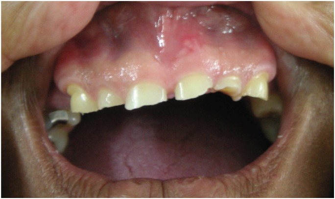
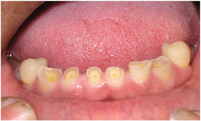
A thorough examination is needed both extra-orally and intra-orally. Extra-oral examination may reveal facial signs of alcoholism such as facial flushing and spider angiomas and enlarged parotid glands which can also be an indicator of autoimmune disease or anorexia. Masseteric muscle hypertrophy may also indicate a clenching or grinding habit (bruxism). Intra-oral examination may reveal signs of salivary hypofunction such as dry mouth with a reduced amount of saliva or saliva that is foamy, viscous or ropy. Shiny facets or wear on the teeth or restorations may also be observed. Clinical features of erosive lesions include: broad concavities within smooth tooth enamel or loss of enamel surface anatomy, cupping out of occlusal surfaces with dentine exposure. The eroded enamel stands higher than the underlying dentine as the dentine is less mineralized compared to enamel and wears away faster once exposed. In the anterior teeth, there is increased incisal translucency, incisal chipping and, in moderate to severe cases, cupping out of the incisal edges (Figs. 1 and 2). Erosion caused by vomiting typically affects the palatal (inner) surfaces of the upper teeth but can be due to dietary acids as well.
Fig. 1. Upper anterior teeth showing moderate tooth wear.
Fig. 2. Lower anterior teeth showing cupping out of incisal edges due to erosion.
Management
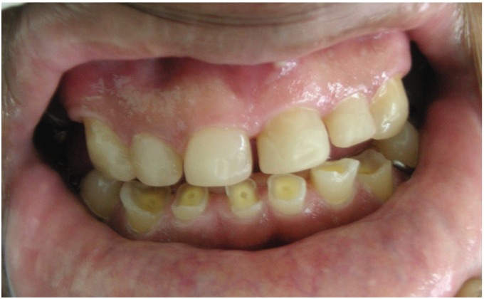
The provision of restorative dental care requires a multi-disciplinary approach and may encompass treatment ranging from simple restorations to comprehensive full mouth rehabilitation. Management includes monitoring and prevention and it is necessary to establish a diagnosis, but even if a definitive diagnosis is not clear initially, general preventive suggestions can be made such as: diet advice, use of chewing gums to increase salivary flow, therapy, counselling and/or referral to medical or dental practitioner, treatment of tooth sensitivity with toothpastes and mouth rinses with fluoride, decreasing abrasive forces eg use of soft tooth brush, provision of splint or mouthguard to reduce bruxism and use of straws to drink thereby avoiding exposure of teeth to acidic beverages. More active restorative treatment involving simple restorations (Fig. 3), removable prosthodontics (dentures), fixed prosthodontics (veneers, onlays, crowns and bridges) and implants, is indicated if patients have problems with aesthetics, function, teeth sensitivity or loss of tooth structure which has weakened the tooth. The description of the dental restorative management is outside the scope of this article.
Fig. 3. Upper anterior teeth restored by bonding tooth-coloured composite dental materials.
REFERENCES
- 1.Eccles JD. Tooth surface loss from abrasion, attrition and erosion. Dent Update. 1982;35:373–381. [PubMed] [Google Scholar]
- 2.Rafeek RN, Marchan S, Eder A, Smith WAJ. Tooth surface loss in adult subjects attending a university dental clinic in Trinidad. Int Dent J. 2006;56:181–186. doi: 10.1111/j.1875-595x.2006.tb00092.x. [DOI] [PubMed] [Google Scholar]
- 3.Kelleher M, Bishop K. Tooth surface loss: an overview. Br Dent J. 1999;186:61–66. doi: 10.1038/sj.bdj.4800020a2. [DOI] [PubMed] [Google Scholar]
- 4.Smith BGN, Knight JK. A comparison of patterns of tooth wear with aetiological factors. Br Dent J. 1984;157:16–19. doi: 10.1038/sj.bdj.4805401. [DOI] [PubMed] [Google Scholar]
- 5.Bartlett DW. The role of erosion in tooth wear: aetiology, prevention and management. Int Dent J. 2005;55:277–284. doi: 10.1111/j.1875-595x.2005.tb00065.x. [DOI] [PubMed] [Google Scholar]
- 6.Milosevic A, Young PJ, Lennon MA. The prevalence of toothwear in 14-year old school children in Liverpool. Comm Dent Health. 1994;11:83–86. [PubMed] [Google Scholar]
- 7.Dahl BL, Krogstad O, Karlsen K. An alternative treatment in cases with advanced localized attrition. J Oral Rehabil. 1975;2:209–214. doi: 10.1111/j.1365-2842.1975.tb00914.x. [DOI] [PubMed] [Google Scholar]
- 8.Smith BGN. Tooth wear: aetiology and diagnosis. Dent Update. 1989;16:204–212. [PubMed] [Google Scholar]
- 9.Mair LH. Wear in dentistry – current terminology. J Dent. 1992;20:140–144. doi: 10.1016/0300-5712(92)90125-v. [DOI] [PubMed] [Google Scholar]
- 10.Milosevic A. Tooth wear: aetiology and presentation. Dent Update. 1998;25 6–1. [PubMed] [Google Scholar]
- 11.Robb ND, Smith BG, Geidrys-Leeper E. The distribution of erosion in the dentitions of patients with eating disorders. Br Dent J. 1995;178:171–175. doi: 10.1038/sj.bdj.4808695. [DOI] [PubMed] [Google Scholar]


