SUMMARY
Thermogenic brown and beige adipose tissues dissipate chemical energy as heat, and their thermogenic activities can combat obesity and diabetes. Herein the functional adaptations to cold of brown and beige adipose depots are examined using quantitative mitochondrial proteomics. We identify arginine/creatine metabolism as a beige adipose signature and demonstrate that creatine enhances respiration in beige fat mitochondria when ADP is limiting. In murine beige fat, cold exposure stimulates mitochondrial Creatine Kinase activity and induces coordinated expression of genes associated with creatine metabolism. Pharmacological reduction of creatine levels decreases whole body energy expenditure after administration of a β3-agonist and reduces the adipose metabolic rate. Genes of creatine metabolism are compensatorily induced when UCP1-dependent thermogenesis is ablated, and creatine reduction in Ucp1-deficient mice reduces core body temperature. These findings link a futile cycle of creatine metabolism to adipose tissue energy expenditure and thermal homeostasis.
INTRODUCTION
Non-shivering thermogenesis primarily takes place in brown and beige adipose tissues. The ability of these depots to dissipate chemical energy has led to interest in their ability to combat obesity and diabetes. The thermogenic property of brown and beige fat relies predominantly on the actions of Uncoupling Protein 1 (UCP1) (Cannon and Nedergaard, 2004). This protein resides in the mitochondrial inner membrane and stimulates thermogenesis by dissipating the protonmotive force (Δp) and activating the rate of substrate flux through the mitochondrial respiratory chain.
It is now appreciated that there are at least two distinct UCP1-expressing cell types. Classical brown adipocytes are derived from a Myf5+ lineage, and are located primarily in developmentally formed depots in the interscapular region of rodents and human infants (Seale et al., 2008). Beige fat cells arise primarily from a Myf5- lineage and generally accumulate in white fat depots upon cold challenge or with the application of a number of different adrenergic stimuli, hormones and peptide factors. Much (but probably not all) of the “brown fat” of adult humans in the neck and supraclavicular areas has the molecular characteristics of beige fat, rather than the classical brown fat of rodents (Shinoda et al., 2015; Wu et al., 2012). On the other hand, the interscapular depots of human infants do indeed resemble the classical brown fat of rodents (Lidell et al., 2013).
Ablation experiments in mice have shown that UCP1+ cells, taken as a whole, protect mice from the metabolic effects of high fat feeding. Diminution of UCP1+ cells via transgenic expression of a toxigene first established the key role of these cells in the regulation of metabolic health (Lowell et al., 1993). Mice with Ucp1 deletion also develop obesity, although in this case obesity is only observed at thermoneutrality (Feldmann et al., 2009). Recently, beige fat function has been ablated in mice, with classical brown fat function left largely intact (Cohen et al., 2014); these animals develop moderate obesity and insulin resistance centered on the liver.
The realization that mammals have two distinct thermogenic cell types raises questions regarding their similarities and their differences. Questions concerning fuel preferences, hormone sensitivities and other key thermogenic pathways and functions are largely unexplored. We have performed quantitative proteomics, comparing highly purified mitochondria from brown and beige fat depots. The results indicate that beige fat cells have a thermogenic mechanism built around a creatine-driven substrate cycle.
RESULTS
Mitochondrial Purification from Cold-exposed Brown and Beige Fat
We set out to compare the proteomic and bioenergetic properties of mitochondria isolated from beige and brown adipose tissue upon induction of thermogenesis through cold exposure. To this end, we exposed mice to 4°C, which is sufficient to drive thermogenesis in subcutaneous inguinal white adipose tissue (iWAT) and classical interscapular brown adipose tissue (BAT). We evaluated the purity of our mitochondrial preparations by Western blotting. As expected, mitochondrial proteins were enriched, while contaminating components of the cytoplasm and endoplasmic reticulum were largely removed during the purification procedure (Figures S1A and S1B). Mitochondrial yield increased substantially (beige: 10-fold and brown: 2-fold) following cold exposure (Figure S1C). These mitochondria from both sources displayed properties indicative of UCP1+ organelles, including the requirement for BSA and purine nucleotides to acquire respiratory control (Figure S1D). These data are in line with previous reports (Shabalina et al., 2013) and indicate that beige fat mitochondria from iWAT are functionally thermogenic following cold exposure.
Quantitative Mitochondrial Proteomics Identifies Arginine/creatine Metabolism as a Signature of Beige Adipose Tissue
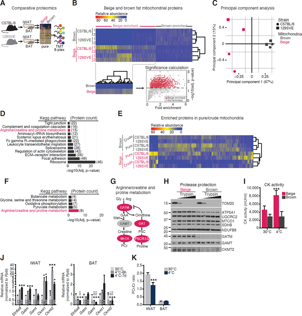
We used tandem mass spectrometry after isobaric peptide tagging to identify protein species exhibiting differential abundance between beige and brown adipose mitochondria from two strains (C57BL/6 and 129SVE) of cold exposed mice (Figure 1A and Table S1). As shown by the global heatmap (Figure 1B) and the principal component analysis (Figure 1C), brown fat mitochondria showed marginal strain-dependent variance, while beige fat mitochondria demonstrated greater diversity across strains. Next, beige and brown fat mitochondrial proteomes were stratified according to differential relative abundance (Figure 1B), followed by identification of beige fat-selective biological pathways (Figure 1D). Because mitochondrial proteins made up a larger percentage of the material after isolating pure organelles through a sucrose gradient, relative to crude mitochondria obtained by differential centrifugation alone (Figures S1A and S1B), we defined bona fide mitochondrial proteins to be those with higher abundance in the pure fraction, relative to the crude fraction. Thus, protein abundance between crude and pure preparations of mitochondria from beige and brown fat was examined by mass spectrometry (Figure 1E and Table S2). Proteins that had higher abundance in pure mitochondria, relative to crude mitochondria were cross-referenced to the initial proteomics inventory (Table S1). Pathway analysis demonstrated that components of arginine-dependent creatine and proline metabolism were reproducibly enriched in beige-fat, relative to brown fat mitochondria (Figure 1D and Figure 1F). A total of 14 proteins were identified that could be assigned to this pathway (Table S3). Enzymes with the ability to synthesize creatine and remove ornithine, an inhibitor creatine biosynthesis (Sipila, 1980), showed beige fat-selectivity (Figure 1G). There was a strong correlation between Western blotting and proteomic quantification for beige- and brown-enriched mitochondrial proteins (Figures S1E – S1G). Increased protein abundance in beige fat mitochondria was observed for components of arginine-dependent creatine and proline metabolism, such as GATM and CKMT2, as well as the majority of ATP Synthase subunits (Figure 1H and Table S1). Beige fat mitochondria contained higher levels of creatine kinase (CK) activity relative to brown fat mitochondria in both C57BL/6 and 129SVE strains of mice following cold exposure (Figure S1H). Moreover, mitochondrial CK activity was cold inducible (~2-fold) in beige fat (Figure 1I).
Figure 1. Characterization of Mitochondria from Brown and Beige Adipose Tissues.
(A) Schematic of mitochondrial purification and quantitative proteomics workflow.
(B) Heatmap of beige fat and brown fat mitochondrial proteomics data (from Table S1). Beige-enriched proteins are shown in the subset heatmap.
(C) Principal component analysis of the mitochondrial proteomics dataset.
(D) Kegg pathway analysis of beige fat-selective mitochondrial proteins from Figure 1B.
(E) Heatmap of beige fat and brown fat mitochondrial proteomics data (from Table S2) after selecting proteins on the basis of an expression ratio greater than 1 in pure/crude mitochondria.
(F) Kegg pathway analysis of significantly enriched beige fat mitochondrial proteins after cross-referencing Table S1 and S2.
(G) Schematic of creatine synthesis and byproduct removal proteins identified by mass spectrometry. Red circles, proteins identified by mass spectrometry; grey circles, proteins not identified. Gly, glycine; Arg, arginine; Met, methionine; PCr, phosphocreatine; P5C, 1-Pyrroline-5-carboxylic acid; Mi-CK, mitochondrial creatine kinase.
(H) Western blot after treatment of beige and brown fat mitochondria with trypsin (0, 10, 25, 50, and 100 µg ml−1).
(I) Creatine Kinase activity of mitochondria from 129SVE mice housed at 30°C or 4°C for 7 days.
(J) Quantitative RT-PCR (qRT-PCR) from C57BL/6 mice housed at 30°C or 4°C for 6 hours (4°C-6h) or 7 days (4°C-7d), n = 3 to 4 mice per group.
(K) PCr to creatine (Cr) ratio in iWAT and BAT from 129SVE mice housed at 30°C or 4°C for 7 days. Data are presented as mean ± SEM. *p < 0.05, ***p < 0.01.
Since creatine metabolism was found to be a distinct feature of thermogenic beige adipocytes at the protein level, we monitored changes in the mRNA expression of genes involved in creatine metabolism following 6 hours and 1 week exposure to 4°C. Transcript levels of th ese genes were coordinately elevated in response to cold in iWAT, but not in BAT (Figure 1J). Gatm and Ckmt1 transcript abundance was similar between iWAT and BAT. However, GATM and CKMT1 protein expression was higher in beige than brown fat mitochondria (Table S3). In contrast, Ckmt2 transcript levels were greater in BAT compared to iWAT. However, the expression level of CKMT2 protein was found to be greater in beige than brown fat mitochondria (Figure 1H and Table S3). The discordance between Ckmt2 mRNA from whole tissue lysates and protein abundance from isolated mitochondria is likely due to a higher mitochondrial content in BAT than iWAT.
We next investigated the levels of creatine and phosphocreatine (PCr) in iWAT and BAT from mice housed at 30°C or 4°C. Creat ine levels in iWAT were elevated two-fold following cold exposure (Figure S1I). In contrast, while higher steady-state creatine levels were observed in BAT, cold exposure had no detectable effect (Figure S1I). There was no difference in PCr levels in either iWAT or BAT in response to cold (Figure S1J), although a modest trend towards lower PCr levels in BAT was observed; these observations are in line with a recent report (Grimpo et al., 2014). As a consequence of these measurements, it is clear that the PCr/creatine ratio in iWAT was reduced significantly in cold-exposed animals (Figure 1K), suggesting increased creatine metabolism in beige fat. These changes in creatine levels were not observed in skeletal muscle of the same animals (Figure S1K).
Creatine Stimulates Respiration in Beige Fat Mitochondria When ADP is Limiting
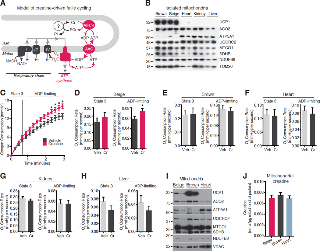
Based on our identification of creatine metabolism as a signature of beige fat mitochondria and due to the functional coupling of mitochondrial CK (Mi-CK) to oxidative phosphorylation through the ATP/ADP carrier (AAC) (Jacobus and Lehninger, 1973; Wyss and Kaddurah-Daouk, 2000), we posited that creatine could dissipate the mitochondrial ATP pool to drive ADP-dependent respiration in beige fat mitochondria. Such a pathway would require creatine and CK-mediated hydrolysis of ATP to drive a catalytic mechanism that stimulated cycling of ATP production and consumption (Figure 2A). We therefore tested whether creatine could stimulate substrate cycling and increase ADP-dependent respiration in beige fat mitochondria.
Figure 2. Creatine Stimulates Respiration in Beige Fat Mitochondria When ADP is Limiting.
(A) Model of creatine-based substrate cycling. IMS, intermembrane space; AAC, ADP/ATP Carrier.
(B) Western blot of mitochondrial proteins. N = 2 mitochondrial preparations, 15 mice per cohort.
(C) Oxygen consumption by beige fat mitochondria treated with and without creatine (0.01 mM) in the presence of 0.2 mM ADP. Vertical dashed line, State 3 to State 4 transition. N = 9 mitochondrial preparations, 15 mice per cohort.
(D) State 3 and ADP-limiting Oxygen consumption rate (OCR) of beige fat mitochondria treated with and without creatine in the presence of 0.2 mM ADP. N = 9 mitochondrial preparations, 15 mice per cohort.
(E) State 3 and ADP-limiting OCR of brown fat mitochondria treated as in D. N = 3 mitochondrial preparations, 15 mice per cohort.
(F) State 3 and ADP-limiting OCR of heart mitochondria treated as in D. N = 3 mitochondrial preparations, 8 mice per cohort.
(G) State 3 and ADP-limiting OCR of kidney mitochondria treated as in D. N = 3 mitochondrial preparations, 15 mice per cohort.
(H) State 3 and ADP-limiting OCR of liver mitochondria treated as in D. N = 3 mitochondrial preparations, 2 mice per cohort.
(I) Western blot of mitochondrial proteins from beige fat, brown fat, and heart. N = 2 mitochondrial preparations, 15 mice per cohort.
(J) Mitochondrial creatine concentration in beige fat, brown fat, and heart. Data are presented as mean ± SEM. *p < 0.05.
Striated muscle tissues are understood to utilize creatine metabolism such that mitochondrial ATP and creatine generate PCr and ADP in a 1:1 stoichiometry (Wyss and Kaddurah-Daouk, 2000). The resulting PCr pool is used to drive substrate-level phosphorylation of ADP during times of ATP deficit. Thus, a direct prediction of this classical metabolic utilization of PCr is that addition of a quantity of creatine to oxidatively coupled mitochondria will result in a molar equivalent production of ADP and PCr through CK-mediated phosphotransferase activity (Jacobus and Lehninger, 1973). Alternatively, if creatine drives futile substrate cycling, addition of a given amount of creatine would instead drive the release of a molar excess of ADP with respect to creatine, thus resulting in a surplus of oxygen consumption. In this case, the relationship of creatine to ADP liberation would be substoichiometric.
The stoichiometric relationship between creatine and ATP synthase-coupled respiration was examined in mitochondria isolated from a panel of tissues. Western blotting demonstrated similar mitochondrial yields from brown fat, beige fat, heart, kidney and liver (Figure 2B). Mitochondria were respired on pyruvate and malate in the presence of varying ADP concentrations. These organelles exhibited the expected behavior on addition of sub-saturating amounts of ADP (0.01 – 0.2 mM), such that respiration transitioned to State 4 as ADP became limiting (Figure S2A and S2B). In contrast, a saturating amount of ADP (1 mM) resulted in State 3 respiration with no transition to State 4, as ADP was no longer limiting over the course of the incubation (Figure S2A).
In the presence of sub-saturating levels of ADP, addition of a substoichiometric amount of creatine (0.01 mM) stimulated a ~30% increase in respiration in beige fat mitochondria. Specifically, creatine did not significantly affect the initial (State 3) ADP-stimulated rate of oxygen consumption (Figure 2C and Figure 2D, left panel), but enhanced the respiration rate when ADP levels became limiting (Figure 2C and Figure 2D, right panel). This action of creatine in beige fat mitochondria did not occur in the absence of exogenous ADP (Figure S2C) nor did it occur in the presence of the AAC inhibitor, carboxyatracyloside (Figure S2D). These data suggest that creatine-mediated respiration required export of mitochondrial matrix ATP through the AAC upon addition of exogenous ADP (Figure 2A). This feature of creatine-dependent respiration is in agreement with the established functional coupling of AAC with Mi-CK isoforms (Jacobus and Lehninger, 1973; Wyss and Kaddurah-Daouk, 2000). Furthermore, incubation of beige fat mitochondria with saturating ADP concentrations (Figure S2E) precluded the respiration stimulating effects of creatine (Figure S2F), further confirming that the stimulatory effects occurred only under ADP-limiting conditions.
On the basis of the mitochondrial P/O ratio (Watt et al., 2010) we calculated the amount of additional ATP that was synthesized by addition of creatine to beige fat mitochondria (Figure 2C). Creatine drove an increase in oxygen consumption equivalent to a 7.82-fold (±2.12) excess phosphorylation of ADP. These results indicate that in beige adipose mitochondria, creatine acts substoichiometrically (with respect to ADP phosphorylation) to increase mitochondrial ATP synthesis and stimulate substrate flux through the mitochondrial respiratory chain. Addition of sub-stoichiometric amounts of creatine to mitochondria isolated from brown fat, heart, kidney or liver (Figure 2B) had no effect on ADP-dependent respiration (Figure S2G and 2E–2H).
Striated muscle contains a large quantity of creatine (Fitch and Chevli, 1980), and so endogenous mitochondrial creatine levels in beige and brown fat were compared to heart following purification of a comparable yield of organelles from these tissues (Figure 2I). Figure 2J demonstrates that mitochondrial creatine levels were similar between tissues, indicating that the lack of a respiration-enhancing effect in heart mitochondria was not due to a high amount of endogenous creatine. Together, these data indicate that creatine stimulates respiration in beige fat mitochondria in a substoichiometric manner with respect to ADP, specifically when ADP is limiting; this is consistent with a model of creatine-driven substrate cycling (Figure 2A).
To examine thermogenesis by creatine-driven substrate cycling, we employed Differential Scanning Calorimetry (Ricquier et al., 1979). As expected, chemical uncoupling by FCCP induced thermogenesis, and this signal was inhibited by rotenone and antimycin; the signal was similar to that obtained when mitochondria were omitted from the reaction (Figure S2H). Importantly, addition of creatine to beige fat mitochondria drove ADP-dependent thermogenesis by ~30% relative to organelles incubated with ADP alone (Figure S2I). This is a direct demonstration of thermogenesis through creatine metabolism in beige fat mitochondria.
Creatine Metabolism in Adipose Tissue Contributes to Energy Expenditure and Thermal Homeostasis in vivo
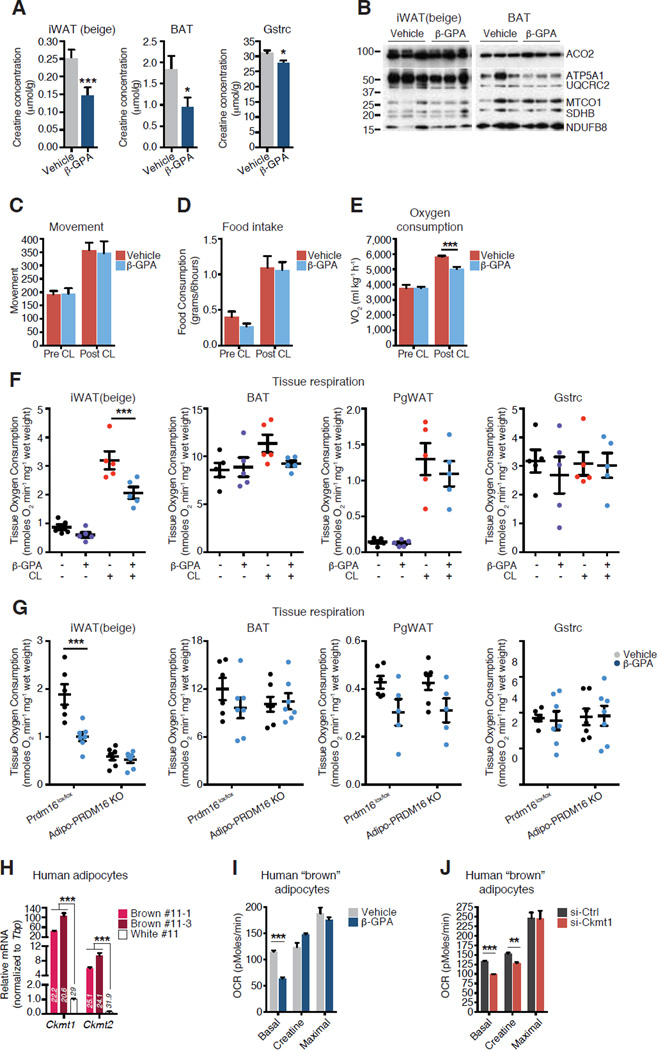
As the role of creatine metabolism in thermogenic fat tissues is essentially unexplored in vivo, we systematically assessed its role in beige and brown adipose metabolic functions. First, we examined if creatine regulates oxidative metabolism in beige and brown fat. To this end, we utilized β-guanidinopropionic acid (β-GPA), a creatine analog that is well established to inhibit creatine transport and to reduce creatine levels in cultured cells and tissues (Fitch and Chevli, 1980). A diet supplemented with a typical dose of β-GPA (2%), resulted in reductions in food intake and weight loss, as previously reported (Oudman et al., 2013); this compromised subsequent metabolic analyses. However, when mice were given daily intraperitoneal injections with a lowered dose of β-GPA (0.4 g kg−1) for four days during cold exposure, body weight and fat pad weight were unaffected (Figure S3A and S3B). Creatine levels were reduced by approximately 50% in iWAT and BAT, and by 15% in gastrocnemius muscle (Figure 3A). Mitochondrial respiratory chain protein expression was not altered (Figure 3B). To determine the contribution of creatine metabolism in brown/beige adipose to whole body energy expenditure, we utilized the β3-adrenergic receptor agonist, CL 316,243 (CL), a well-known activator of adipose thermogenesis (Bloom et al., 1992; Granneman et al., 2003). Movement, food intake, and oxygen consumption were monitored in CL-treated mice that were co-treated with either vehicle or β-GPA. There was no difference in ambulatory movement, food intake or fuel utilization between vehicle- or β-GPA-treated animals (Figures 3C, 3D and S3C). As expected, we detected a 54% increase in the metabolic rate of mice following CL injection. Strikingly, a reduction in creatine through the administration of β-GPA diminished the CL-induced increase in whole body oxygen consumption by ~40% compared to vehicle treatment (Figure 3E).
Figure 3. Creatine Metabolism in Adipose Tissue Contributes to Energy Expenditure and Thermal Homeostasis in vivo.
(A) Creatine concentration in iWAT (beige), BAT, and Gastrocnemius muscle (Gstrc) from cold-exposed C57BL/6 mice treated with four daily injections of vehicle or β-GPA (0.4 g kg−1), n = 5 mice per group.
(B) Western blot of iWAT (beige) and BAT from animals treated as in A, n = 3 mice per group.
(C) Movement, n= 8 mice per group. CL (0.2 mg kg−1) was co-injected intraperitonealy with vehicle (saline) or β-GPA (0.4 g kg−1).
(D) Food intake, n = 8 mice per group, treated as in C.
(E) Oxygen consumption, n = 8 mice per group, treated as in C.
(F) OCR of minced tissues following four daily injections of vehicle, β-GPA (0.4 g kg−1), CL (0.2 mg kg−1), or β-GPA + CL, n = 5 mice per group.
(G) OCR of minced tissues from Prdm16lox/lox and Adipo-PRDM16 KO mice after 7 days cold exposure. β-GPA was administered on the last four days of cold exposure, n = 5 to 7 mice per group.
(H) qRT-PCR of Ckmt1 and Ckmt2 in two human brown adipocyte lines (#11-1 and 11-3) and one human white adipocyte line (#11). Raw ct values are embedded in the bars.
(I) OCR of human brown adipocytes treated with vehicle, β-GPA (2 mM), and creatine (5 mM).
(J) OCR of human brown adipocytes transfected with si-Ctrl or si-Ckmt1. Creatine used at 5 mM. Data are presented as mean ± SEM. *p < 0.05, **p < 0.01, ***p < 0.0001.
The reduction in whole body oxygen consumption prompted an examination of which tissues were most affected by β-GPA, in terms of whole tissue respiration. Thus, another cohort of mice, housed at 23°C, was separated into four groups: vehicle, β-GPA, CL, or CL + β-GPA. β-GPA had no effect on the respiration of any tissue examined in the absence of a browning stimulus (Figure 3F). CL-treatment provided a powerful browning/beiging stimulus, resulting in a five-fold increase in iWAT oxygen consumption, a 33% increase in BAT respiration, a nine-fold elevation in PgWAT respiration, and no effect on Gstrc respiration (Figure 3F). Reducing creatine levels with β-GPA had a profound and significant effect on beige iWAT, resulting in a 34% reduction in oxygen consumption (Figure 3F). BAT oxygen consumption was reduced by 18% following β-GPA treatment, PgWAT oxygen consumption was decreased by 15% and no detectable effect on Gstrc tissue respiration was detected (Figure 3F). Although the β-GPA-dependent reduction in BAT respiration did not reach statistical significance, the absolute decrease was larger than any other tissue examined, and so in addition to iWAT, BAT may well contribute to the effects of β-GPA observed in vivo.
We next examined the contribution of beige adipocytes within iWAT to creatine-dependent oxidative metabolism. Mice with an adipose-selective deletion of Prdm16 (Adipo-PRDM16 KO) have disrupted beige adipose function (Cohen et al., 2014). Following 7 days cold exposure, creatine reduction with β-GPA significantly decreased iWAT oxidative metabolism from control Prdm16lox/lox mice, but had no further effect on the already reduced respiratory rate of iWAT from Adipo-PRDM16 KO mice (Figure 3G). No significant effect of creatine reduction was detected in any other tissue in either genotype, although again, there was a modest reduction in the respiration of BAT from Prdm16lox/lox mice (Figure 3G). Therefore, using two models of browning/beiging, these data demonstrate that creatine reduction attenuates the oxidative metabolism of thermogenic adipose tissues in vivo.
Respiration of Cultured Human Brown Adipocytes is Regulated by Creatine
Much, though not all, of the brown fat observed in adult humans has characteristics of murine beige adipocytes (Lidell et al., 2013; Shinoda et al., 2015; Wu et al., 2012). Thus, it was of interest to examine whether creatine contributed to oxidative metabolism in the recently cloned human brown adipocytes (Shinoda et al., 2015). Both Ckmt1 and Ckmt2 mRNA levels were higher in two human clonal brown adipocyte lines compared to human white adipocytes (Figure 3H). When human brown adipocytes were treated with β-GPA, basal respiration was significantly reduced by 45%; this reduction was completely rescued by creatine supplementation (Figure 3I), demonstrating the specificity of β-GPA. Next, RNAi-mediated knockdown was used to assess the function of Ckmt1 in these adipocytes. Transfection of siRNAs targeting Ckmt1 resulted in a ~40% reduction in Ckmt1 mRNA levels, relative to control siRNA-transfected cells (Figure S3D). Knockdown of Ckmt1 resulted in a 26% reduction in basal respiration, and creatine supplementation partially rescued this effect (Figure 3J). These data demonstrate that creatine and CKMT1 regulate oxidative properties of isolated human brown adipocytes.
Compensatory Regulation of Creatine Metabolism Components and UCP1 in Adipose Tissues in vivo
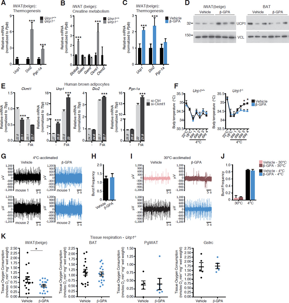
The ability of creatine to stimulate thermogenesis in an apparently substoichiometric manner with respect to ADP-dependent respiration strongly suggested that this creatine-based futile cycle could be an important mechanism of thermoregulation in vivo, independent of UCP1. To begin to test this supposition, we re-examined the longstanding and important observation that mice lacking Ucp1 can maintain thermal homeostasis at 4°C if they are gradually acclimated to this temperature (Golozoubova et al., 2001). These data demonstrated that alternative thermogenic pathway(s) compensate for the lack of UCP1 (Ukropec et al., 2006). We therefore gradually acclimated Ucp1-deficient (Ucp1−/−) mice to the cold over a period of 21 days. In addition, we also reduced creatine levels by administration of β-GPA to both Ucp1+/+ and Ucp1−/− animals. As shown previously (Ukropec et al., 2006), cold-exposed Ucp1−/− mice had elevated expression of classical thermogenic genes in iWAT, such as Dio2 and Pgc-1α, compared to Ucp1+/+ mice (Figure 4A). Pgc-1α was also induced in BAT from Ucp1−/− mice (Figure S4A). These data suggest a strong induction of a thermogenic phenotype in the iWAT of Ucp1−/− mice. Slc6a8 and Ckmt1 transcripts were elevated in iWAT of cold-exposed Ucp1−/− mice, relative to Ucp1+/+ littermates (Figure 4B), while Slc6a8 and Ckmt2 transcript levels were higher in BAT from the same animals (Figure S4B).
Figure 4. Creatine Metabolism Regulates UCP1-Independent Thermal Homeostasis in vivo.
(A) qRT-PCR from iWAT of 4°C-acclimated Ucp1+/+ and Ucp1−/− mice, n = 5 mice per group.
(B) qRT-PCR of creatine metabolism genes from same samples as A.
(C) qRT-PCR from iWAT of 4°C-acclimated mice treate d with four daily injections of vehicle or β-GPA (0.4 g kg−1), n = 5 mice per group.
(D) Western blot from iWAT and BAT from mice treated as in C. Vinculin (VCL), loading control, n = 3 mice per group.
(E) qRT-PCR of clonal human brown adipocytes, (line #11-1). Small interfering RNAs (siRNAs) targeted against control (si-Ctrl) or Ckmt1 (si-Ckmt1). Forskolin was used at 10 µM for 4 hours. (The data showing si-Ctrl and si- Ckmt1, without forskolin treatment, is the same data shown in Figure S3D), n = 3 per group.
(F) Body temperature of Ucp1+/+ and Ucp1−/− mice treated with vehicle or β-GPA (0.4 g kg−1), n = 7 to 8 mice per group.
(G) Representative electromyogram (EMG) traces, measured at 4°C, of 4°Cacclimated Ucp1−/− mice treated as in F.
(H) Frequency of shivering bursts quantified from data in G.
(I) Representative EMG traces, at 30°C and followin g 15 – 45 minutes at 4°C, of 30°C-acclimated wild type C57BL/6 mice treated a s in F.
(J) Frequency of shivering bursts quantified from data in I.
(K) OCR of minced tissues from 4°C-acclimated Ucp1−/− mice treated as in F. N = 16 to 17 mice per group (iWAT and BAT), n = 5 to 6 mice per group (PgWAT and Gstrc). Data are presented as mean ± SEM. *p < 0.05, ***p < 0.0001.
As the induction of creatine metabolism genes was elevated in Ucp1−/− mice, classical thermogenic genes, such as Ucp1 and Dio2, were significantly elevated in iWAT of cold-exposed mice after creatine reduction with β-GPA (Figure 4C). A modest increase in Ucp1 and Pgc-1α mRNA was observed in BAT of cold-exposed Ucp1+/+ mice following creatine reduction with β-GPA (Figure S4C). UCP1 protein levels were also elevated in response to β-GPA in iWAT of Ucp1+/+ mice, but did not increase in the BAT of the same animals (Figure 4D).
We also examined the compensatory regulation between creatine metabolism genes and Ucp1 in human brown adipocytes. In these cell lines, Ckmt1 was more abundant than Ckmt2 (Figure 3H). Transfection of siRNAs targeting human Ckmt1 resulted in a ~40% reduction in Ckmt1 mRNA levels, relative to control siRNA-transfected cells (Figure 4E). Strikingly, Ucp1 mRNA was elevated 2.5-fold in cells transfected with Ckmt1-targeted siRNAs relative to control siRNA transfected cells. Forskolin treatment of human brown fat cells resulted in a near 8-fold induction of Ucp1 mRNA (Figure 4E), and combining forskolin treatment with Ckmt1 knockdown resulted in a 15-fold elevation in Ucp1 transcript abundance above non-treated si-Ctrl cells (Figure 4E). Furthermore, si-Ckmt1 transfected cells had an elevated abundance of Dio2 and Pgc-1α, relative to si-Ctrl transfected cells, following forskolin treatment (Figure 4E). Taken together, these data demonstrate that genes involved in creatine metabolism are increased in the absence of UCP1, and expression of classical thermogenic genes is elevated when creatine metabolism is disrupted. These data suggest a compensatory relationship between UCP1- and creatine-dependent bioenergetics in mice and humans.
Creatine Regulates UCP1-Independent Thermal Homeostasis in vivo
To test the role of creatine metabolism in adaptive thermogenesis in vivo, we examined the body temperature of cold-acclimated Ucp1+/+ and Ucp1−/− mice that were treated with either β-GPA or vehicle for four days. Ucp1−/− mice weighed slightly less than Ucp1+/+ mice (Figure S4D). Body temperature of Ucp1−/− mice was higher than Ucp1+/+ animals at 23°C and 4°C (Figure 4F) ( Ucp1+/+: 34.4± 0.11°C and Ucp1−/−: 34.7± 0.14°C, at 4°C). This temperature differenc e between genotypes is consistent with previous work (Liu et al., 2003). Strikingly, body temperature of the Ucp1−/− animals was significantly lower when endogenous creatine levels were reduced with β-GPA (Figure 4F), suggesting that in the absence of UCP1, creatine regulates a larger proportion of cold-induced thermogenesis than when UCP1 is present.
Creatine contributes to skeletal muscle metabolism, and so it was critical to determine whether the effect of β-GPA on body temperature was due to reductions in shivering thermogenesis. Thus, we used electromyography (EMG) to monitor shivering directly. Mice housed at 30°C showed minimal EMG activity. However, within 15 to 30 minutes of exposure to 4°C , bursts of shivering were detected (Figure S4E). Next, we used EMG on Ucp1−/− mice that had been acclimated to 4°C, and similar to previous work (Golozoubova et al., 2001), we detected robust shivering activity (Figure S4E). These measurements were confirmed as shivering bursts because they were abolished with the nicotinic acetylcholine receptor inhibitor, D-tubocurarine (Figure S4E). The capacity for shivering of cold-acclimated Ucp1−/− mice was not altered by β-GPA treatment (Figure 4G and Figure 4H). We next examined shivering thermogenesis and body temperature in wild type mice that had been acclimated to 30°C and acutely cold challenged at 4°C following administration of vehicle or β-GPA for four days. Under these conditions, reduced creatine levels did not alter shivering capacity (Figure 4I and Figure 4J) or body temperature (Figure S4F). Analysis of serum CK activity was used as an independent method to monitor shivering. Although serum CK activity was elevated in Ucp1−/− mice at 18°C, CK activity was elevated to a similar extent in Ucp1+/+ and Ucp1−/− mice when ambient temperature was reduced to 10°C and 4°C (Figure S4G). Importantly, β-GPA administration had no effect on serum CK activity in either genotype. Together, these results indicate that the reduced body temperature of β-GPA-treated Ucp1−/− mice was not due to aberrant shivering, and that the metabolic and thermogenic adaptations that exist in the absence of UCP1 cannot be explained solely by shivering thermogenesis. Examination of respiration by isolated tissues indicated that despite a lower respiratory capacity of iWAT from Ucp1−/− mice, relative to Ucp1+/+ mice (compare Figure 4K to S4H), β-GPA treatment reduced oxygen consumption substantially in this depot from Ucp1−/− mice by 40% (Figure 4K). No significant effect on respiration due to creatine reduction was observed in any other tissue examined; however, a modest reduction was observed in BAT (Figure 4K and S4H).
A Screen of Mitochondrial Phosphatases to Identify Candidate Regulators of a Creatine-driven Substrate Cycle
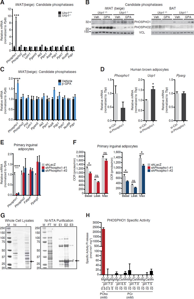
Taken together, our data indicated that creatine drives high-energy phosphate-dependent substrate cycling, which requires ADP-stimulated respiration. This led us to consider candidate molecules in adipose mitochondria that could carry out this process. To this end, we mined our mitochondrial proteomics inventory for putative phosphatases/hydrolases, and identified 11 candidate phosphatases. Since several known members of creatine metabolism had elevated expression in Ucp1−/− mice (Figure 4B), we posited that the expression of a key enzyme(s) with phosphatase activity might also be elevated in the absence of UCP1. Transcript levels for all 11 of these phosphatases were measured in iWAT and BAT of cold-exposed Ucp1+/+ and Ucp1−/− mice. One gene - Phospho1 (Phosphatase orphan 1) - was dramatically altered at the mRNA level, with an 8- fold increase in iWAT of Ucp1−/− animals (Figure 5A). In contrast, none of the candidates were increased in BAT at the mRNA level (Figure S5A). The consistent identification of PHOSPHO1 in our mitochondrial proteomic experiments suggested that PHOSHPO1 is at least partly associated with adipose mitochondria. Strikingly, protein expression of PHOSPHO1 was dramatically higher in the iWAT and BAT of Ucp1−/− mice, relative to Ucp1+/+ mice (Figure 5B). PHOSPHO2, the closest homolog of PHOSPHO1, displayed the opposite expression pattern (Figure 5B). Proteomic quantification of PHOSPHO1 from iWAT and BAT of cold-acclimated Ucp1+/+ and Ucp1−/− mice also demonstrated similar results as Western blotting (Figure S5B). Interestingly, creatine reduction with β-GPA resulted in a small increase in Phospho1 mRNA levels in iWAT from cold exposed Ucp1+/+ mice (Figure 5C), and a modest trend in the same direction was detected in BAT (Figure S5C). We also examined the compensatory regulation between Phospho1 and Ucp1 in human brown adipocytes. Transfection of siRNAs targeting human Phospho1 resulted in a ~50% reduction in Phospho1 mRNA levels, relative to control siRNA-transfected cells (Figure 5D). Ucp1 mRNA was significantly elevated 1.5-fold in cells transfected with Phospho1-targeted siRNAs relative to control siRNA transfected cells, and no change was observed for the differentiation marker Pparg (Figure 5D). Therefore, based on the reciprocal expression pattern between Phospho1 and Ucp1 in mouse tissue and human cultured cells, PHOSPHO1 was considered as a candidate for involvement in a creatine-driven substrate cycle.
Figure 5. A Screen of Mitochondrial Phosphatases with Ucp1-deficient Mice Identifies PHOSPHO1 as a Regulator of Adipocyte Respiration.
(A) qRT-PCR of candidate phosphatases in iWAT of 4°C-acclimated Ucp1+/+ and Ucp1−/− mice, n = 5 mice per group.
(B) Western blot of PHOSPHO1 and PHOSPHO2 from 4°C- acclimated Ucp1+/+ and Ucp1−/− mice, treated with vehicle or β-GPA (0.4 g kg−1). Vinculin (VCL), loading control.
(C) qRT-PCR of candidate phosphatases in iWAT of 4°C-ac climated wild type mice treated as in B, n = 5 mice per group.
(D) qRT-PCR of clonal human brown adipocytes, (line #11-1) treated with si-Ctrl or si-Phospho1, n = 3 per group.
(E) qRT-PCR of primary mouse inguinal adipocytes after Phospho1 knockdown (shPhospho1-#1 and shPhospho1-#2), n = 4 to 5 per group.
(F) OCR of primary mouse inguinal adipocytes treated as in E, n = 5 to 7 per group.
(G) Coomassie stain of SDS-PAGE demonstrating PHOSPHO1 protein expression and purification. M, molecular weight marker; NI, non-induced; I, induced; FT, flow-through; W, wash; E1 – E3, elutions 1 – 3. Arrows indicate recombinant PHOSPHO1.
(H) Specific activity of PHOSPHO1 toward PCho (0.5 mM) and PCr (0.5 to 10 mM) exposed to various buffer pH and divalent metals. Data are presented as mean ± SEM. *p < 0.05, ***p < 0.0001.
PHOSPHO1 Regulates Oxidative Metabolism in Primary Inguinal Adipocytes
Phospho1 displayed an expression pattern that would be predicted for a factor involved in an alternative, UCP1-independent pathway of energy expenditure. At the mRNA level, its steady-state expression was modestly higher in murine primary inguinal adipoyctes, relative to primary brown fat cells, whereas Ucp1 demonstrated the opposite expression pattern (Figure S5D). Therefore, the role of PHOSPHO1 in adipocyte bioenergetics was examined. Using adenoviral-mediated shRNA delivery, we achieved ~90% knockdown of Phospho1 at the mRNA level in primary inguinal cells, with no mRNA changes detected for its homolog, Phospho2 or the differentiation markers Fabp4 and Pparg2 (Figure 5E). Reduced PHOSPHO1 levels were also observed at the protein level (Figure S5E). Both shRNAs used to knockdown PHOSPHO1 significantly reduced basal oxygen consumption but had no effect on proton leak; the second shRNA also reduced maximal respiration (Figure 5F). Knockdown of Phospho1 in primary brown adipocytes (Figure S5F) resulted in a modest reduction in basal respiration, with no effect on proton leak or maximal respiration (Figure S5G). Thus, reduced Phospho1 levels attenuated oxygen consumption in primary inguinal and brown adipocytes, although the relative effect was greater in inguinal cells. The more pronounced effect on basal respiration (largely regulated by ATP demand) than proton leak is consistent with a role for PHOSPHO1 in ATP-coupled oxygen consumption. One interpretation of this data was that PHOSPHO1 regulates substrate cycling by liberating the high-energy phosphate from PCr. Therefore, we purified recombinant PHOSPHO1 (Figure 5G) in order to examine whether PCr can be directly hydrolyzed by PHOSPHO1 in vitro. PHOSPHO1 specific activity towards phosphocholine was similar to a prior report (Roberts et al., 2004). However, there was limited phosphatase activity towards PCr; this was done under various buffer conditions (Figure 5H). Taken together, these data suggest that if PHOSPHO1 is involved in a creatine-driven substrate cycle, it likely regulates high-energy phosphate metabolism downstream of the phosphotransfer event that utilizes PCr (Figure 6).
Figure 6. Models of Creatine-Driven Futile Substrate Cycling.
(A) Model of creatine-driven futile substrate cycling based on direct hydrolysis of PCr.
(B) Model of creatine-driven futile substrate cycling based on multiple phosphotransfer events catalyzed by multiple enzymes (Enz1 – Enzn).
DISCUSSION
While UCP1 is critical for optimal thermogenesis, Ucp1−/− animals can survive cold temperatures if gradually acclimated (Golozoubova et al., 2001; Meyer et al., 2010; Ukropec et al., 2006). Furthermore, it has been shown with adrenergic stimulation and peptide factors that the thermogenic responses of Ucp1+/+ and Ucp1−/− mice are similar (Granneman et al., 2003; Grimpo et al., 2014; Veniant et al., 2015). These findings imply the presence of UCP1-independent thermogenic mechanisms. Glycerol-3-phosphate shuttle activation and lipid turnover (Flachs et al., 2011; Grimpo et al., 2014) have been posited to act independently of UCP1. Calcium cycling has been proposed to be an additional source of thermogenesis in BAT (Ukropec et al., 2006) and is a well-established thermogenic mechansim in the extraocular heater muscle cells of certain fish and in mammalian skeletal muscle (Bal et al., 2012; Block et al., 1994). Interestingly, large reductions in creatine levels have previously been linked to deregulated thermal homeostasis in rats (Wakatsuki et al., 1996; Yamashita et al., 1995), through unknown mechanisms.
The stoichiometric relationships observed between creatine, ADP and oxygen consumption suggest a creatine-driven substrate cycle. By liberating a molar excess of ADP, with respect to the amount of added creatine, beige fat mitochondria enhance respiration by maintaining a state of ATP synthesis, as shown by the increased rate of oxygen consumption when ADP was limiting. Reducing creatine levels by even 50% in beige and brown fat was associated with a blunted response to β3-agonism at the level of whole body energy expenditure. While not statistically significant, these data also suggest that classical brown fat may also execute this creatine futile cycle. In fact, a role for creatine in brown adipose tissue metabolism was posited nearly four decades ago (Berlet et al., 1976).
Gene expression analyses demonstrated a clear compensatory regulation between classical thermogenic genes, and the genes involved in creatine metabolism in murine tissues and human adipocytes. Consistent with this reciprocal relationship, a reduction in creatine levels attenuated oxygen consumption of iWAT from Ucp1−/− mice and perturbed thermal homeostasis of these animals without diminishing shivering. The expression of Phospho1 was elevated at the mRNA and protein level in Ucp1-deficient animals, and thus became the focus of examination. Although the effect of PHOSPHO1 knockdown on adipocyte bioenergetics is consistent with a role for it in high-energy phosphate metabolism, PHOSPHO1 does not hydrolyze PCr in vitro, at least under any conditions we tested. Interestingly, phosphoethanolamine (PEtn), a direct PHOSPHO1 substrate (Roberts et al., 2004), has been demonstrated to inhibit ADP-stimulated mitochondrial respiration (Gohil et al., 2013), and so PHOSPHO1 could effect oxygen consumption by regulating PEtn levels.
The data presented here indicate that creatine metabolism plays an important role in adipose energy expenditure in vivo. Most likely, creatine facilitates the regeneration of ADP through futile hydrolysis of PCr. This could occur in a single enzymatic step, whereby PCr would be the direct substrate of a so-called PCr phosphatase (Figure 6A), or via multiple phosphotransfer reactions prior to phosphate hydrolysis from a currently unknown phosphometabolite (Figure 6B)
Similar to what was shown in the recently cloned human brown adipocytes (Shinoda et al., 2015), CKMT1 and CKMT2 expression is enriched in human BAT (Svensson et al., 2011). If creatine metabolism plays a substantial role in thermogenesis in humans, as suggested by the work here with isolated cells, it could open up possibilities to manipulate energy expenditure in patients with metabolic diseases by new drugs or even with dietary supplementation.
EXPERIMENTAL PROCEDURES
Sucrose Gradient-Purified Mitochondria
Sucrose was dissolved to a concentration of 1M and 1.5M in Gradient Buffer (10 mM HEPES, pH 7.8, 5 mM EDTA, and 2 mM DTT), and layered in a polyallomer centrifuge tube. Mitochondrial samples was loaded on top of the gradient and ultracentrifuged at 32,000 rpm for 1h at 4°C. Intac t organelles that banded at the interface of the sucrose cushion were carefully extracted, washed twice in SHE buffer, and stored at −80°C until LC-MS/MS or Weste rn blot analyses.
Mitochondrial Respiration
Mitochondrial respiration was determined using an XF24 Extracellular Flux Analyzer (Seahorse Bioscience) using 15µg mitochondrial protein in a buffer containing 50mM KCl, 4mM KH2PO4, 5mM HEPES, and 1mM EGTA, 4% BSA, 10mM Pyruvate, 5mM Malate, 1mM GDP. Mitochondria were plated and centrifuged 2,000 g for 20 minutes to promote adherence to the XF24 V7 cell culture microplate. Uncoupled and maximal OCR was determined using Oligomycin (14µM) and FCCP (10µM). Rotenone and Antimycin A (4µM each) were used to inhibit Complex 1- and Complex 3-dependent respiration.
Primary Inguinal Adipocyte Differentiation
Primary inguinal preadipocytes were counted and plated in the evening at 20,000 cells per well of a seahorse plate. The following morning inguinal preadipocytes were induced to differentiate with 1µM rosiglitazone, 0.5mM isobutylmethylxanthine, 1µM dexamethasone, and 5 µg ml−1 insulin. Cells were re-fed every 48 hours with 1µM rosiglitazone and 5µg ml−1 insulin. Cells were fully differentiated by day 5 post-induction.
Primary Brown Adipocyte Differentiation
Primary brown preadipocytes were counted and plated in the evening at 15,000 cells per well of a seahorse plate. The following morning brown preadipocytes were induced to differentiate with 1µM rosiglitazone, 0.5mM IBMX, 5µM dexamethasone, 0.114µg ml−1 insulin, 1nM T3, and 125µM Indomethacin. Cells were re-fed every 48 hours with 1µM rosiglitazone and 0.5µg ml−1 insulin. Cells were fully differentiated by day 5 post-induction
Supplementary Material
ACKNOWLEDGEMENTS
We are grateful to Marc W. Kirschner, Mike P. Murphy, Bé Wieringa, and members of the Spiegelman lab for helpful discussions. pBAD-TOPO-TA/hPhospho1 was a kind gift from Colin Farquharson. The Biophysical Instrumentation Facility (NSF-0070319) is acknowledged for help with DSC. This work was supported by a Canadian Institutes of Health Research postdoctoral fellowship to L.K., by a Human Frontier Science Program postdoctoral fellowship to E.T.C., and by NIH grant (DK031405) and the JPB foundation to B.M.S.
Footnotes
Publisher's Disclaimer: This is a PDF file of an unedited manuscript that has been accepted for publication. As a service to our customers we are providing this early version of the manuscript. The manuscript will undergo copyediting, typesetting, and review of the resulting proof before it is published in its final citable form. Please note that during the production process errors may be discovered which could affect the content, and all legal disclaimers that apply to the journal pertain.
Supplemental information includes Experimental Procedures, 5 figures, and 3 tables.
AUTHOR CONTRIBUTIONS
Conceptualization, L.K. and B.M.S.; Methodology, L.K., E.T.C., B.M.S.; Investigation, L.K., E.T.C., M.P.J., B.K.E., K.S., G.Z.L.; Resources, D.B.L., P.C., R.V., S.C.H.; Writing – Original Draft, L.K., E.T.C., B.M.S.; Writing – Review & Editing, L.K., E.T.C., P.C., B.M.S.; Funding Acquisition, L.K., E.T.C., S.K., S.P.G., B.M.S.; Supervision, S.K., S.P.G., B.M.S.
REFERENCES
- Bal NC, Maurya SK, Sopariwala DH, Sahoo SK, Gupta SC, Shaikh SA, Pant M, Rowland LA, Bombardier E, Goonasekera SA, et al. Sarcolipin is a newly identified regulator of muscle-based thermogenesis in mammals. Nature medicine. 2012;18:1575–1579. doi: 10.1038/nm.2897. [DOI] [PMC free article] [PubMed] [Google Scholar]
- Berlet HH, Bonsmann I, Birringer H. Occurrence of free creatine, phosphocreatine and creatine phosphokinase in adipose tissue. Biochimica et biophysica acta. 1976;437:166–174. doi: 10.1016/0304-4165(76)90358-5. [DOI] [PubMed] [Google Scholar]
- Block BA, O'Brien J, Meissner G. Characterization of the sarcoplasmic reticulum proteins in the thermogenic muscles of fish. The Journal of cell biology. 1994;127:1275–1287. doi: 10.1083/jcb.127.5.1275. [DOI] [PMC free article] [PubMed] [Google Scholar]
- Bloom JD, Dutia MD, Johnson BD, Wissner A, Burns MG, Largis EE, Dolan JA, Claus TH. Disodium (R,R)-5-[2-[[2-(3-chlorophenyl)-2-hydroxyethyl]-amino] propyl]-1,3-benzodioxole-2,2-dicarboxylate (CL 316,243). A potent beta-adrenergic agonist virtually specific for beta 3 receptors. A promising antidiabetic and antiobesity agent. Journal of medicinal chemistry. 1992;35:3081–3084. doi: 10.1021/jm00094a025. [DOI] [PubMed] [Google Scholar]
- Cannon B, Nedergaard J. Brown adipose tissue: function and physiological significance. Physiological reviews. 2004;84:277–359. doi: 10.1152/physrev.00015.2003. [DOI] [PubMed] [Google Scholar]
- Cohen P, Levy JD, Zhang Y, Frontini A, Kolodin DP, Svensson KJ, Lo JC, Zeng X, Ye L, Khandekar MJ, et al. Ablation of PRDM16 and beige adipose causes metabolic dysfunction and a subcutaneous to visceral fat switch. Cell. 2014;156:304–316. doi: 10.1016/j.cell.2013.12.021. [DOI] [PMC free article] [PubMed] [Google Scholar]
- Feldmann HM, Golozoubova V, Cannon B, Nedergaard J. UCP1 ablation induces obesity and abolishes diet-induced thermogenesis in mice exempt from thermal stress by living at thermoneutrality. Cell metabolism. 2009;9:203–209. doi: 10.1016/j.cmet.2008.12.014. [DOI] [PubMed] [Google Scholar]
- Fitch CD, Chevli R. Inhibition of creatine and phosphocreatine accumulation in skeletal muscle and heart. Metabolism: clinical and experimental. 1980;29:686–690. doi: 10.1016/0026-0495(80)90115-8. [DOI] [PubMed] [Google Scholar]
- Flachs P, Ruhl R, Hensler M, Janovska P, Zouhar P, Kus V, Macek Jilkova Z, Papp E, Kuda O, Svobodova M, et al. Synergistic induction of lipid catabolism and anti-inflammatory lipids in white fat of dietary obese mice in response to calorie restriction and n-3 fatty acids. Diabetologia. 2011;54:2626–2638. doi: 10.1007/s00125-011-2233-2. [DOI] [PubMed] [Google Scholar]
- Gohil VM, Zhu L, Baker CD, Cracan V, Yaseen A, Jain M, Clish CB, Brookes PS, Bakovic M, Mootha VK. Meclizine inhibits mitochondrial respiration through direct targeting of cytosolic phosphoethanolamine metabolism. The Journal of biological chemistry. 2013;288:35387–35395. doi: 10.1074/jbc.M113.489237. [DOI] [PMC free article] [PubMed] [Google Scholar]
- Golozoubova V, Hohtola E, Matthias A, Jacobsson A, Cannon B, Nedergaard J. Only UCP1 can mediate adaptive nonshivering thermogenesis in the cold. FASEB journal : official publication of the Federation of American Societies for Experimental Biology. 2001;15:2048–2050. doi: 10.1096/fj.00-0536fje. [DOI] [PubMed] [Google Scholar]
- Granneman JG, Burnazi M, Zhu Z, Schwamb LA. White adipose tissue contributes to UCP1-independent thermogenesis. American journal of physiology Endocrinology and metabolism. 2003;285:E1230–E1236. doi: 10.1152/ajpendo.00197.2003. [DOI] [PubMed] [Google Scholar]
- Grimpo K, Volker MN, Heppe EN, Braun S, Heverhagen JT, Heldmaier G. Brown adipose tissue dynamics in wild-type and UCP1- knockout mice: in vivo insights with magnetic resonance. Journal of lipid research. 2014;55:398–409. doi: 10.1194/jlr.M042895. [DOI] [PMC free article] [PubMed] [Google Scholar]
- Jacobus WE, Lehninger AL. Creatine kinase of rat heart mitochondria. Coupling of creatine phosphorylation to electron transport. The Journal of biological chemistry. 1973;248:4803–4810. [PubMed] [Google Scholar]
- Lidell ME, Betz MJ, Dahlqvist Leinhard O, Heglind M, Elander L, Slawik M, Mussack T, Nilsson D, Romu T, Nuutila P, et al. Evidence for two types of brown adipose tissue in humans. Nature medicine. 2013;19:631–634. doi: 10.1038/nm.3017. [DOI] [PubMed] [Google Scholar]
- Liu X, Rossmeisl M, McClaine J, Riachi M, Harper ME, Kozak LP. Paradoxical resistance to diet-induced obesity in UCP1-deficient mice. The Journal of clinical investigation. 2003;111:399–407. doi: 10.1172/JCI15737. [DOI] [PMC free article] [PubMed] [Google Scholar]
- Lowell BB, V SS, Hamann A, Lawitts JA, Himms-Hagen J, Boyer BB, Kozak LP, Flier JS. Development of obesity in transgenic mice after genetic ablation of brown adipose tissue. Nature. 1993;366:740–742. doi: 10.1038/366740a0. [DOI] [PubMed] [Google Scholar]
- Meyer CW, Willershauser M, Jastroch M, Rourke BC, Fromme T, Oelkrug R, Heldmaier G, Klingenspor M. Adaptive thermogenesis and thermal conductance in wild-type and UCP1-KO mice. American journal of physiology Regulatory, integrative and comparative physiology. 2010;299:R1396–R1406. doi: 10.1152/ajpregu.00021.2009. [DOI] [PMC free article] [PubMed] [Google Scholar]
- Oudman I, Clark JF, Brewster LM. The effect of the creatine analogue beta-guanidinopropionic acid on energy metabolism: a systematic review. PloS one. 2013;8:e52879. doi: 10.1371/journal.pone.0052879. [DOI] [PMC free article] [PubMed] [Google Scholar]
- Ricquier D, Gaillard JL, Turc JM. Microcalorimetry of isolated mitochondria from brown adipose tissue. Effect of guanosine-di-phosphate. FEBS letters. 1979;99:203–206. doi: 10.1016/0014-5793(79)80279-3. [DOI] [PubMed] [Google Scholar]
- Roberts SJ, Stewart AJ, Sadler PJ, Farquharson C. Human PHOSPHO1 exhibits high specific phosphoethanolamine and phosphocholine phosphatase activities. The Biochemical journal. 2004;382:59–65. doi: 10.1042/BJ20040511. [DOI] [PMC free article] [PubMed] [Google Scholar]
- Seale P, Bjork B, Yang W, Kajimura S, Chin S, Kuang S, Scime A, Devarakonda S, Conroe HM, Erdjument-Bromage H, et al. PRDM16 controls a brown fat/skeletal muscle switch. Nature. 2008;454:961–967. doi: 10.1038/nature07182. [DOI] [PMC free article] [PubMed] [Google Scholar]
- Shabalina IG, Petrovic N, de Jong JM, Kalinovich AV, Cannon B, Nedergaard J. UCP1 in brite/beige adipose tissue mitochondria is functionally thermogenic. Cell reports. 2013;5:1196–1203. doi: 10.1016/j.celrep.2013.10.044. [DOI] [PubMed] [Google Scholar]
- Shinoda K, Luijten IH, Hasegawa Y, Hong H, Sonne SB, Kim M, Xue R, Chondronikola M, Cypess AM, Tseng YH, et al. Genetic and functional characterization of clonally derived adult human brown adipocytes. Nature medicine. 2015;21:389–394. doi: 10.1038/nm.3819. [DOI] [PMC free article] [PubMed] [Google Scholar]
- Sipila I. Inhibition of arginine-glycine amidinotransferase by ornithine. A possible mechanism for the muscular and chorioretinal atrophies in gyrate atrophy of the choroid and retina with hyperornithinemia. Biochimica et biophysica acta. 1980;613:79–84. doi: 10.1016/0005-2744(80)90194-1. [DOI] [PubMed] [Google Scholar]
- Svensson PA, Jernas M, Sjoholm K, Hoffmann JM, Nilsson BE, Hansson M, Carlsson LM. Gene expression in human brown adipose tissue. International journal of molecular medicine. 2011;27:227–232. doi: 10.3892/ijmm.2010.566. [DOI] [PubMed] [Google Scholar]
- Ukropec J, Anunciado RP, Ravussin Y, Hulver MW, Kozak LP. UCP1-independent thermogenesis in white adipose tissue of cold-acclimated Ucp1−/− mice. The Journal of biological chemistry. 2006;281:31894–31908. doi: 10.1074/jbc.M606114200. [DOI] [PubMed] [Google Scholar]
- Veniant MM, Sivits G, Helmering J, Komorowski R, Lee J, Fan W, Moyer C, Lloyd DJ. Pharmacologic Effects of FGF21 Are Independent of the "Browning" of White Adipose Tissue. Cell metabolism. 2015;21:731–738. doi: 10.1016/j.cmet.2015.04.019. [DOI] [PubMed] [Google Scholar]
- Wakatsuki T, Hirata F, Ohno H, Yamamoto M, Sato Y, Ohira Y. Thermogenic responses to high-energy phosphate contents and/or hindlimb suspension in rats. The Japanese journal of physiology. 1996;46:171–175. doi: 10.2170/jjphysiol.46.171. [DOI] [PubMed] [Google Scholar]
- Watt IN, Montgomery MG, Runswick MJ, Leslie AG, Walker JE. Bioenergetic cost of making an adenosine triphosphate molecule in animal mitochondria. Proceedings of the National Academy of Sciences of the United States of America. 2010;107:16823–16827. doi: 10.1073/pnas.1011099107. [DOI] [PMC free article] [PubMed] [Google Scholar]
- Wu J, Bostrom P, Sparks LM, Ye L, Choi JH, Giang AH, Khandekar M, Virtanen KA, Nuutila P, Schaart G, et al. Beige adipocytes are a distinct type of thermogenic fat cell in mouse and human. Cell. 2012;150:366–376. doi: 10.1016/j.cell.2012.05.016. [DOI] [PMC free article] [PubMed] [Google Scholar]
- Wyss M, Kaddurah-Daouk R. Creatine and creatinine metabolism. Physiological reviews. 2000;80:1107–1213. doi: 10.1152/physrev.2000.80.3.1107. [DOI] [PubMed] [Google Scholar]
- Yamashita H, Ohira Y, Wakatsuki T, Yamamoto M, Kizaki T, Oh-ishi S, Ohno H. Increased growth of brown adipose tissue but its reduced thermogenic activity in creatine-depleted rats fed beta-guanidinopropionic acid. Biochimica et biophysica acta. 1995;1230:69–73. doi: 10.1016/0005-2728(95)00067-s. [DOI] [PubMed] [Google Scholar]
Associated Data
This section collects any data citations, data availability statements, or supplementary materials included in this article.


