Abstract
MicroRNA (miR)-145 is the most abundant miR in vascular smooth muscle cells (VSMCs). However, the effect of hyperglycemia on the regulation of miR-145 is unknown. We hypothesized that the hyperglycemic condition activates a proinflammatory response that mediates the expression of miR-145 in VSMCs. We investigated whether miR-145 serves as a critical regulator to regulate the downstream proliferation factors (including Kruppel-like factor 4 [Klf4] and myocardin) in VSMCs under hyperglycemic conditions. Human coronary artery smooth muscle cells (HCASMCs) were cultured under high glucose conditions. Sustained high glucose at 25 mmol/L significantly decreased the expression of miR-145 in HCASMCs. High glucose significantly increased angiotensin II (Ang II) secretion from HCASMCs and Ang II suppressed miR-145 expression in HCASMCs. Ang II repression of miR145 expression resulted in increased Klf4 and decreased myocardin expression under conditions of high glucose. Overexpression of miR-145 significantly decreased Klf4 and increased myocardin expression and inhibited HCASMC proliferation and migration induced by a high glucose state. Balloon injury of the carotid artery in diabetic rats was performed to investigate miR-145, Klf and myocardin expression. The expression of miR-145 was maximally increased at 7 d after carotid injury and gradually declined thereafter. Overexpression of miR-145 and treatment with valsartan reversed Klf4 and myocardin protein expression induced by balloon injury and improved vascular injury. In conclusion, our study reveals that Ang II downregulates miR-145 to regulate Klf4 and myocardin expression in HCASMCs under high glucose conditions. Ang II plays a critical role in the regulation of miR-145 under hyperglycemic conditions.
INTRODUCTION
The incidence of type 2 diabetes mellitus is increased significantly worldwide and diabetes remains a great challenge for health care because vascular complications, including microvascular and macrovascular diseases, occur frequently (1). To prevent or delay the vascular complications caused by diabetes mellitus, a better understanding of the mechanisms inducing vascular complications is important to provide novel therapeutic approaches.
MicroRNA (miR) is a newly identified class of small (around 20 to 25 nucleotides) noncoding RNA emerging as crucial players in the pathogenesis of hyperglycemia-induced vascular damage (2,3). Several studies have demonstrated a high expression of miR-145 in vascular walls and the critical role of miR-145 in vascular smooth muscle cell (VSMC) phenotype switching (4,5). The miR-145 gene may lie downstream of the serum response factor and/or myocardin in the developmental program regulating VSMC differentiation (6). Xin et al. have demonstrated that both the serum response factor and myocardin upregulate the expression of miR-145 in VSMCs (7). miR-145 promotes VSMC differentiation in part by increasing myocardin expression and functioning in feed-forward reinforcement of its own expression by the serum response factor–myocardin complex (8). Further, miR-145 represses Kruppel-like factor 4 (Klf4), which was shown to be involved in multiple events including neointimal proliferation. Klf4 plays a key role in regulating the VSMC phenotype because it facilitates VSMC migration and downregulates VSMC differentiation marker genes (9).
Boettger et al. have reported that miR-143/145 knockout mice exhibited reduced systolic blood pressure in response to angiotensin II (Ang II) stimulation (4). Ang II seems to be involved in the pathogenesis of insulin resistance. Insulin resistance occurs in a wide variety of pathological states and is commonly associated with obesity, type 2 diabetes, accelerated atherosclerosis and hypertension (10). The effect of Ang II on miR-145 expression under a hyperglycemic state is yet to be determined.
The concept of VSMCs in the pathophysiology of progressive proatherogenic process in the hyperglycemic condition has been proposed and no data have been presented to verify the effect of hyperglycemic condition on the regulation of miR-145 in VSMCs. Therefore, we hypothesized that the hyperglycemic condition activates a proinflammatory response that mediates the expression of miR-145 in VSMCs. We also sought to investigate whether miR-145 serves as a critical regulator to regulate the downstream proliferation factor (such as Klf4 and myocardin) in VSMCs under hyperglycemic conditions.
MATERIALS AND METHODS
Primary Human Coronary Artery Smooth Muscle Cell (HCASMC) Culture
Human coronary artery smooth muscle cells (HCASMCs) were a PromoCell GmbH product. The cells were cultured in smooth muscle cell growth medium supplemented with 10% fetal bovine serum, 100 U/mL penicillin and 100 μg/mL streptomycin at 37°C in a humidified atmosphere of 5% CO2 in air. Cells were grown to 80% to 90% confluence in 100-mm culture dishes and were subcultured in the ratio of 1:2 Ang II antibody at 5 μg/mL (Santa Cruz Biotechnology Inc.) or Ang II receptor (Ang II-R) antibody at 5 μg/mL was added 30 min before high glucose stimulation to block the effect of humoral factors.
Quantification of MicroRNAs
Total RNA from cultured cells or arterial tissue was isolated using TRIzol Reagent (Invitrogen Corporation) according to the manufacturer’s instruction. TaqMan MicroRNA Assays were used to quantitate miRs in all of our studies according to the manufacturer’s instruction as described previously (11). The expression levels of target miRs were normalized to U6.
Construction and Delivery of miR-145 Expression Vector
A 88bp miR-145 precursor construct was generated in the following manner. Genomic DNA was amplified with forward primer AGAGAACTCCAGCTG and reverse primer GGCAACTGTGGGGTG. The 199-bp amplified product was digested with EcoRI and BamHI restriction enzymes and ligated into pmR-ZsGreen1 plasmid vector (coexpression miR-145 and green fluorescent protein) (Clontech Laboratories) digested with the same enzymes. Antagomir-145 and mutant-miR-145 precursor construct was generated in pmR-ZsGreen1 plasmid vector. The mutant miR-145 precursor sequence was mutated from GGGGG of the miR-145 precursor construct to CCCCC. The constructed plasmid was transfected into cells using a low pressure-accelerated gene gun (Bioware Technologies) following the protocol from the manufacturer. In brief, 2 μg of plasmid DNA was suspended in 20 μL of PBS and was delivered to the cultured cells at a helium pressure of 15 psi.
Western Blot Analysis
Western blot was performed as described previously (12). Proteins of interest were revealed with specific antibodies, including Klf4 and myocardin, as indicated (1:200 dilution) for 1 h at room temperature followed by incubation with a 1:5000 dilution of horseradish peroxidase-conjugated polyclonal anti-rabbit antibody for 1 h at room temperature. Equal protein loading of the samples was further verified by staining mouse anti-tubulin monoclonal antibody (Santa Cruz Biotechnology Inc.).
Real-Time Polymerase Chain Reaction
The real-time polymerase chain reaction (PCR) was performed as described previously (12). The primers used were as follows: Klf4, 5′-ATGCAGGCTG GGCAAAAC-3′ (forward) and 5′-CAGCC GTCCCAGTCACAGT-3′ (reverse); myocardin, 5′-CAGCTGGCTAACCAA GGCTT-3′ (forward) and 5′-TCAGA GAGTCTTCAGTCTTGGCACTA-3′; GAPDH, 5′-CATCACCATCTTCCA GGAGC-3′ (forward) and 5′-GGATG ATGTTCTGGGCTGCC-3′ (reverse).
Measurement of Ang II Concentration
Conditioned media from cells subjected to treatment and those from control (nontreatment) cells were collected for Ang II measurement. The level of Ang II was measured by a quantitative sandwich enzyme immunoassay kit (Phoenix Pharmaceuticals Inc.). The lower limit of detection of Ang II was 0.07 ng/mL. Both the intraobserver and interobserver coefficient of variance were < 10%.
RNA Interference
Cultured HCASMCs were transfected with 800 ng Klf4 and myocardin small interfering RNA (siRNA) oligonucleotide (Thermo Scientific). Klf4 and myocardin siRNA is a target-specific 20- to 25-nt siRNA designed to knock down Klf4 and myocardin gene expression, respectively. Klf4 siRNA sequences were CUGUG GUAGUGGGCCCUA[dT][dT],(forward) and UAGGGCGCCACUACCACAG [dT][dT] (reverse), respectively. Myocardin siRNA sequences were UGCAACUGCA GAAGCAGAA[dT][dT] (forward) and UUCUGCUUCUGCAGUUGCA[dT][dT] (reverse), respectively. Angiotensin receptor type 1 (ATR1) siRNA sequences were CACUCAAGCCUGUCUACGA [dT][dT] (forward) and UCGUAGACAGGCUUGAGUG[dT][dT] (reverse), respectively. HCASMC were transfected with siRNA oligonucleotides using Effectene Transfection Reagent as suggested by the manufacturer (Qiagen Inc.). The transfection efficiency of this method when using fluorescent oligo was 71%.
Proliferation Assay
The proliferation of HCASMCs was determined by Alamar blue assay (AbD) (Serotec-BIORAD). Prior to the cell proliferation assay, HCASMCs were seeded on ViewPlate (Packard Instrument) at a density of 5 × 10³ cells/well in serum-free medium for 24 h. Then cells were seeded in 96-well plates at a density of 5 × 10³ in 200 μl of M199 medium supplemented with 5% FBS at 37°C for 2 and 6 h with or without hyperglycemia. In brief, Alamar-Blue was added aseptically in an amount equal to 10% of the volume in the well. Plates were read at dual wavelength (570 and 595 nm) in an enzyme-linked immunosorbent assay (ELISA) plate reader (BioTek) after required incubation.
Migration Assay
The migration activity of HCASMCs was determined using the growth factor-reduced Matrigel invasion system (BD Biosciences), following the protocol provided by the manufacturer. Migration assay was performed as described previously (13). 5 × 10⁴ HCASMCs were seeded on top of ECMatrix gel (Chemicon International Inc.) prepared as described previously (14). Cells were then incubated at 37°C for 18 h. Three different phase-contrast microscopic high-power fields per well were photographed. The migratory VSMCs with positive stain were counted and the observer was blind to the experiment.
Balloon Injury of Carotid Artery in Diabetic Rats
Male Wistar rats (220 to 250 g), aged 15 wks, were injected with a single intraperitoneal injection of streptozotocin (STZ) at 90 mg/kg (Sigma-Aldrich) to become diabetic. Adult Wistar rats were anesthetized with isoflurane (3%) and subjected to balloon catheter injury of the right carotid artery after confirming a fully anaesthetized state as described previously (15). Briefly, a 2F Forgarty balloon catheter (Biosensors International Inc.) was inserted through the right external carotid artery, inflated and passed three times along the length of the isolated segment (1.5 to 2 cm in length), then the catheter was removed. miR-145 expression vector (pmR-ZsGreen1) (Clontech) was injected into the segment and electric pulses using CUY21-EDIT Square Wave Electroporator (Nepa Gene) were administered with five pulses and five opposite polarity pulses at 250 V/cm, 50-ms duration, 75-ms interval using Parallel fixed platinum electrode (CUY610P2–1, 1-mm tip, 2-mm gap). The detailed experiment and procedure of the animal study is described in Supplementary Methods. Animal experiments were approved by the Institutional Animal Care and Use Committee of Shin Kong Wu Ho-Su Memorial Hospital and carried out in accordance with the Guide for the Care and Use of Laboratory Animals (16).
Statistical Analysis
All results were expressed as mean ± SEM. Statistical significance was evaluated with analysis of variance (ANOVA) (GraphPad Software Inc.). Dunnett test was used to compare multiple groups to a single control group. Tukey–Kramer comparison was used for pairwise comparisons between multiple groups after the ANOVA. A value of P < 0.05 was considered to denote statistical significance.
All supplementary materials are available online at www.molmed.org.
RESULTS
Sustained High Glucose Decreases miR-145 Expression in Vascular Smooth Muscle Cells
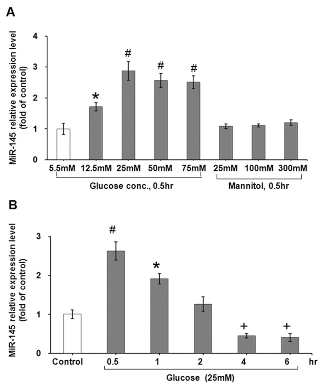
To explore the effect of high glucose concentration on miR-145 expression, different concentrations of glucose were added to the culture medium for 0.5 h. As shown in Figure 1A, miR-145 was increased significantly from 12.5 mmol/L to 75 mmol/L, with 25 mmol/L high glucose having the maximal effect. Therefore, 25 mmol/L high glucose was used for the following experiments. To exclude the osmotic effect of high glucose on miR-145 expression, three concentrations of mannitol were added. Mannitol did not effect miR-145 expression. As shown in Figure 1B, 25 mmol/L high glucose increased miR-145 expression maximally at 0.5 h and decreased gradually and reached significantly less than the control level after 4 to 6 h. High glucose stimulation for 0.5 h significantly increased reactive oxygen species production as compared with control cells (Supplementary Figure 1A). The addition of N-acetylcysteine (NAC), a free radical scavenger, at 500 μmol/L could reverse the miR-145 expression induced by high glucose and 2,2′-Dipyridyl–N, N-dimethylsemicarbazone (Dp44mT), an iron chelator to generate reactive oxygen species (Supplementary Figure 1B). These results indicate that hyperglycemia via ROS regulates miR-145 expression at 0.5 h in HCASMC.
Figure 1.
Effect of glucose level on miR-145 expression in cultured vascular smooth muscle cells. (A) Treatment of different glucose level for 0.5 h. Mannitol did not affect the miR-145 expression. (B) Treatment of glucose level at 25 mmol/L for different periods of time. *P < 0.05 versus control. #P < 0.01 versus control. +P < 0.01 versus control. n = 4 per group.
Angiotensin II Suppresses miR-145 Expression in Smooth Muscle Cells
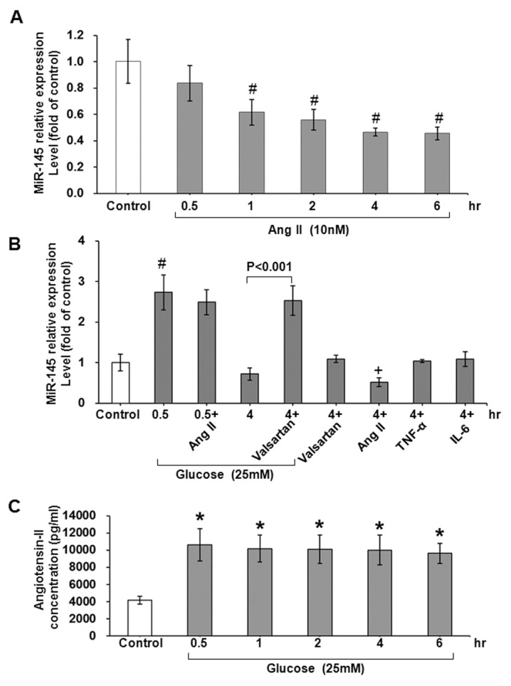
To evaluate the effect of Ang II on miR-145 expression in HCASMCs, Ang II (Bachem) at 10 nmol/L was added to the culture medium for 0.5 h to 6 h. As shown in Figure 2A, Ang II significantly inhibited miR-145 expression from 1 h to 6 h. Exogenous administration of tumor necrosis factor-α (PeproTech Inc.) at 1 ng/mL and interleukin-6 (R&D Systems Inc.) at 10 ng/mL did not affect the miR-145 expression (Figure 2B). Addition of angiotensin II receptor antagonist valsartan (Sigma-Aldrich) at 10 μmol/L significantly increased miR-145 expression in the high glucose state as compared with high glucose stimulation for 4 h. Addition of valsartan in cells not treated with high glucose did not affect miR-145 expression. High glucose did not significantly affect miR-145 expression after 0.5 h in cells pretreated with Ang II. High glucose stimulation also increased secretion of Ang II from cultured HCASMCs from 0.5 to 6 h (Figure 2C). The increased expression of Ang II mRNA was confirmed by real time-PCR in HCASMCs and in balloon-injured carotid artery in diabetic and wild-type (WT) rats (Supplementary Figure 2). These results indicate that Ang II plays a crucial role in miR-145 expression in HCASMCs under high glucose stimulation. Recently, Ang II has been shown to downregulate miR-145 via extracellular signal-regulated kinase1/2 in VSMCs (17).
Figure 2.
Effect of angiotensin 2 (Ang II) on miR-145 expression in cultured vascular smooth muscle cells. (A) Exogenous administration of Ang II at 10 nmol/L in usual culture medium without extra glucose treatment for different periods of time. (B) Exogenous administration of different cytokine proteins and valsartan for 4 h. #P < 0.01 versus control. +P < 0.05 versus control. n = 4 per group. (C) Effect of high glucose on secretion of angiotensin II from cultured vascular smooth muscle cells. Ang II was measured by immunosorbent assay. *P < 0.01 versus control. n = 3 per group.
High Glucose Increases Klf4 and Decreases Myocardin Expression in Smooth Muscle Cells
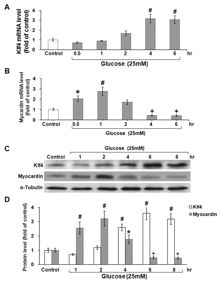
Since Klf4 and myocardin are the target genes of miR-145 (18,19), we sought to investigate the effect of high glucose on Klf4 and myocardin expression in HCASMCs. High glucose initially did not have an effect on Klf4 expression as shown in Figure 3A. However, Klf4 began to increase after 2 h of high glucose stimulation and Klf4 significantly increased messenger RNA and protein expression after 4 to 8 h (Figures 3A, C, D). In contrast to the Klf4 expression, high glucose significantly increased myocardin messenger RNA and protein expression for 0.5 to 1 h, followed by a gradual decrease after 2 to 8 h (Figures 3B–D).
Figure 3.
Effect of high glucose level at 25 mmol/L on Klf4 and myocardin mRNA and protein expression in cultured vascular smooth muscle cells. Quantitative analysis of Klf4 (A) and myocardin (B) mRNA levels. The values from VSMCs under high glucose treatment have been normalized to matched GAPDH measurement and then expressed as a ratio of normalized values to mRNA in control group. *P < 0.05 versus control. #P < 0.01 versus control. +P < 0.01 versus control. (n = 4 per group. (C) Representative Western blots for Klf4 and myocardin in VSMCs after high glucose at 25 mmol/L for various periods of time. (D) Quantitative analysis of Kfl4 and myocardin protein levels. The values from VSMCs after high glucose treatment have been normalized to matched α-tubulin measurement and then expressed as a ratio of normalized values to protein in control group. *P < 0.05 versus control. #P < 0.01 versus control. +P < 0.01 versus control. n = 4 per group.
Angiotensin II Mediates Klf4 and Myocardin Expression Induced by High Glucose Stimulation
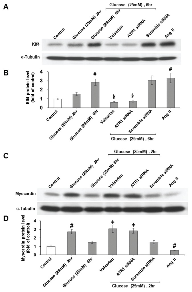
As shown in Figures 4A and B, high glucose significantly increased Klf4 protein expression at 6 h and exogenous addition of valsartan and angiotensin receptor type1 (ATR1) siRNA significantly inhibited the Klf4 protein expression induced by high glucose. High glucose significantly increased myocardin protein expression at 2 h (Figures 4C, D) but did not affect myocardin protein expression at 6 h. Exogenous addition of valsartan and ATR1 siRNA significantly increased myocardin expression in the high glucose state for 6 h. Exogenous addition of scramble siRNA did not have an effect on Klf4 and myocardin expression induced by high glucose stimulation. Exogenous addition of Ang II at 10 nmol/L in usual culture medium had a similar effect on Klf4 and myocardin protein expression in high glucose stimulation (Figure 4). Exogenous addition of angiotensin-converting enzyme inhibitor enalaprilat dehydrate (Sigma-Aldrich) at 10 μmol/L before high glucose stimulation also reversed Klf4 and myocardin expression induced by high glucose stimulation (Supplementary Figure 3). These data indicate that Ang II mediates the Klf4 and myocardin expression in a high glucose state.
Figure 4.
Angiotensin II mediates the increase of Klf4 and myocardin protein expression by high glucose in vascular smooth muscle cells. (A and C) Representative Western blots for Klf4 and myocardin protein levels in VSMCs subjected to high glucose stimulation for 6 h in the absence or presence of valsartan, angiotensin II receptor (ATR1) siRNA or angiotensin II (Ang II). (B and D) Quantitative analysis of Klf4 and myocardin protein levels. The values from stimulated VSMCs have been normalized to values in control cells. #P < 0.01 versus control. +P < 0.01 versus 6 h. §P < 0.01 versus 6 h. n = 4 per group.
miR-145 Decreases Klf4 and Increases Myocardin Protein Expression in High Glucose State
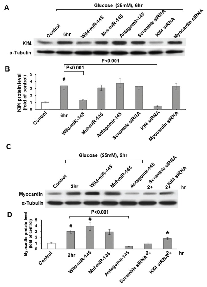
To evaluate the relationship between miR-145 and Klf4 and myocardin, WT miR-145, antagomir-145 and a mutant type of miR-145 were added to the cultured cells. As shown in Figures 5A and B, high glucose significantly increased Klf4 protein expression at 6 h and overexpression of WT miR-145 significantly attenuated the Klf4 protein expression induced by high glucose. The addition of antagomir-145 and the overexpression of mutant miR-145 did not affect the Klf4 protein expression under high glucose stimulation. Overexpression of WT miR-145 significantly increased myocardin protein expression as compared with the control group. (Figures 5C, D). The addition of antagomir-145 significantly inhibited the myocardin protein expression induced by high glucose stimulation. Klf4 siRNA significantly inhibited Klf4 protein expression and significantly increased myocardin protein expression, while myocardin siRNA did not affect Klf4 protein expression (Figure 5), indicating that Klf4 is upstream of myocardin and downregulates myocardin expression. To investigate whether Klf4 is a direct target of miR-145, we performed a luciferase assay to measure Klf4 promoter activity. As shown in Supplementary Figure 4, high glucose stimulation for 4 h and Ang II alone for 4 h without high glucose stimulation significantly increased Klf4 promoter activity as compared with control cells. Overexpression of miR-145, addition of valsartan and enalaprilat significantly attenuated the promoter activity induced by high glucose. When the conserved site of miR-145 in the promoter area of Klf4 was mutated, the increased promoter activity induced by high glucose and Ang II was abolished. Luciferase assay also showed that miR-145 is able to bind Klf4 and myocardin 3′-UTR and induced the opposite expression as shown in Supplementary Figure 5. High glucose stimulation for 0.5 h decreased Klf4–3′ UTR luciferase activity and increased myocardin–3′ UTR luciferase activity. Sustained high glucose stimulation for 4 h reversed the luciferase activity of Klf4 3′ UTR and myocardin 3′ UTR. Overexpression of miR-145 under high glucose stimulation attenuated Klf4–3′ UTR luciferase activity and increased myocardin–3′ UTR luciferase activity at 4 h.
Figure 5.
Effect of miR-145 on Klf4 and myocardin protein expression under high glucose stimulation in cultured VSMCs. (A) Representative Western blots for Klf4 protein levels in VSMCs subjected to high glucose stimulation for 6 h in the absence or presence of WT miR-145, antagomir-145, mutant miR-145 and Klf4 and myocardin siRNA. (B) Quantitative analysis of Klf4 protein levels. The values from stimulated VSMCs have been normalized to values in control cells. #P < 0.01 versus control. n = 4 per group. (C) Representative Western blots for myocardin protein levels in VSMCs subjected to high glucose stimulation for 6 h in the absence or presence of WT miR-145, mutant miR-145, antogomir-145, scramble and Klf4 siRNA. (D) Quantitative analysis of myocardin protein levels. The values from stimulated VSMCs have been normalized to values in control cells. #P < 0.01 versus control. *P < 0.05 versus control. n = 4 per group.
miR-145 Inhibits HCASMCs Proliferation and Migration Induced by High Glucose
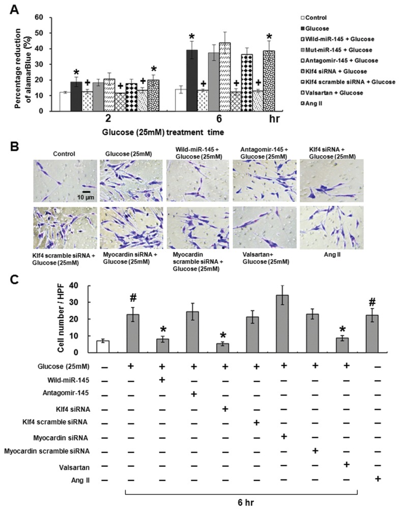
High glucose at 25 mmol/L and Ang II significantly increased HCASMCs proliferation and migration at 6 h as compared with the control group (Figure 6). Over-expression of WT miR-145 and the addition of Klf4 siRNA and valsartan reversed the proliferation and migration induced by high glucose stimulation for 6 h. Over-expression of mutant miR-145 and the addition of antagomir-145 and scramble siRNA did not decrease the proliferation and migration induced by high glucose. The migration of HCASMCs in myocardin silenced cell by myocardin siRNA did not change in high glucose stimulation. High glucose significantly decreased smooth muscle (SM)-specific contractile protein expression (SM-α actin) and increased synthetic protein (nonmuscle myosin heavy chain-B) expression. Overexpression of miR-145 reversed the effect of high glucose on these protein expressions (Supplementary Figure 6). Klf 5 plays an essential role in VSMC phenotype and vascular neointimal lesion formation (20). High glucose significantly increased Klf5 protein expression and the overexpression of miR-145 significantly attenuated the increase of Klf5 induced by high glucose. Knockdown of Klf5 by Klf5 siRNA reversed the effect of high glucose on VSMC phenotype (Supplementary Figure 6).
Figure 6.
Effect of high glucose and angiotensin II on HCASMCs proliferation and migration. (A) DNA synthesis by Alamar blue assay. *P < 0.01 versus control. +P < 0.01 versus hyperglycemia (25 mmol/L glucose). n = 4 per group. (B) HCASMCs migrated through filter were stained. HCASMCs treated with hyperglycemia and Ang II for 6 h in the presence or absence of WT miR-145, antagomir-145, Klf4 siRNA, or valsartan. (C) Migration of HCASMCs was quantified by staining and counting the number of cells that migrated to the bottom of the filter in five fields under a 400× high-power field (HPF). (n = 4 per group). #P < 0.001 versus control (lane 1). *P < 0.001 versus hyperglycemia (lane 2).
Balloon Injury Induces miR-145 Expression
The blood sugar in the streptozotocin-induced diabetic rat at 2 wks was 196 ± 9 mg/dl and 67 ± 16 mg/dl in the WT Wistar rat. As shown in Supplementary Figure 7, miR-145 expression in arterial tissue began to increase at 3 d, reached a maximal level at 7 d and maintained elevation up to 28 d after balloon injury of carotid artery in diabetic and WT rats. The miR-145 expression in the arterial tissue was significantly higher in the diabetic group than it was in the WT group from 3 d to 14 d. Klf4 protein expression was increased significantly after carotid artery balloon injury from 3 d to 28 d (Supplementary Figure 8). Myocardin protein expression also was increased significantly after balloon injury from 3 to 28 d, but the myocardin expression was maximal at 7 d and declined gradually up to 28 d after injury.
miR-145 Decreases Klf4 and Increases Myocardin Protein Expression in Arterial Tissue after Carotid Artery Balloon Injury
Treatment with WT miR-145 and valsartan significantly decreased the Klf4 protein expression and increased myocardin protein expression after balloon injury as compared with diabetes rats without balloon injury (Supplementary Figure 9). The Klf4 protein expression was significantly higher and myocardin protein expression was significantly lower in the antagomir-145 treatment group than in the WT miR-145 treatment group after balloon injury. Balloon injury increased the intimal area and decreased the lumen size of the carotid artery, while treatment with WT miR-145 and valsartan decreased the intimal area and increased the lumen size. Treatment with antagoir-145 and mutant type miR-145 did not change either the intimal area and or the lumen size as compared with balloon injury at 14 d (Supplementary Figure 10). Masson staining was used to assess the extent of vascular neointimal lesion formation in balloon-injured rat carotid arteries (Supplementary Figure 11).
DISCUSSION
A miR is a small, 20- to 25-nucleotide non-protein-coding RNA that usually inhibits transcription or translation by interacting with the 3′ untranslated regions of target mRNA and promoting target mRNA degradation (gene silencing) (21). Many miRs have been investigated in diabetic complications (20). However, rare miR was studied in diabetic vascular remodeling (2,22). miR-21 and miR143/145 have been shown to be associated with vascular remodeling (23). miR-143/145 also affected the angiotensin signaling and therefore affected blood pressure (4). Several miRs, including miR-21, miR-221, miR-222, miR-143 and miR-145 play a role in VSMC differentiation. miR-145 is the most abundant miR in VSMCs (6,24). Transition of cell type in VSMCs plays an important role in the pathogenesis of diabetic vasculopathy (25). Recently, miR-145 has been shown to restore contractile vascular muscle phenotype and coronary collateral growth in rats with metabolic syndrome (26). However, the effect of hyperglycemia on the regulation of miR-145 expression is currently unknown. In the present study, we found that high glucose significantly increased miR-145 expression briefly (within 1 h) and sustained hyperglycemia-inhibited miR-145 expression in HCASMCs. The upregulation of miR-145 expression immediately after hyperglycemia (0.5 h) may reflect a protective response.
Ang II was found to be enhanced by high glucose stimulation (27). In this study, we found that miR-145 was significantly inhibited by Ang II and that other cytokines such as TNF-α and IL-6 did not have the inhibitory effect on miR-145 expression. High glucose conditions also enhanced Ang II secretion from HCASMCs. The addition of valsartan, an Ang II receptor antagonist and enalaprilat dehydrate significantly upregulated miR-145 expression in high glucose stimulation for 4 h. This finding indicates that Ang II via angiotensin receptor downregulates miR-145 in VSMCs under high glucose conditions. Our study is the first one to demonstrate that valsartan has a direct effect on mediating the expression of miR-145 in VSMCs under hyperglycemic conditions. Hyperglycemia has been demonstrated to upregulate AT1 receptor expression in VSMCs and renal mesangial cells (28,29). The increased AT1 receptor in hyperglycemia condition is through oxidative stress (29).
Klf4 and myocardin are targets of miR-145 (7,19). The miR-145 binding sites in the 3′ untranslated region of KLf4 and myocardin could mediate Klf4 and myocardin expression. miR-145 downregulates Klf4 but upregulates myocardin expression. In the present study, we found that sustained high glucose increased Klf4 mRNA and protein expression while brief exposure to high glucose increased myocardin mRNA and protein expression but sustained high-glucose-inhibited myocardin mRNA and protein expression. Valsartan can reverse the effect of high glucose on Klf4 and myocardin expression. Ang II alone had a similar effect on Klf4 and myocardin expression as that under sustained high glucose stimulation. These results indicate that Klf4 and myocardin expression in a high glucose state is mediated by Ang II. Type 2 diabetic mellitus in practical medicine is not only the hyperglycemic condition but also the insulin resistance condition. The insulin resistance is an important mechanism to induce the undifferentiation and the proliferation of VSMCs. Hutcheson et al. have demonstrated that miR-145 downregulates Klf4 and upregulates myocardin expression in SMCs of metabolic syndrome rat (26). Recently, Wen et al. have demonstrated that miR-145 plays a role in the development of resistin-induced insulin resistance in HepG2 cell (30).
In the present study, we found that hyperglycemia and Ang II alone significantly increased VSMC proliferation by Alamar blue assay. Overexpression of miR-145 and the addition of Klf4 siRNA significantly attenuated the proliferation induced by hyperglycemia. The addition of valsartan before high glucose stimulation also significantly attenuated VSMC proliferation induced by hyperglycemia. The findings of the study confirm that miR-145 is an important vasculoprotective miR. Actually, miR-143/145 deficiency impairs vascular function (31). Restoration of miR-145 expression can limit neointimal formation in response to vascular injury by promoting Klf4 down-regulation (6). Taken together, these data demonstrate the importance of physiological levels of miR-145 for normal vascular function.
The expression of miR-145 and myocardin was maximally increased at 7 d after carotid injury and gradually declined thereafter in diabetic rats and Klf4 protein expression was gradually increased from 3 to 28 d after balloon injury. This result had a similar trend as in the in vitro study. There is a trend that when the expression of Klf4 increased after balloon injury, the expression of miR145 decreased. Overexpression of miR-145 and treatment with valsartan could reverse the Klf4 and myocardin protein expression induced by balloon injury. The intimal area and lumen size after balloon injury was reversed after overexpression of WT miR-145, confirming that miR-145 is an important vasculoprotective miR. Our in vivo results are contradictory to the result reported by Cheng et al. (6). Different animal species and injury procedure may explain the discrepancy.
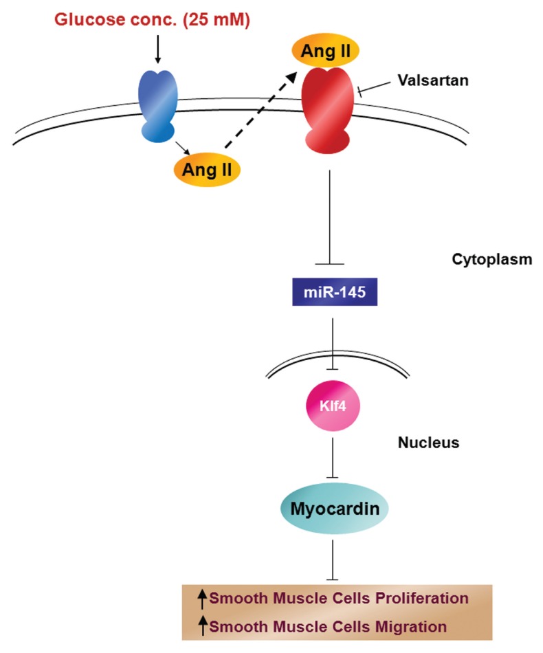
In summary, the present study reveals the molecular regulation of miR-145 in the downstream gene (such as Klf4 and myocardin) expression of HCASMCs under hyperglycemic condition. Ang II plays a critical role in the regulation of miR-145 under hyperglycemia conditions. The decreased miR-145 will increase the expression of its target gene KLf4. The increased Klf4 will then decrease myocardin to induce proliferation and migration of VSMCs. The pathway of effect of high glucose on Ang II and miR-145 in vascular smooth muscle cell is summarized in Figure 7. miR-145 has been considered to be a therapeutic target to reduce atherosclerosis in apolipoprotein E knockout mice (32). The better understanding of the detailed mechanisms of therapeutic miR-145 under hyperglycemic condition will provide us a new insight into therapeutic development for atherosclerosis that is frequently encountered in patients suffering from diabetes mellitus.
Figure 7.
Proposed signal pathway of high glucose on the regulation of miR-145 and Klf4 and myocardin for proliferation and migration in vascular smooth muscle cells. High glucose increases Ang II expression and Ang II through angiotensin receptor downregulates miR-145. miR-145 downregulates Klf4 and Klf4 downregulates myocardin expression, finally causing smooth muscle cell proliferation and migration to promote atherogenesis.
Supplemental Data
ACKNOWLEDGMENTS
This study was supported by grants from Ministry of Science and Technology, Taiwan and Shin Kong Wu Ho-Su Memorial Hospital, Taipei, Taiwan.
Footnotes
Online address: http://www.molmed.org
DISCLOSURE
The authors declare that they have no competing interests as defined by Molecular Medicine, or other interests that might be perceived to influence the results and discussion reported in this paper.
Cite this article as: Shyu K-G, et al. (2015) Angiotensin II downregulates microRNA-145 to regulate Kruppel-like factor 4 and myocardin expression in human coronary arterial smooth muscle cells under high glucose conditions. Mol. Med. 21:616–25.
REFERENCES
- 1.Paneni F, Beckman JA, Creager MA, Cosentino F. Diabetes and vascular disease: pathophysiology, clinical consequences, and medical therapy: part 1. Eur Heart J. 2013;34:2436–46. doi: 10.1093/eurheartj/eht149. [DOI] [PMC free article] [PubMed] [Google Scholar]
- 2.Shntikumarn S, Caporali A, Emanueli C. Role of microRNAs in diabetes and its cardiovascular complications. Cardiovasc Res. 2012;93:583–93. doi: 10.1093/cvr/cvr300. [DOI] [PMC free article] [PubMed] [Google Scholar]
- 3.Zampetaki A, Mayr M. MicroRNAs in vascular and metabolic disease. Circ Res. 2012;110:508–22. doi: 10.1161/CIRCRESAHA.111.247445. [DOI] [PubMed] [Google Scholar]
- 4.Boettger T, et al. Acquisition of the contractile phenotype by murine arterial smooth muscle cells depends on the Mir143/145 gene cluster. J Clin Invest. 2009;119:2634–47. doi: 10.1172/JCI38864. [DOI] [PMC free article] [PubMed] [Google Scholar]
- 5.Cordes KR, et al. miR-145 and miR-143 regulate smooth muscle cell fate and plasticity. Nature. 2009;460:705–10. doi: 10.1038/nature08195. [DOI] [PMC free article] [PubMed] [Google Scholar]
- 6.Cheng Y, et al. MicroRNA-145, a novel smooth muscle cell phenotypic marker and modulator, controls vascular neointimal lesion formation. Circ Res. 2009;105:158–66. doi: 10.1161/CIRCRESAHA.109.197517. [DOI] [PMC free article] [PubMed] [Google Scholar]
- 7.Xin M, et al. MicroRNAs miR-143 and miR-145 modulate cytoskeletal dynamics and responsiveness of smooth muscle cells to injury. Genes Dev. 2009;23:2166–78. doi: 10.1101/gad.1842409. [DOI] [PMC free article] [PubMed] [Google Scholar]
- 8.Wang Z, et al. Myocardin and ternary complex factors compete for SRF to control smooth muscle gene expression. Nature. 2004;428:185–9. doi: 10.1038/nature02382. [DOI] [PubMed] [Google Scholar]
- 9.Garvey SM, Sinden DS, Schoppee Bortz PD, Wamhoff BR. Cyclosporine up-regulates Kruppel-like factor-4 (KLF4) in vascular smooth muscle cells and drives phenotypic modulation in vivo. J Pharmacol Exp Ther. 2010;333:34–42. doi: 10.1124/jpet.109.163949. [DOI] [PMC free article] [PubMed] [Google Scholar]
- 10.Zanella MT, Kohlmann O, Jr, Ribeiro AB. Treatment of obesity, hypertension and diabetes syndrome. Hypertension. 2001;38:705–8. doi: 10.1161/01.hyp.38.3.705. [DOI] [PubMed] [Google Scholar]
- 11.Shyu KG, Wang BW, Wu GJ, Lin CM, Chang H. Mechanical stretch via transforming growth factor-β1 activates microRNA208a to regulate endoglin expression in cultured rat cardiac myoblasts. Eur J Heart Fail. 2013;15:36–45. doi: 10.1093/eurjhf/hfs143. [DOI] [PubMed] [Google Scholar]
- 12.Cheng WP, Hung HF, Wang BW, Shyu KG. The molecular regulation of GADD153 in apoptosis of cultured vascular smooth muscle cells by cyclic mechanical stretch. Cardiovasc Res. 2008;77:551–9. doi: 10.1093/cvr/cvm057. [DOI] [PubMed] [Google Scholar]
- 13.Wang BW, Chang H, Lin S, Kuan P, Shyu KG. Induction of matrix metalloproteinase-14 and -2 by cyclical mechanical stretch is mediated by tumor necrosis factor-α in cultured human umbilical vein endothelial cells. Cardiovasc Res. 2003;59:460–9. doi: 10.1016/s0008-6363(03)00428-0. [DOI] [PubMed] [Google Scholar]
- 14.Chang H, et al. GL-331 inhibits HIF-1α expression in a lung cancer model. Biochem Biophys Res Commun. 2003;302:95–100. doi: 10.1016/s0006-291x(03)00111-6. [DOI] [PubMed] [Google Scholar]
- 15.Shyu KG, Wang BW, Kuan P, Chang H. RNA Interference for discoidin domain receptor 2 attenuates neointimal formation in balloon injured rat carotid artery. Arterioscler Thromb Vasc Biol. 2008;28:1447–53. doi: 10.1161/ATVBAHA.108.165993. [DOI] [PubMed] [Google Scholar]
- 16.Committee for the Update of the Guide for the Care and Use of Laboratory Animals, Institute for Laboratory Animal Research, Division on Earth and Life Studies, National Research Council of the National Academies. Guide for the Care and Use of Laboratory Animals. 8th edition. Washington (DC): National Academic Press; 2011. [Google Scholar]
- 17.Hu B, et al. Mechanical stretch suppresses microRNA-145 expression by activating extracellular signal-regulated kinase1/2 and upregulating angiotensin-converting enzyme to alter vascular smooth muscle cell phenotype. PLoS One. 2014;9:e96338. doi: 10.1371/journal.pone.0096338. [DOI] [PMC free article] [PubMed] [Google Scholar]
- 18.Davies-Dusenbery BN, et al. Down-regulation of Kruppel-like factor-4 (KLF4) by microRNA-143/145 is critical for modulation of vascular smooth muscle cell phenotype by transforming growth factor-β and bone morphogenetic protein 4. J Biol Chem. 2011;286:28097–110. doi: 10.1074/jbc.M111.236950. [DOI] [PMC free article] [PubMed] [Google Scholar]
- 19.Rangrez AY, Massy ZA, Meuth VML, Metzinger L. miR-143 and miR-145: Molecular keys to switch the phenotype of vascular smooth muscle cells. Circ Cardiovasc Genet. 2011;4:197–205. doi: 10.1161/CIRCGENETICS.110.958702. [DOI] [PubMed] [Google Scholar]
- 20.Shu HJ, Wen JK, Miao SB, Liu Y, Zheng B. KLF5 and hhLIM cooperatively promote proliferation of vascular smooth muscle cells. Mol Cell Biochem. 2012;367:185–94. doi: 10.1007/s11010-012-1332-9. [DOI] [PubMed] [Google Scholar]
- 21.Bartel DP. MicroRNAs: genomics, biogenesis, mechanism, and function. Cell. 2004;116:281–97. doi: 10.1016/s0092-8674(04)00045-5. [DOI] [PubMed] [Google Scholar]
- 22.Natarajan R, Putta S, Kato M. MicroRNAs and diabetic complication. J Cardiovasc Trans Res. 2012;5:413–22. doi: 10.1007/s12265-012-9368-5. [DOI] [PMC free article] [PubMed] [Google Scholar]
- 23.Small EM, Olson EN. Pervasive roles of microRNAs in cardiovascular biology. Nature. 2007;469:36–342. doi: 10.1038/nature09783. [DOI] [PMC free article] [PubMed] [Google Scholar]
- 24.Ji R, et al. MicroRNA expression signature and antisense-mediated depletion reveal an essential role of microRNA in vascular neointimal lesion formation. Circ Res. 2007;100:1579–88. doi: 10.1161/CIRCRESAHA.106.141986. [DOI] [PubMed] [Google Scholar]
- 25.Rzucidlo EM, Martin KA, Powell RJ. Regulation of vascular smooth muscle cell differentiation. J Vasc Surg. 2007;45:A25–32. doi: 10.1016/j.jvs.2007.03.001. [DOI] [PubMed] [Google Scholar]
- 26.Hutcheson R, et al. MicroRNA-145 restores contractile vascular smooth muscle phenotype and coronary collateral growth in the metabolic syndrome. Arterioscler Thromb Vasc Biol. 2013;33:727–36. doi: 10.1161/ATVBAHA.112.301116. [DOI] [PMC free article] [PubMed] [Google Scholar]
- 27.Natarajan R, Scott S, Bai W, Yerneni KKV, Nadler J. Angiotensin II signaling in vascular smooth muscle cells under high glucose conditions. Hypertension. 1999;33:378–84. doi: 10.1161/01.hyp.33.1.378. [DOI] [PubMed] [Google Scholar]
- 28.Sodhi CP, Kanwar YS, Sahai A. Hypoxia and high glucose upregulate AT1 receptor expression and potentiate ANG II-induced proliferation in VSM cells. Am J Physiol Heart Circ Physiol. 2003;284:H846–52. doi: 10.1152/ajpheart.00625.2002. [DOI] [PubMed] [Google Scholar]
- 29.Xue, et al. H2S inhibits hyperglycemia-induced intrarenal renin-angiotensin system activation via attenuation of reactive oxygen species generation. PLoS One. 2013;8:e74336. doi: 10.1371/journal.pone.0074366. [DOI] [PMC free article] [PubMed] [Google Scholar]
- 30.Wen, et al. miRNA-145 is involved in the development of resistin-induced insulin resistance in HepG2 cells. Biochem Biophys Res Commun. 2014;445:517–23. doi: 10.1016/j.bbrc.2014.02.034. [DOI] [PubMed] [Google Scholar]
- 31.Norta GD, et al. MicroRNA 143–145 deficiency impairs vascular function. Int J Immunol Pharmacol. 2012;25:467–84. doi: 10.1177/039463201202500216. [DOI] [PubMed] [Google Scholar]
- 32.Lovren F, et al. MicroRNA-145 targeted therapy reduces atherosclerosis. Circulation. 2012;126(11 Suppl 1):S81–90. doi: 10.1161/CIRCULATIONAHA.111.084186. [DOI] [PubMed] [Google Scholar]
Associated Data
This section collects any data citations, data availability statements, or supplementary materials included in this article.


