Abstract
CRISPR-Cas systems, which obstruct both viral infection and incorporation of mobile genetic elements by horizontal transfer, are a specific immune response common to prokaryotes. Antiviral protection by CRISPR-Cas comes at a cost, as horizontally-acquired genes may increase fitness and provide rapid adaptation to habitat change. To date, investigations into the prevalence of CRISPR have primarily focused on pathogenic and clinical bacteria, while less is known about CRISPR dynamics in commensal and environmental species. We designed PCR primers and coupled these with DNA sequencing of products to detect and characterize the presence of cas1, a universal CRISPR-associated gene and proxy for the Type II CRISPR1-Cas system, in environmental and non-clinical Enterococcus isolates. CRISPR1-cas1 was detected in approximately 33% of the 275 strains examined, and differences in CRISPR1 carriage between species was significant. Incidence of cas1 in E. hirae was 73%, nearly three times that of E. faecalis (23.6%) and 10 times more frequent than in E. durans (7.1%). Also, this is the first report of CRISPR1 presence in E. durans, as well as in the plant-associated species E. casseliflavus and E. sulfureus. Significant differences in CRISPR1-cas1 incidence among Enterococcus species support the hypothesis that there is a tradeoff between protection and adaptability. The differences in the habitats of enterococcal species may exert varying selective pressure that results in a species-dependent distribution of CRISPR-Cas systems.
Introduction
Bacteria and Archaea possessing CRISPR-Cas systems trade off horizontally-acquired adaptation to a changing environment for protection against lethal virus infection. CRISPRs are clustered regularly interspaced short palindromic repeats of DNA; Cas refers to CRISPR-associated proteins. Together, they comprise a uniquely prokaryotic multi-step adaptive immune response that provides defense against bacteriophage infection [1]. In the process, incorporation of transmissible genetic elements is interrupted, including plasmids and DNA with potential advantages for the host cell, such as those conferring antibiotic resistance [2]. Briefly, fragments of non-self DNA called protospacers are acquired by Cas proteins, and incorporated as spacers between the DNA repeats of the CRISPR array. These repeat-spacer modules are transcribed and expressed as crRNAs, a small interference-type RNA. If invading nucleic acid has a short sequence with perfect complementarity to the spacer region of the crRNA, a sequence-specific cleavage event is initiated, degrading the foreign nucleic acids [3,4]. CRISPR arrays are widespread among Bacteria and Archaea, in approximately 90% of archaeal and 40% of bacterial genomes examined [5,6]. The diversity of CRISPR systems is extensive. CRISPRs may be broadly divided into those lacking cas genes, thus consisting solely of repeat-spacer arrays (also referred to as orphan CRISPRs), and those comprised of both an array and associated functional genes (CRISPR-Cas). CRISPR-Cas systems are further divided into types and subtypes, defined by presence of subtype-specific Cas proteins [7]. Several Cas proteins are considered universal, with orthologs appearing in every active subtype. One of these is Cas1 [7,8]. Encoded by a single gene (cas1), the ubiquity of Cas1 makes it a suitable marker for the presence of a potentially active CRISPR-Cas system.
We focused on CRISPR1 systems in the genus Enterococcus, a clade of commensal bacteria common to animal and human gut microflora. Enterococci emerged as a cause of multidrug resistant hospital acquired infection in the 1970s, and presently represent one of the most prevalent causes of nosocomial infections in the United States [9]. Two species–E. faecalis and E. faecium–are primarily responsible for these infections [10]. They are also the predominant enterococcal human gastrointestinal (GI) commensals [11]. Mobile elements, including plasmids, pathogenicity islands, and antibiotic resistance genes, comprise as much as 25% of the genomes of hospital-adapted lineages of both species [12,13,14]. Palmer and Gilmore (15) showed that multiple drug resistance and incidence of CRISPR-Cas are negatively correlated in E. faecalis and E. faecium. That is, their results suggest that there is a tradeoff between acquisition of drug resistance and CRISPR-mediated protection from foreign DNA. Three Type II CRISPRs have been identified in human GI E. faecalis: two with associated cas genes (CRISPR1-Cas and CRISPR3-Cas) and one orphan repeat-spacer array (CRISPR2) [15]. CRISPR2 is present in 95% of E. faecalis isolates; as many as half of these strains contain CRISPR1-Cas, and CRISPR3-Cas has been detected in four E. faecalis genomes to date [15,16]. This suggests that species under different selective pressures may vary significantly in their incidence of CRISPR.
Several studies have investigated CRISPR in clinical and virulent enterococci, but few have addressed the prevalence of these systems in environmental and commensal strains [16,17,18,19]. Additionally, CRISPR content in E. faecalis and E. faecium has been extensively reported, but a comprehensive survey including other Enterococcus species is lacking [15,17,18,20,21]. Since antiviral protection by CRISPR-Cas also prevents incorporation of potentially beneficial genes, retention of a CRISPR locus represents a tradeoff between protection and adaptability. To test the hypothesis that different habitats affect this tradeoff and thus the prevalence of CRISPR, our objective was to determine the frequency of active Type II CRISPR1 systems in Enterococcus species. Environmental, non-clinical enterococci were screened for presence of the conserved CRISPR1-cas1 gene, as a marker for the active CRISPR locus most commonly detected in this genus. CRISPR1-cas1 was detected in multiple Enterococcus species, including several not previously characterized as containing CRISPR systems. Significant differences in cas1 incidence between species were also observed.
Methods
Enterococcus strains
Enterococcus isolates were cultured from activated sludge, oxygenated wastewater from residential and industrial sources, including storm runoff. Other samples included soil and sediment, compost, vegetation, marine and freshwater sources, and canine, feline, and avian fecal specimens (S1 Table). No permits were required for the described study, which complied with all relevant regulations. Water, soil, sediment, plant clippings, and fecal samples were taken from public properties where permission was not required, or from private property with permission of the owners. Activated sludge samples were supplied by water treatment plant supervisory personnel.
Activated sludge was diluted to 1:1000, and 10 mL of the dilution was filtered through 0.22-μm pore-size membrane filters, then incubated on mEnteroccocus agar (Difco) at 35°C for 24 hours. Isolated colonies were selected from the agar, and streaked for isolation of pure cultures on Enterococcosel agar (BBL). Environmental and fecal samples were enriched by incubation in azide dextrose broth for 24 hours at 35°C, followed by isolation of pure cultures on Enterococcosel agar. Additional Enterococcus strains from beach sand were isolated as previously described [22].
Enterococcus faecalis OG1RF (ATCC 47077), which contains a CRISPR1 locus, was selected as a positive control [20]. The strain was purchased from the American Type Culture Collection (Manassas, VA).
All isolates were Gram-positive, catalase-negative cocci. Species identity of all isolates was determined by 16S rRNA sequence match in the Ribosomal Database Project (http://rdp.cme.msu.edu/index.jsp), and identities were verified by 16S rRNA phylogenetic analysis. Isolate cas1 sequences were confirmed to be Enterococcus cas1 genes by BLASTn (NCBI) sequence match against the nucleotide collection (nr/nt) database.
Identification of CRISPR components in available genome sequences
Enterococcus cas1 genes for primer design were identified by BLASTn of the NCBI nucleotide collection (nr/nt) database, using the E. faecalis OG1RF cas1 sequence (accession number CP002621.1) as the query (Fig A in S1 Text). CRISPR repeat-spacer arrays, and cas genes in proximity to the arrays, were investigated in 13 available Enterococcus genomes in CRISPRdb (E. casseliflavus EC20, accession number CP004856.1; E. faecalis 62, CP002491.1; E. faecalis D32, CP003726.1; E. faecalis OG1RF, CP002621.1; E. faecalis str. Symbioflor 1, HF558530.1; E. faecalis V583, AE016830.1; E. faecium Aus0004, CP003351.1; E. faecium Aus0085, CP006620.1; E. faecium DO, CP003583.1; E. faecium NRRL B-2354, CP004063.1; E. hirae ATCC 9790, CP003504.1; E. mundtii QU 25, AP013036.1; Enterococcus sp. 7L76, FP929058.1 [5]. Additional draft genomes (E. durans ATCC 6056, accession number GCA_000406985.1; E. faecium FB129-CNAB-4, GCA_000315405.1; E. durans IPLA 655, GCA_000350465.1) were downloaded from GenBank and analyzed for CRISPR content using CRISPRfinder (Table A in S1 Text) [6].
PCR and sequencing
Nucleic acid extractions were performed using the MoBio UltraClean® Microbial DNA Isolation Kit. The variable region of the 16S rRNA gene was amplified using universal bacterial DNA primers, forward, 5'-CCTACGGGAGGCAGCAG-3'; reverse, 5'-ATTACCGCGGCTGCTGG-3' [23].
To screen isolates for CRISPR1-cas1, primers amplifying a 212-bp internal region of the cas1 gene (forward, 5’-ATGGGCTGGCGAAC-3’; reverse, 5’- CGCTTRTCATCGCAA-3’) were used. Multiple alignment of Enterococcus CRISPR1-cas1 nucleotide sequences available at that time (E. faecalis OG1RF, accession number CP002621.1; E. faecalis D32, CP003726.1; E. hirae ATCC 9790, CP003504.1) was performed by MUSCLE [24,25] to locate conserved regions of the cas1 homologs. Primers were designed manually, and their compatibility was confirmed using Primer3 (http://bioinfo.ut.ee/primer3/) [26,27]. Primers were deemed compatible, as Tm differed by 0.75°C, and no complementarity (self, pair, and primer hairpin) was detected. Target specificity of the primer set was further confirmed by Primer BLAST (http://www.ncbi.nlm.nih.gov/tools/primer-blast/) against all Enterococcus (taxid: 1350), using all variations of the reverse primer, which contains a degenerate base. The primer set amplified in silico in E. faecalis OG1RF, E. faecalis D32, and E. hirae ATCC 9790. Amplification was optimized for the following program: 2 minutes at 94°C, 30 cycles of [1 minute at 94°C, 1 minute at 48.9°C, 1 minute at 72°C], 10 minutes at 72°C.
PCR products were submitted to Massachusetts General Hospital DNA Sequencing Core Facility or Eton Biosciences, Boston, MA for sequencing. Sequences were curated manually, and 16S rRNA gene sequences were deposited in GenBank (S1 Table).
Analysis and phylogeny
To test whether CRISPR1-cas1 distribution significantly differed by species or source, data were analyzed by Chi square and Fisher’s exact tests (Tables 1–3).
Table 1. Detection of CRISPR1-cas1 in all Enterococcus strains, by source of isolate.
Differences between sources are not significant, P value = 0.6598.
| Source | cas1-positive | No. of isolates | % cas1- positives |
|---|---|---|---|
| Activated sludge | 40 | 131 | 30.5 |
| Environmental samples | 38 | 113 | 33.6 |
| Animal fecal | 12 | 31 | 38.7 |
Table 3. Detection of CRISPR1-cas1 in E. hirae strains, by source of isolate.
Differences between sources are not significant, P value = 0.3302.
| Source | cas1-positive | No. of isolates | % cas1 positives |
|---|---|---|---|
| Activated sludge | 29 | 39 | 74.4 |
| Environmental samples | 16 | 25 | 64.0 |
| Animal fecal | 12 | 14 | 85.7 |
Table 2. Detection of CRISPR1-cas1 in E. faecalis strains, by source of isolate.
Differences between sources are not significant, P value = 0.6166.
| Source | cas1-positive | No. of isolates | % cas1- positives |
|---|---|---|---|
| Activated sludge | 9 | 38 | 23.7 |
| Environmental samples | 17 | 69 | 24.6 |
| Animal fecal | 0 | 3 | - |
Phylogeny was constructed using SeaView 4 (http://doua.prabi.fr/software/seaview). Multiple sequence alignment was performed within Seaview 4 using MUSCLE [25], and gap-only sites were removed. A maximum likelihood tree (PhyML) was generated, using the GTR model and aLRT branch support, with all other parameters set to default (nucleotide equilibrium frequencies: empirical; Ts/Tv ratio: fixed, 4.0; invariable sites: none; across site rate variation: optimized; tree searching operations: NNI; starting tree: BioNJ, optimized tree topology). FigTree 1.4.2 (http://tree.bio.ed.ac.uk/software/figtree) was used for tree visualization. Two of the sequences used to design the cas1 primers were used as reference sequences in the cas1 phylogenetic tree; E. faecalis D32 was omitted, as it is identical to that of E. faecalis OG1RF.
Results
The predominant Enterococcus species isolated were E. faecalis (40.0% of 275 total isolates), E. hirae (28.4%), E. durans (20.4%), and E. faecium (5.1%). Additional enterococcal species were isolated less frequently, and include E. casseliflavus, E. sulfureus, E. mundtii, E. malodoratus, E. termitis, and E. sanguinicola (Table 4).
Table 4. Detection of CRISPR1-cas1 in Enterococcus, by species.
Differences in CRISPR1-cas1 detection between E. faecalis, E. hirae, and E. durans isolates are significant, P value < 0.0001. Species for which a low number of strains were isolated are indicated in italics.
| Species | Cas1-positives | Total isolates | Percent cas1 positive |
|---|---|---|---|
| E. faecalis | 26 | 110 | 23.6 |
| E. hirae | 57 | 78 | 73.1 |
| E. durans | 4 | 56 | 7.1 |
| E. faecium | 1 | 14 | 7.1 |
| E. casseliflavus | 1 | 7 | 14.3 |
| E. sulfureus | 1 | 2 | 50.0 |
| E. mundtii | 0 | 2 | 0.0 |
| E. sanguinicola | 0 | 1 | 0.0 |
| E. malodoratus | 0 | 4 | 0.0 |
| E. termitis | 0 | 1 | 0.0 |
| Total | 90 | 275 | 32.7 |
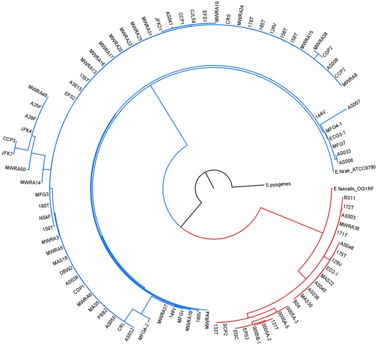
The CRISPR1-cas1 gene was detected in 32.7% of all Enterococcus isolates (Table 4). Within the three most predominant species isolated, frequency of cas1 detection varied significantly. The incidence of CRISPR1-cas1 genes between E. faecalis, E. durans, and E. hirae is significantly different (Table 4; p < 0.0001). The frequency of remaining species was not considered in this analysis due to small sample size. CRISPR1-cas1 was detected in 23.6% of E. faecalis isolates, while 73.1% of E. hirae and 7.1% of E. durans strains contain the gene. Cas1 was also detected in isolates of E. faecium, E. casseliflavus and E. sulfureus. The few strains of E. malodoratus, E. sanguinicola, E. mundtii, and E. termitis that were isolated did not contain cas1 (Table 4). The origin of the bacterial strain and presence of a CRISPR1-cas1 gene were not significantly correlated. This observation was consistent for all Enterococcus species analyzed, as well as intraspecific analyses of the two most commonly isolated species, E. faecalis and E. hirae (Tables 1–3). A phylogenetic tree of partial cas1 sequences formed two strongly distinct clusters around the E. faecalis OG1RF and the E. hirae ATCC 9790 cas1 reference sequences (Fig 1). All but 4 of the 26 E. faecalis cas1 genes clustered with the E. faecalis OG1RF-like cas1 gene. The remaining four strains of E. faecalis (MWRA37, MWRA22, 176T, and 158T) contained an E. hirae-like cas1 homolog. All identified E. hirae strains possess a cas1 homolog similar to that of E. hirae ATCC 9790. Cas1 sequences for E. casseliflavus, E. faecium, and E. sulfureus share identity with the E. hirae gene. E. durans strains contained cas1 genes homologous to both the E. faecalis OG1RF and E. hirae ATCC9790 cas1 types.
Fig 1. Phylogenetic tree of CRISPR1-cas1 partial sequences.
Red branches represent the E. faecalis-like cas1 cluster; blue branches represent the E. hirae cas1 cluster.
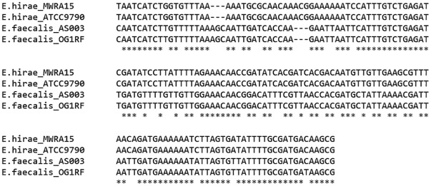
Cas1 sequences are conserved in the region amplified in this study, and the E. hirae and E. faecalis homologs are distinctly different from each other, perhaps reflecting species-level evolution. Within this region, the sequences differ by 16 transitions, 20 transversions, and a 3 bp indel, and not a continuum of differences between the two clusters (Fig 2). E. faecalis strains usually contain an E. faecalis cas1 homolog, and E. hirae-like cas1 genes typically appear in strains identified as E. hirae. Additionally, three of four E. durans cas1-positive strains contain E. hirae homologs, but one contains an E. faecalis-like gene. Horizontal transfer of CRISPR components in enterococci has yet to be demonstrated.
Fig 2. Comparison of partial CRISPR1-cas1 sequences.
Representative isolates (E. hirae MWRA15 and E. faecalis AS003) and reference strains (E. hirae ATCC 9790 and E. faecalis OG1RF) were aligned using MUSCLE. Bases conserved between all analyzed sequences are indicated with asterisks; spaces denote transitions and transversions, and dashes represent indel regions.
Discussion
Incidence of cas1 in Enterococcus
This study is the first systematic analysis of Type II CRISPR1-Cas incidence in non-clinical enterococci. The incidence of CRISPR1-associated cas1 in E. hirae (73.1%) is significantly higher than in E. faecalis (Table 4). E. faecalis is a human commensal species, and selective pressure for antibiotic resistance may be high [15]. If the selective pressure for adapting to antibiotics in the human gut environment is higher than the selective pressure by lytic bacteriophage, then lower incidence of CRISPR-Cas is expected for species in habitats with higher phage pressure. The phage pressure in the typical habitats of E. hirae are not characterized, but E. hirae is primarily associated with animals, including birds, household pets, and livestock. Although E. hirae is implicated in animal disease, it is very rarely pathogenic to humans [28]. As CRISPR presence is inversely correlated with acquisition of traits such as antibiotic resistance in enterococci, widespread distribution of CRISPR1-cas1 within this species may correspond with its lack of virulence.
An effect of the source of isolated enterococci was not observed; however, activated sludge contains wastewater influent from a variety of sources, including household, commercial, and clinical sewers, as well as storm drain runoff. Therefore, differentiating bacterial isolates by host species origin from a common source is problematic. Additionally, environmental samples, such as the beach sand and sediment used in this study, may be influenced by human or animal presence, and should not be considered autochthonous [29]. Thus, it is not possible to conclusively compare strain origin or source and CRISPR-Cas presence in the current study. This remains an area for future research.
Cas1 phylogeny indicates horizontal transfer of CRISPR loci
The tight clustering of the partial cas1 sequence phylogeny was striking. Therefore, the four strains of E. faecalis that clustered with the E. hirae cas1 sequences indicates horizontal transfer of CRISPR elements between Enterococcus species (Fig 1). CRISPR1-cas1 genes identified in E. sulfureus, E. casseliflavus, and E. faecium cluster with the cas1 homologs in E. hirae strains (Fig 1). This is further indication of horizontal transfer, or CRISPR-Cas systems may be conserved with high levels of sequence similarity between these species. A more comprehensive description of the CRISPR-Cas systems in E. durans, E. faecium, E. casseliflavus, and E. sulfureus is needed to answer this question, as well as to shed light on differences in cas genes and array content that may explain interspecific CRISPR diversity.
CRISPR1 in E. durans, E. casseliflavus, and E. sulfureus
This is the first report of the presence of CRISPR1-Cas systems in E. durans, E. casseliflavus, and E. sulfureus. E. durans is a minor component of human and animal gut flora, and is also found in food of animal origin, especially dairy products [11,28]. Lack of virulence genes, including those that confer antibiotic resistance, indicate a probiotic role for E. durans [30]. CRISPR1 incidence in E. durans is low, but phage pressure in typical habitats of this species are not well characterized. E. casseliflavus and E. sulfureus are primarily plant-associated species [31,32,33]. Recent studies of E. casseliflavus have implicated the bacterium in human infection; however, these cases remain infrequent [34,35,36,37]. Reports implicating E. sulfureus in human disease could not be found in scientific literature. The rarity with which these species are pathogenic suggests an inverse correlation between virulence and CRISPR content, as was demonstrated in Escherichia coli [15,38,39]. Accurate frequencies of CRISPR1 loci in these species will require more comprehensive testing. In this study, only a few isolates of these species were cultured and screened for the cas1 gene.
CRISPR1-cas1 was not detected in isolates of E. mundtii and E. malodoratus. CRISPR loci have not been reported in two E. mundtii genomes previously analyzed, and incidence in E. malodoratus has also not been reported [40,41]. However, these sample sizes are too small to conclude that these species do not possess CRISPR1 loci. Additionally, the Type II-specific cas1 primers used in this study are unlikely to amplify all cas1 genes within Enterococcus, as species may contain CRISPR-Cas systems of different types [42]. With the three additional species reported here to contain CRISPR1-cas1, six species of Enterococcus are reported to possess CRISPR. But, as many as 40 other Enterococcus species have yet to be investigated [43]. Although more thorough characterization is warranted, the presence of cas genes in the species reported here indicates that CRISPR1-Cas systems may be widespread among the Enterococcus genus. The primers designed here successfully amplified a conserved region of the cas1 gene in multiple enterococcal species, making it an efficient marker for screening for CRISPR1 loci. Furthermore, widespread incidence of active CRISPRs and omnipresence of the clade in many environments make Enterococcus an ideal model for investigation of CRISPR dynamics.
Conclusions
Immunity against lytic phages is a recognizable evolutionary benefit for a bacterium, demonstrated both experimentally and in mathematical models of CRISPR-Cas/phage interaction [1,44]. Often considered as beneficial, indiscriminate insertion of foreign genetic elements, such as genomic islands, prophages and plasmids, on the other hand, can result in disruption of essential gene function and incorrect regulation of acquired genes [45]. CRISPR-mediated prevention of these detrimental insertions may also confer an evolutionary advantage [2]. However, horizontally-acquired genes may increase fitness by conferring habitat adaptations. Such adaptations in Enterococcus include antibiotic resistance, enhanced biofilm formation, resistance to metal toxicity, and expanded metabolic capacity [46,47]. Maintaining a functional CRISPR-Cas system also incurs an energetic cost for the organism [45]. Thus, for the bacterium possessing CRISPR loci, there is a tradeoff between adaptability and protection. Significant differences in Type II CRISPR1-cas1 incidence seen here indicate that selective pressures exerted by this tradeoff may influence CRISPR-Cas distribution in a species-dependent manner. The nature of this selection remains an area of future research.
Supporting Information
(TIF)
(DOCX)
(DOCX)
Acknowledgments
We are grateful to Wogenie Tessema and Laura LeBlanc for contributing data to this study, and Steven Bryant and Sarah Feinman for providing additional bacterial strains. The work was supported in part by NSF (IOS -0847691 8), NSF REU Program (DBI-1062748) and the NIH Bridges to the Baccalaureate Program (2R25GM075306).
Data Availability
All relevant data are within the paper and its Supporting Information files.
Funding Statement
This work was supported by the National Science Foundation (IOS -0847691 8) (http://www.nsf.gov), the National Science Foundation Research Experiences for Undergraduates (DBI-1062748) (http://www.nsf.gov), and the National Institutes of Health Bridges to the Baccalaureate Program (2R25GM075306) (http://www.nih.gov). The funders had no role in study design, data collection and analysis, decision to publish, or preparation of the manuscript.
References
- 1. Barrangou R, Fremaux C, Deveau H, Richards M, Boyaval P, Moineau S, et al. (2007) CRISPR provides acquired resistance against viruses in prokaryotes. Science 315: 1709–1712. [DOI] [PubMed] [Google Scholar]
- 2. Marraffini LA, Sontheimer EJ (2008) CRISPR interference limits horizontal gene transfer in staphylococci by targeting DNA. Science 322: 1843–1845. 10.1126/science.1165771 [DOI] [PMC free article] [PubMed] [Google Scholar]
- 3. Bhaya D, Davison M, Barrangou R (2011) CRISPR-Cas systems in bacteria and archaea: versatile small RNAs for adaptive defense and regulation. Annual review of genetics 45: 273–297. 10.1146/annurev-genet-110410-132430 [DOI] [PubMed] [Google Scholar]
- 4. Horvath P, Barrangou R (2010) CRISPR/Cas, the immune system of bacteria and archaea. Science 327: 167–170. 10.1126/science.1179555 [DOI] [PubMed] [Google Scholar]
- 5. Grissa I, Vergnaud G, Pourcel C (2007) The CRISPRdb database and tools to display CRISPRs and to generate dictionaries of spacers and repeats. BMC bioinformatics 8: 172 [DOI] [PMC free article] [PubMed] [Google Scholar]
- 6. Grissa I, Vergnaud G, Pourcel C (2007) CRISPRFinder: a web tool to identify clustered regularly interspaced short palindromic repeats. Nucleic acids research 35: W52–57. [DOI] [PMC free article] [PubMed] [Google Scholar]
- 7. Makarova KS, Haft DH, Barrangou R, Brouns SJ, Charpentier E, Horvath P, et al. (2011) Evolution and classification of the CRISPR-Cas systems. Nature reviews Microbiology 9: 467–477. 10.1038/nrmicro2577 [DOI] [PMC free article] [PubMed] [Google Scholar]
- 8. Haft DH, Selengut J, Mongodin EF, Nelson KE (2005) A guild of 45 CRISPR-associated (Cas) protein families and multiple CRISPR/Cas subtypes exist in prokaryotic genomes. PLoS Computational Biology 1: e60 [DOI] [PMC free article] [PubMed] [Google Scholar]
- 9. Magill SS, Edwards JR, Bamberg W, Beldavs ZG, Dumyati G, Kainer MA, et al. (2014) Multistate point-prevalence survey of health care-associated infections. The New England journal of medicine 370: 1198–1208. 10.1056/NEJMoa1306801 [DOI] [PMC free article] [PubMed] [Google Scholar]
- 10. Gilmore MS, Lebreton F, van Schaik W (2013) Genomic transition of enterococci from gut commensals to leading causes of multidrug-resistant hospital infection in the antibiotic era. Current opinion in microbiology 16: 10–16. 10.1016/j.mib.2013.01.006 [DOI] [PMC free article] [PubMed] [Google Scholar]
- 11. Blaimont B, Charlier J, Wauters G (1995) Comparative distribution of Enterococcus species in faeces and clinical samples. Microbial ecology in health and disease 8: 87–92. [Google Scholar]
- 12. Leavis HL, Willems RJ, van Wamel WJ, Schuren FH, Caspers MP, Bonten MJ (2007) Insertion sequence-driven diversification creates a globally dispersed emerging multiresistant subspecies of E. faecium . PLoS pathogens 3: e7 [DOI] [PMC free article] [PubMed] [Google Scholar]
- 13. Paulsen IT, Banerjei L, Myers GS, Nelson KE, Seshadri R, Read TD, et al. (2003) Role of mobile DNA in the evolution of vancomycin-resistant Enterococcus faecalis . Science 299: 2071–2074. [DOI] [PubMed] [Google Scholar]
- 14. Shankar N, Baghdayan AS, Gilmore MS (2002) Modulation of virulence within a pathogenicity island in vancomycin-resistant Enterococcus faecalis . Nature 417: 746–750. [DOI] [PubMed] [Google Scholar]
- 15. Palmer KL, Gilmore MS (2010) Multidrug-resistant enterococci lack CRISPR-cas. mBio 1. [DOI] [PMC free article] [PubMed] [Google Scholar]
- 16. Katyal I, Chaban B, Ng B, Hill JE (2013) CRISPRs of Enterococcus faecalis and E. hirae isolates from pig feces have species-specific repeats but share some common spacer sequences. Microbial ecology 66: 182–188. 10.1007/s00248-013-0217-0 [DOI] [PubMed] [Google Scholar]
- 17. Burley KM, Sedgley CM (2012) CRISPR-Cas, a prokaryotic adaptive immune system, in endodontic, oral, and multidrug-resistant hospital-acquired Enterococcus faecalis . Journal of endodontics 38: 1511–1515. 10.1016/j.joen.2012.07.004 [DOI] [PubMed] [Google Scholar]
- 18. Lindenstrauss AG, Pavlovic M, Bringmann A, Behr J, Ehrmann MA, Vogel RF (2011) Comparison of genotypic and phenotypic cluster analyses of virulence determinants and possible role of CRISPR elements towards their incidence in Enterococcus faecalis and Enterococcus faecium . Systematic and applied microbiology 34: 553–560. 10.1016/j.syapm.2011.05.002 [DOI] [PubMed] [Google Scholar]
- 19. Qin X, Galloway-Pena JR, Sillanpaa J, Roh JH, Nallapareddy SR, Chowdhury S, et al. (2012) Complete genome sequence of Enterococcus faecium strain TX16 and comparative genomic analysis of Enterococcus faecium genomes. BMC microbiology 12: 135 10.1186/1471-2180-12-135 [DOI] [PMC free article] [PubMed] [Google Scholar]
- 20. Bourgogne A, Garsin DA, Qin X, Singh KV, Sillanpaa J, Yerrapragada S, et al. (2008) Large scale variation in Enterococcus faecalis illustrated by the genome analysis of strain OG1RF. Genome biology 9: R110 10.1186/gb-2008-9-7-r110 [DOI] [PMC free article] [PubMed] [Google Scholar]
- 21. van Schaik W, Top J, Riley DR, Boekhorst J, Vrijenhoek JE, Schapendonk CM, et al. (2010) Pyrosequencing-based comparative genome analysis of the nosocomial pathogen Enterococcus faecium and identification of a large transferable pathogenicity island. BMC genomics 11: 239 10.1186/1471-2164-11-239 [DOI] [PMC free article] [PubMed] [Google Scholar]
- 22. Yasuda M, Paar J, Doolittle M, Brochi J, Pancorbo OC, Tang RJ, et al. (2010) Enterococcus species composition determined by capillary electrophoresis of the groESL gene spacer region DNA. Water research 44: 3982–3992. 10.1016/j.watres.2010.05.007 [DOI] [PubMed] [Google Scholar]
- 23. Muyzer G, de Waal EC, Uitterlinden AG (1993) Profiling of complex microbial populations by denaturing gradient gel electrophoresis analysis of polymerase chain reaction-amplified genes coding for 16S rRNA. Applied and environmental microbiology 59: 695–700. [DOI] [PMC free article] [PubMed] [Google Scholar]
- 24. Edgar RC (2004) MUSCLE: a multiple sequence alignment method with reduced time and space complexity. BMC bioinformatics 5: 113 [DOI] [PMC free article] [PubMed] [Google Scholar]
- 25. Edgar RC (2004) MUSCLE: multiple sequence alignment with high accuracy and high throughput. Nucleic acids research 32: 1792–1797. [DOI] [PMC free article] [PubMed] [Google Scholar]
- 26. Untergasser A, Cutcutache I, Koressaar T, Ye J, Faircloth BC, Remm M, et al. (2012) Primer3—new capabilities and interfaces. Nucleic acids research 40: e115 [DOI] [PMC free article] [PubMed] [Google Scholar]
- 27. Rozen S, Skaletsky H (2000) Primer3 on the WWW for general users and for biologist programmers. Methods in molecular biology 132: 365–386. [DOI] [PubMed] [Google Scholar]
- 28. Devriese LA, Vancanneyt M, Descheemaeker P, Baele M, Van Landuyt HW, Gordts B, et al. (2002) Differentiation and identification of Enterococcus durans, E. hirae and E. villorum. Journal of applied microbiology 92: 821–827. [DOI] [PubMed] [Google Scholar]
- 29. Halliday E, Gast RJ (2011) Bacteria in beach sands: an emerging challenge in protecting coastal water quality and bather health. Environmental science & technology 45: 370–379. [DOI] [PMC free article] [PubMed] [Google Scholar]
- 30. Pieniz S, de Moura TM, Cassenego APV, Andreazza R, Frazzon APG, de Oliveira Camargo FA, et al. (2015) Evaluation of resistance genes and virulence factors in a food isolated Enterococcus durans with potential probiotic effect. Food Control 51: 49–54. [Google Scholar]
- 31. Martinez-Murcia AJ, Collins MD (1991) Enterococcus sulfureus, a new yellow-pigmented Enterococcus species. FEMS microbiology letters 64: 69–74. [DOI] [PubMed] [Google Scholar]
- 32. Mundt JO, Graham WF (1968) Streptococcus faecium var. casseliflavus, nov. var. Journal of bacteriology 95: 2005–2009. [DOI] [PMC free article] [PubMed] [Google Scholar]
- 33. Vaughan DH, Riggsby W, Mundt JO (1979) Deoxyribonucleic acid relatedness of strains of yellow-pigmented, group D streptococci. International journal of systematic bacteriology 29: 204–212. [Google Scholar]
- 34. Holzel C, Bauer J, Stegherr EM, Schwaiger K (2014) Presence of the vancomycin resistance gene cluster vanC1, vanXYc, and vanT in Enterococcus casseliflavus . Microbial drug resistance 20: 177–180. 10.1089/mdr.2013.0108 [DOI] [PubMed] [Google Scholar]
- 35. Low JR, Teoh CS, Chien JM, Huang EH (2015) Enterococcus casseliflavus endophthalmitis due to metallic intraocular foreign body. Eye. [DOI] [PMC free article] [PubMed] [Google Scholar]
- 36. Narciso-Schiavon JL, Borgonovo A, Marques PC, Tonon D, Bansho ET, Maggi DC, et al. (2015) Enterococcus casseliflavus and Enterococcus gallinarum as causative agents of spontaneous bacterial peritonitis. Annals of hepatology 14: 270–272. [PubMed] [Google Scholar]
- 37. Liu Y, Wang Y, Dai L, Wu C, Shen J (2014) First report of multiresistance gene cfr in Enterococcus species casseliflavus and gallinarum of swine origin. Veterinary microbiology 170: 352–357. 10.1016/j.vetmic.2014.02.037 [DOI] [PubMed] [Google Scholar]
- 38. Lebreton F, van Schaik W, McGuire AM, Godfrey P, Griggs A, Mazumdar V, et al. (2013) Emergence of epidemic multidrug-resistant Enterococcus faecium from animal and commensal strains. mBio 4. [DOI] [PMC free article] [PubMed] [Google Scholar]
- 39. Garcia-Gutierrez E, Almendros C, Mojica FJ, Guzman NM, Garcia-Martinez J (2015) CRISPR Content Correlates with the Pathogenic Potential of Escherichia coli. PloS one 10: e0131935 10.1371/journal.pone.0131935 [DOI] [PMC free article] [PubMed] [Google Scholar]
- 40. Magni C, Espeche C, Repizo GD, Saavedra L, Suarez CA, Blancato VS, et al. (2012) Draft genome sequence of Enterococcus mundtii CRL1656. Journal of bacteriology 194: 550 10.1128/JB.06415-11 [DOI] [PMC free article] [PubMed] [Google Scholar]
- 41. Shiwa Y, Yanase H, Hirose Y, Satomi S, Araya-Kojima T, Watanabe S, et al. (2014) Complete genome sequence of Enterococcus mundtii QU 25, an efficient L-(+)-lactic acid-producing bacterium. DNA research: an international journal for rapid publication of reports on genes and genomes 21: 369–377. [DOI] [PMC free article] [PubMed] [Google Scholar]
- 42. Duerkop BA, Palmer KL, Horsburgh MJ (2014) Enterococcal Bacteriophages and Genome Defense In: Gilmore MS, Clewell DB, Ike Y, Shankar N, editors. Enterococci: From Commensals to Leading Causes of Drug Resistant Infection. Boston. [Google Scholar]
- 43. Parte AC (2014) LPSN—list of prokaryotic names with standing in nomenclature. Nucleic acids research 42: D613–616. 10.1093/nar/gkt1111 [DOI] [PMC free article] [PubMed] [Google Scholar]
- 44. Levin BR (2010) Nasty viruses, costly plasmids, population dynamics, and the conditions for establishing and maintaining CRISPR-mediated adaptive immunity in bacteria. PLoS genetics 6: e1001171 10.1371/journal.pgen.1001171 [DOI] [PMC free article] [PubMed] [Google Scholar]
- 45. Bondy-Denomy J, Davidson AR (2014) To acquire or resist: the complex biological effects of CRISPR-Cas systems. Trends in Microbiology 22: 218–225. 10.1016/j.tim.2014.01.007 [DOI] [PubMed] [Google Scholar]
- 46. Palmer KL, Kos VN, Gilmore MS (2010) Horizontal gene transfer and the genomics of enterococcal antibiotic resistance. Current opinion in microbiology 13: 632–639. 10.1016/j.mib.2010.08.004 [DOI] [PMC free article] [PubMed] [Google Scholar]
- 47. Santagati M, Campanile F, Stefani S (2012) Genomic diversification of enterococci in hosts: the role of the mobilome. Frontiers in microbiology 3: 95 10.3389/fmicb.2012.00095 [DOI] [PMC free article] [PubMed] [Google Scholar]
Associated Data
This section collects any data citations, data availability statements, or supplementary materials included in this article.
Supplementary Materials
(TIF)
(DOCX)
(DOCX)
Data Availability Statement
All relevant data are within the paper and its Supporting Information files.


