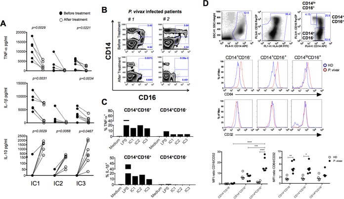
FIG 6 .
Purified ICs induce high levels of proinflammatory cytokines by CD14+ CD16+ monocytes from malaria patients. (A) PBMCs from P. vivax malaria patients (n = 6) before and 30 to 45 days after treatment were isolated and stimulated with 60 µg/ml of ICs for 24 h. The levels of TNF-α, IL-1β, and IL-10 were measured in supernatants by CBA. The P values were determined by a paired t test. (B) Flow cytometric analysis shows an increased frequency of CD14+ CD16+ cells in PBMCs from two P. vivax malaria patients. The frequency of CD14+ CD16+ cells decreased to levels similar to those for healthy donors at 30 to 40 days after treatment and parasitological cure. Results of three additional patients and healthy donors are shown in Fig. S4A in the supplemental material. (C) PBMCs from two different P. vivax malaria patients were isolated and stimulated with 60 µg/ml of ICs from three different patients for 10 h in culture containing brefeldin A and submitted to flow cytometric analysis to measure the expression of intracellular TNF-α and IL-1β in CD14+ CD16− as well as CD14+ CD16+ monocytes. The results are representative of two out of five patients. (D) Top panels show the gating strategy to identify the monocyte subsets: CD14+ CD16−, CD14+ CD16+, and CD14lo CD16+. The middle two panels are representative histograms of CD64 and CD32 expression in P. vivax-infected patients and healthy donors (HD). Bottom panels show the mean fluorescence intensity (MFI) ratios of CD16/CD32 (left) and CD64/CD32 (right) of the three monocyte subsets from P. vivax-infected patients (n = 6) and in healthy donors (HD, n = 4). *, P < 0.05; **, P < 0.01; ***, P < 0.001; ****, P < 0.0001.

