FIG 3 .
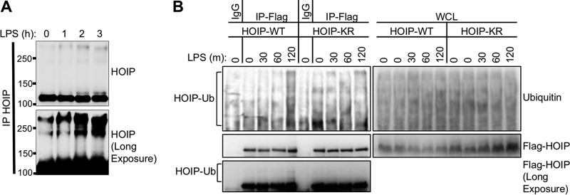
HOIP is ubiquitinated upon TLR4 stimulation. (A) A20.2J mouse B cells were stimulated with LPS and lysed under denaturing conditions. Endogenous HOIP was immunoprecipitated, separated by SDS-PAGE, and analyzed by immunoblotting. High-molecular-weight HOIP bands (>120,000) appeared after LPS stimulation. (B) Complemented mouse A20.2J HOIP−/− cells used for Fig. 2C were stimulated with LPS for the indicated times and lysed under denaturing conditions. Anti-IgG (control)- or anti-Flag (HOIP)-immunoprecipitated samples were prepared as described for panel A. A long exposure of the Flag immunoblot revealed the presence of high-molecular-weight HOIP bands. Data are representative of three independent experiments.

