

Abstract
Acromioclavicular joint dislocations are a common injury particularly among contact sports players. There has been an increasing trend toward arthroscopic management of these injuries. To date, these reconstructions have primarily addressed superoinferior instability by reconstructing the coracoclavicular ligaments. We describe an all-arthroscopic technique for reconstruction of the coracoclavicular ligaments using Arthrex ABS TightRopes (Arthrex, Naples, FL), with additional stabilization of the superior acromioclavicular joint capsule using an anchor-based suture bridge to address anteroposterior instability.
Injury to the acromioclavicular joint (ACJ) is common; however, the true incidence is difficult to measure because many of these injuries are not diagnosed and treated. Athletes, particularly those involved in contact sports, skiing, or cycling, are at increased risk of these injuries.
There is a sequential pattern of injuries to the supporting structures of the ACJ during trauma that was first described by Cadenat.1 With increasing energy, the acromioclavicular (AC) ligaments tear first, followed by the coracoclavicular (CC) ligaments. Finally, the deltoid and trapezius muscles or their attachments fail. This pattern of injury was used as the basis for the classification system of Tossy et al.,2 which was subsequently extended by Rockwood3 in 1984. The Rockwood classification system remains the most common classification in use today.3
The ACJ is a diarthrodial joint with 4 df. It moves in both the anteroposterior and superoinferior planes. A synovial lining surrounds the joint. Its surfaces are coated in hyaline cartilage. An articular disk sits within the joint; however, the actual function of the disk is most likely negligible.
The ACJ is stabilized by the AC ligaments and CC ligaments. These ligaments are further stabilized by the deltoid and trapezius muscles. Of the AC ligaments, the posterosuperior capsule contributes most to stability, primarily resisting horizontal translation.4 Of the 2 CC ligaments, the conoid is primarily responsible for control of vertical translation whereas the trapezoid is mostly responsible for controlling ACJ compression.4
There has been increasing interest in arthroscopic management of the ACJ; however, previously described techniques have principally addressed reconstruction of the CC ligaments alone. Control of the anteroposterior plane mandates reconstruction of the ACJ ligaments, particularly the superior capsule. A partial arthroscopic technique designed to address this structure has been described; however, this uses a mini-open approach with tendon graft passed through the bone tunnels crossing the ACJ.5 We describe an all-arthroscopic technique to address AC and CC ligament stability using extra-articular restraints to horizontal and vertical translation (Table 1).
Table 1.
Tips, Pitfalls, and Indications
| Tips |
| Creating the working space above the ACJ is achieved by blindly inserting the arthroscope and using a sweeping side-to-side motion subcutaneously to open the space. A switching stick is occasionally required as a retractor to hold the space open. |
| Before final tightening of the TightRope, it is best to view the ACJ from above to observe it being reduced as the TightRope is tensioned. |
| Pitfalls |
| The surgeon should check that the ACJ can be easily reduced at the beginning of the case to avoid problems when tensioning the TightRope. |
| The surgeon should take care when using the Arthrex aiming device to ensure that it remains in position. We normally view the position of the device on the undersurface of the coracoid to ensure that it does not displace during drilling. |
| Indications |
| This technique is indicated for grade 4 and 5 ACJ dislocations, as well as selected grade 3 injuries. |
ACJ, acromioclavicular joint.
Technique
Our technique (Video 1) is indicated for repair of acute grade 4 and 5 ACJ dislocations and selected grade 3 injuries. Buttonholing of the lateral clavicle is not a contraindication to performing this arthroscopic repair. We consider injuries presenting within 6 weeks to be acute.
The repair comprises 2 distinct stages. First, reconstruction of the CC ligaments is performed. Once the clavicle is held reduced, reconstruction of the superior AC ligaments is performed.
We position our patients in the beach-chair position on a table with a T-Max shoulder positioner (Smith & Nephew, Andover, MA). The arm is held by the Spider 1 limb-positioning table attachment (Smith & Nephew). The images and video used in this description show reconstruction of a patient's left shoulder.
CC Reconstruction
The portals used in this procedure are named in keeping with the arthroscopic shoulder portals described by Lafosse et al.6 for the arthroscopic Latarjet procedure. Three shoulder portals are used for the arthroscopic ACJ reconstruction. The D portal, placed off the anterolateral corner of the acromion, is the main viewing portal. The C portal, placed off the lateral border of the acromion in line with the posterior border of the clavicle, is used as a working portal to develop the space over the superior aspect of the ACJ and lateral clavicle. The standard anterior portal (E portal) is placed immediately lateral to the tip of the coracoid process. Two further 1-cm incisions are used for prosthesis placement. One incision is positioned over the superior aspect of the clavicle in line with the coracoid process. The other incision is centered over the ACJ superiorly for placement of the 4 anchors used to reconstruct the superior ACJ ligaments.
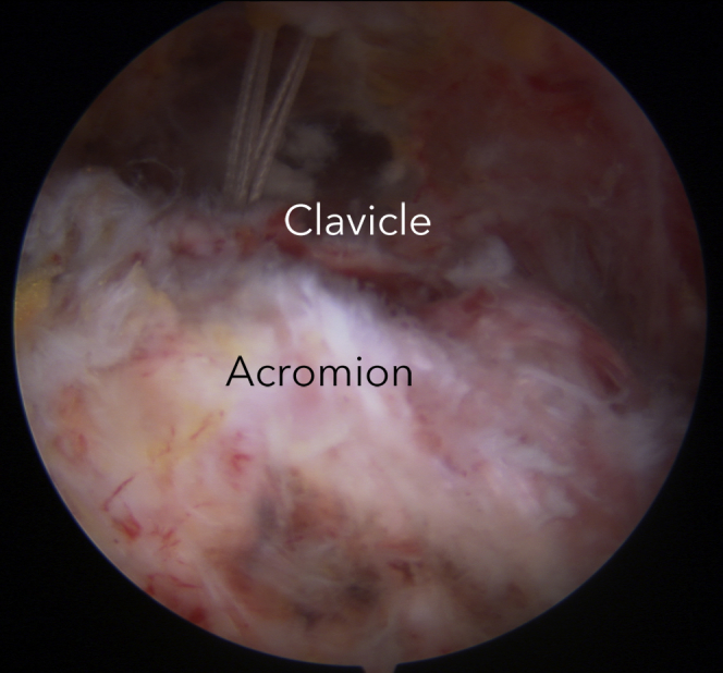
The procedure commences with a 30° arthroscope introduced through the D portal aiming at the coracoid. The anterior E portal is then placed adjacent to the tip of the coracoid, and a chondrotome (we use a 5.5-mm Dyonics incisor-blade chondrotome [Smith & Nephew]) is introduced through the E portal to clear the inferior aspect of the coracoid process (Fig 1). The arthroscope is then withdrawn and reintroduced through the D portal so that it lies superior to the acromion. The working space over the acromion is initially created with a sweeping motion of the arthroscope as it is introduced. A chondrotome is then introduced through the C portal to assist in further developing the space so that the superior aspects of the acromion, ACJ, and lateral clavicle are visualized (Fig 2). The space is extended over the clavicle medial to the level of the coracoid process. The VAPR Coolpulse 90 electrocautery system (DePuy, Warsaw, Poland) is used as required.
Fig 1.
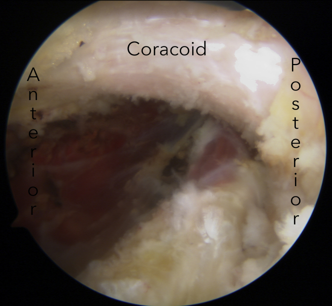
The undersurface of the coracoid is cleared using electrocautery in preparation for drilling of a tunnel connecting the clavicle to the coracoid. This tunnel will be used to pass 2 ABS TightRopes to reconstruct the coracoclavicular ligaments.
Fig 2.
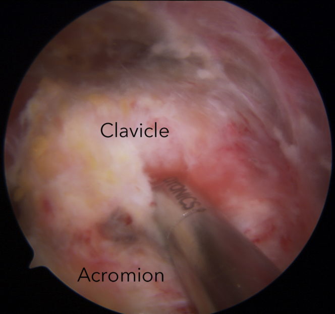
The superior surfaces of the clavicle and acromion are cleared using electrocautery and an arthroscopic 5.5-mm incisor-blade chondrotome. This space is required to visualize reduction of the acromioclavicular joint and to allow the suture bridge repair of the acromioclavicular ligaments.
Reducibility of the ACJ is confirmed while the ACJ is visualized from the space created over the acromion. A 1-cm incision is then placed over the superior aspect of the clavicle immediately in line with the palpated coracoid process. The Arthrex aiming guide (Arthrex, Naples, FL) is introduced through this incision over the clavicle and through the anterior E portal. The arthroscope is moved into the D portal so that the position of the aiming guide under the coracoid process can be visualized (Fig 3). The base of the coracoid process is the preferred position for guide placement. The cannulated 3-mm Arthrex drill is drilled between the clavicle and the coracoid process with the aid of the guide (Fig 4). The central core is removed, and the Arthrex nitinol loop is passed through the cannulated drill from above and retrieved under the coracoid process with an arthroscopic grasper (Fig 5).
Fig 3.
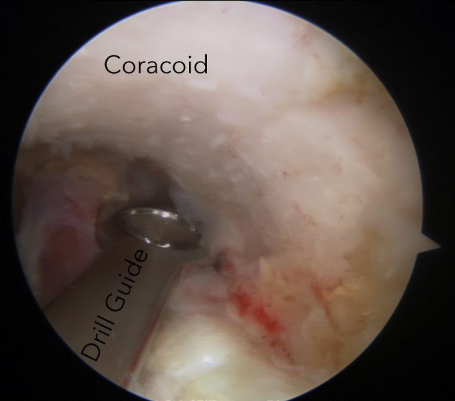
An Arthrex aiming guide is placed under the coracoid near its base. The upper part of the guide is centered on the clavicle by feel. This allows a cannulated drill to be inserted through the clavicle and coracoid.
Fig 4.
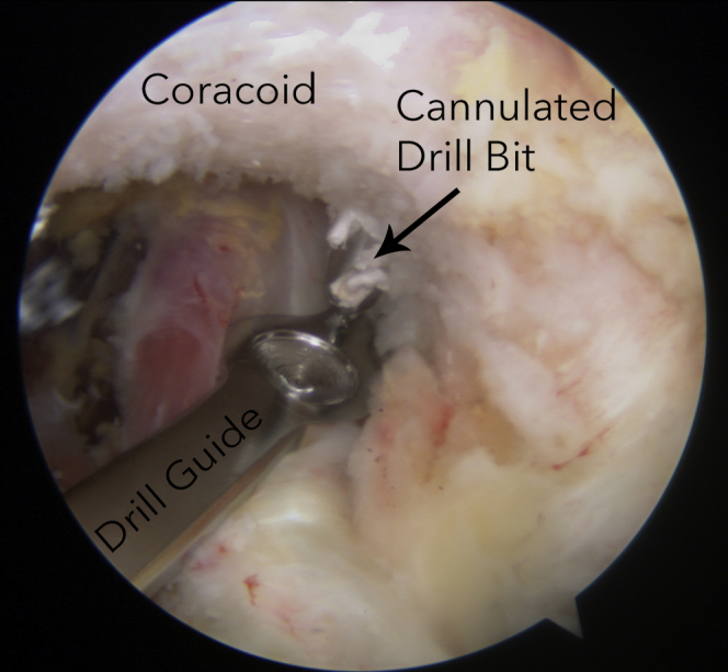
The drill is shown emerging through the coracoid after passing through the clavicle. The central part of the drill is then removed to allow passage of a nitinol suture-retrieving loop through the drill.
Fig 5.
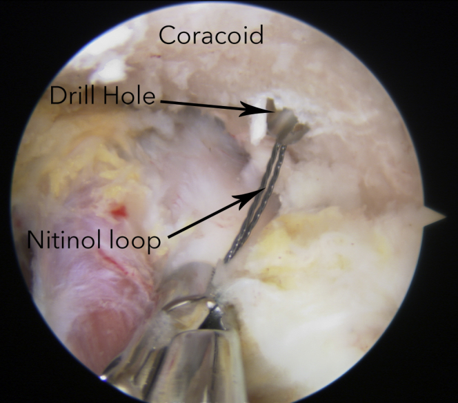
A nitinol suture-retrieving loop is retrieved through the skin using an arthroscopic grasper. This loop passes upward through the coracoid, clavicle, and skin, allowing a TightRope to be pulled upward through the tunnels.
Two Arthrex ABS TightRopes are passed through the coracoid and clavicle on a FiberWire leading suture (Arthrex) (Figs 6 and 7) with a Dog-Bone button (Arthrex) attached so that it comes to lie on the underside of the coracoid (Fig 8). With the 4 strands of the ABS TightRopes retrieved through the skin incision overlying the clavicle, a second Dog-Bone button is attached to the 2 loops. This second Dog-Bone button is then parachuted into position on the superior surface of the clavicle (Fig 9) as the TightRopes are progressively tightened. The reduction and degree of tightening of the TightRopes are determined by watching the reduction of the ACJ with the arthroscope sitting in the D portal (Fig 10). The arthroscope is advanced medially, and the position of the Dog-Bone button on the clavicle is visualized. The ABS TightRopes are self-locking, and therefore no knotting is required. Finally, the sutures of the TightRopes are cut with arthroscopic suture cutters.
Fig 6.
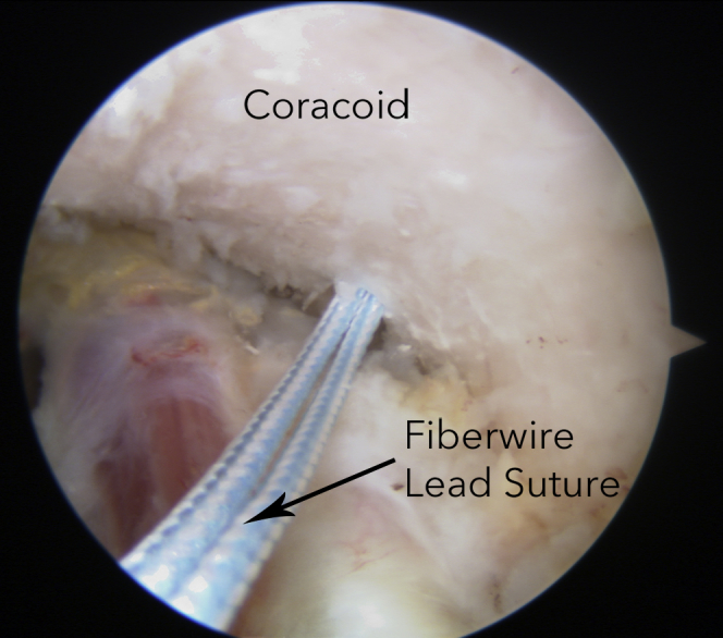
A FiberWire lead suture has been pulled through the coracoid and clavicle drill holes using a nitinol suture-retrieving loop. This lead suture is attached to 2 ABS TightRopes and will be used to pull the TightRopes through the coracoid and clavicle tunnels.
Fig 7.
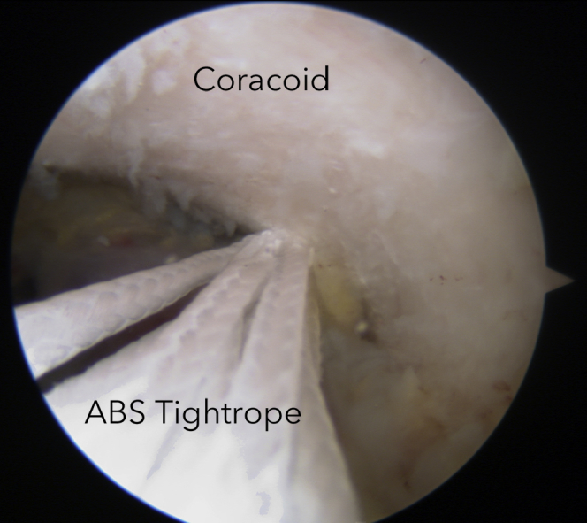
The 2 ABS TightRopes are being pulled through the coracoid and clavicle drill holes on a FiberWire lead suture.
Fig 8.
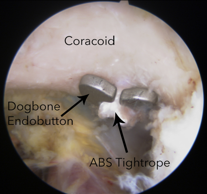
After a tunnel has been drilled between the coracoid and clavicle, 2 ABS TightRopes are passed with an attached Dog-Bone button. As the TightRopes are pulled through the tunnel, the Dog-Bone button is carefully positioned under the coracoid with the aid of arthroscopic graspers if required.
Fig 9.
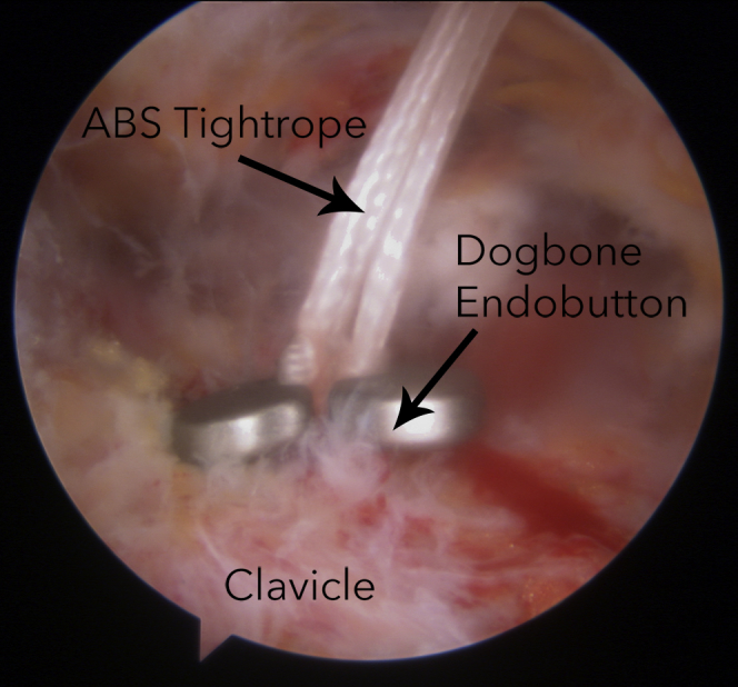
After the 2 ABS TightRopes are passed through the coracoid and clavicle tunnel, with an attached Dog-Bone button seated under the coracoid, another Dog-Bone button is used to secure the TightRopes on top of the clavicle.
Fig 10.
Visualization of the acromion and clavicle allows assessment of the reduction as the coracoclavicular TightRopes are tensioned. The reduction maneuver can be assisted with downward pressure on the clavicle by an assistant. Because the ABS TightRopes are self-locking, care should be taken at this stage because, once tensioned, they cannot easily be loosened.
AC Reconstruction
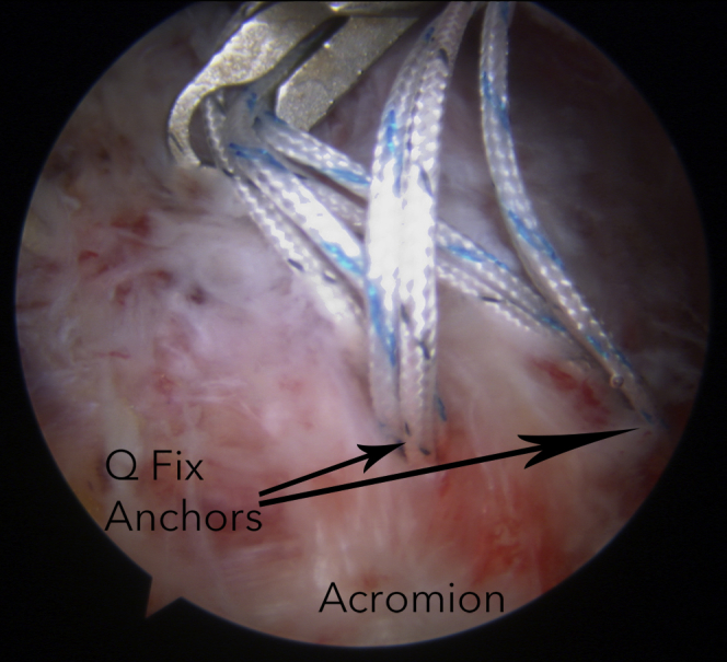
To reconstruct the ACJ, two 2.4-mm double-loaded Q-Fix arthroscopic anchors (ArthroCare, Austin, TX) are inserted into the superior aspect of the acromion adjacent to the ACJ (Fig 11) through a 1-cm incision positioned over the center of the ACJ. All 4 anchors used to repair the ACJ are inserted through this portal. The sutures (8 in total) are divided into 2 bundles, with each bundle containing 2 sutures from each anchor (Fig 12). These 2 bundles of sutures are then anchored to the distal clavicle using two 4.75-mm Healix Advance Knotless anchors (DePuy) (Fig 13) to produce a suture bridge–type construct similar to that described for a double-row rotator cuff repair. This creates an 8-stranded repair of the superior AC ligaments (Fig 14). The final construct is shown diagrammatically in Figure 15.
Fig 11.
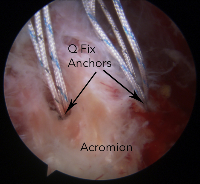
Two Q-Fix anchors are inserted into the superior aspect of the acromion. These anchors will be used to create a suture bridge over the acromioclavicular joint to reconstruct the acromioclavicular ligaments.
Fig 12.
Half of the sutures from each acromial anchor are retrieved through a working portal and will be anchored to the clavicle using a Healix Advance Knotless lateral-row anchor.
Fig 13.
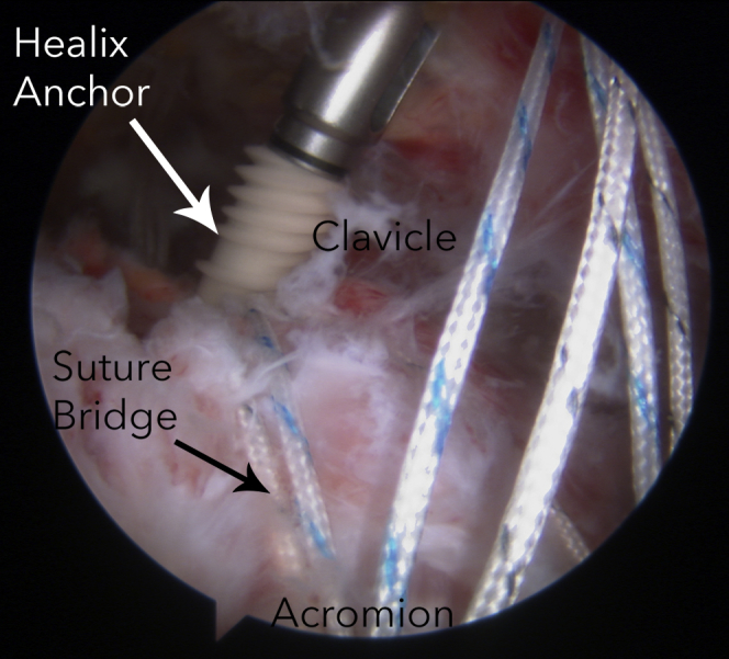
Half of the sutures from each acromial anchor are being secured to the clavicle using a Healix Advance Knotless anchor, creating a suture bridge across the superior aspect of the acromioclavicular joint.
Fig 14.
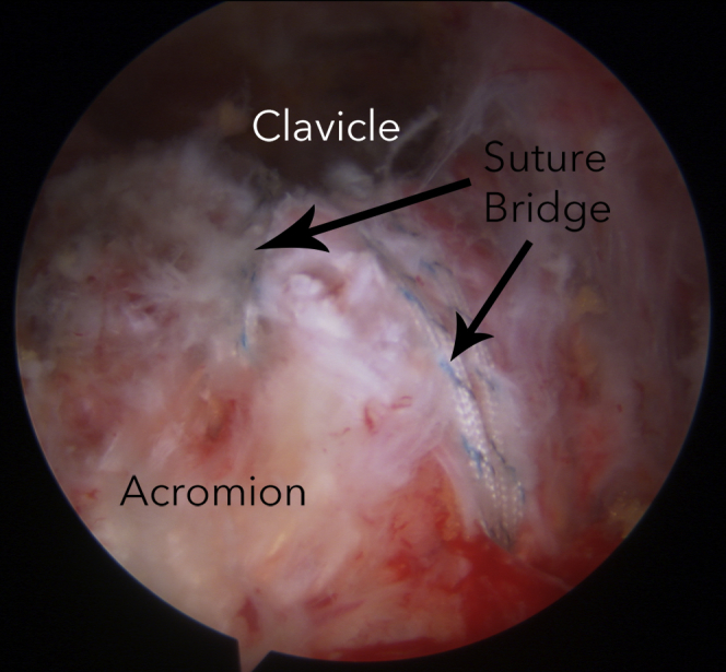
An 8-stranded suture bridge construct is created over the acromioclavicular joint to reconstruct the superior capsule in addition to the coracoclavicular reconstruction achieved with an ABS TightRope and Dog-Bone buttons.
Fig 15.

Biplanar construct with a TightRope between the clavicle and coracoid and an 8-stranded suture bridge between the acromion and clavicle.
Postoperative Regimen
All patients are dressed with a Cold Pad (DonJoy, Vista, CA) and shoulder abduction sling (Ultrasling; DonJoy) postoperatively. Patients are maintained in a sling for 6 weeks. Strengthening commences at 12 weeks. Return to sports is restricted until 6 months.
Discussion
We have performed this reconstruction on 4 patients with grade 3, 4, or 5 ACJ disruptions between February and August 2014. To date, there have been no failures and no problems with prominent hardware, residual ACJ pain, or scarring.
Repair of ACJ dislocations with reconstruction of the CC ligaments alone fails to control anteroposterior translation of the ACJ. The aim of this added repair is to increase the strength of the reconstruction and thereby reduce the failure rate.
All suture anchors anchors were selected for the ACJ repair because their relatively small size avoids inferior penetration of the relatively thin acromion. More conventional anchors would protrude from the undersurface of the acromion, leading to impingement of the cuff.
Because the Dog-Bone buttons can be applied to the TightRopes after they are passed, the tunnels in the clavicle and coracoid can be small (3 mm). A small hole in the coracoid process reduces the possibility of coracoid fracture,7 which is a risk with the larger 6-mm coracoid tunnel used for GraftRope (Arthrex) constructs.5
The reconstruction of the ACJ is surprisingly simple. The anchors are easy to insert. The space over the ACJ is readily developed and relatively bloodless, and the assistance of a switching stick to hold the space open is usually unnecessary. Of the ACJ ligaments, the posterosuperior ligaments are the strongest and therefore the most important when one is considering repair. Our technique serves to repair these superior ligaments. A further advantage of the described technique is that no knots are used. The region over the acromion and the clavicle is subcutaneous, and knots placed in this location may cause irritation to the patient.
As with all procedures, there are some risks. Alignment of the Arthrex aiming guide below the coracoid and above the clavicle has to be performed partially by feel because confirming the location of both of these points under vision requires the use of 2 separate viewing portals. It is also possible for the drill bit to deflect when drilling the tunnel between the clavicle and coracoid. This deflection can be decreased by using minimal antegrade pressure on the drill. Finally, it is worthwhile to carefully assess the inferior surface of the acromion because penetration of the anchors is possible and may lead to irritation of the rotator cuff. We recommend this reconstruction as a readily reproducible technique that increases the strength of previously described arthroscopic techniques to reconstruct the CC ligaments, hopefully leading to lower failure rates for acute ACJ repairs.
Acknowledgment
The authors acknowledge the assistance of Susan Peters, Research Coordinator, Brisbane Hand and Upper Limb Research Institute, for assistance with editing.
Footnotes
The authors report the following potential conflict of interest or source of funding: K.C. receives support from DePuy, Smith & Nephew, and Arthrex. Brisbane Hand and Upper Limb Research Institute received funding from DePuy, Medartis, LMT, and Lima Orthopaedics.
Supplementary Data
Operative technique for all-arthroscopic reconstruction of an acute left acromioclavicular joint (ACJ) dislocation. The patient is in the beach-chair position with the arm held in a Spider limb-positioning device. The repair comprises a coracoclavicular reconstruction using Arthrex ABS TightRopes and an acromioclavicular reconstruction using an 8-stranded suture bridge.
References
- 1.Cadenat F. The treatment of dislocations and fractures of the outer end of the clavicle. Int Clin. 1917;1:145–169. [Google Scholar]
- 2.Tossy J.D., Mead N.C., Sigmond H.M. Acromioclavicular separations: Useful and practical classification for treatment. Clin Orthop Relat Res. 1963;28:111–119. [PubMed] [Google Scholar]
- 3.Rockwood C. Injuries to the acromioclavicular joint. In: Ca R., Green D., editors. Fractures in adults. Ed 2. JB Lippincott; Philadelphia: 1984. pp. 860–910. [Google Scholar]
- 4.Fukuda K., Craig E.V., An K.N., Cofield R.H., Chao E.Y. Biomechanical study of the ligamentous system of the acromioclavicular joint. J Bone Joint Surg Am. 1986;68:434–440. [PubMed] [Google Scholar]
- 5.Jensen G., Kattahagen J.C., Alvarado L., Lill H., Voigt C. Arthroscopically assisted stabilization of chronic AC-joint instabilities in GraftRope technique with an additive horizontal tendon augmentation. Arch Orthop Trauma Surg. 2013;133:841–851. doi: 10.1007/s00402-013-1745-2. [DOI] [PubMed] [Google Scholar]
- 6.Lafosse L., Boyle S., Gutierrez-Aramberri M., Shah A., Meller R. Arthroscopic Laterjet procedure. Orthop Clin North Am. 2010;41:393–405. doi: 10.1016/j.ocl.2010.02.004. [DOI] [PubMed] [Google Scholar]
- 7.Nordin J.S., Aagaard K.E., Lunsjo K. Chronic acromioclavicular joint dislocations treated by the GraftRope device. Acta Ortop. 2015;86:225–228. doi: 10.3109/17453674.2014.976806. [DOI] [PMC free article] [PubMed] [Google Scholar]
Associated Data
This section collects any data citations, data availability statements, or supplementary materials included in this article.
Supplementary Materials
Operative technique for all-arthroscopic reconstruction of an acute left acromioclavicular joint (ACJ) dislocation. The patient is in the beach-chair position with the arm held in a Spider limb-positioning device. The repair comprises a coracoclavicular reconstruction using Arthrex ABS TightRopes and an acromioclavicular reconstruction using an 8-stranded suture bridge.