



Abstract
Introduction:
Lymph node counts have become a surrogate measure for the extent and quality of pelvic lymph node dissection (PLND) at radical cystectomy, but little consideration has been given to the methodology of lymph node processing. We report results from a prospective series comparing a conventional protocol for processing PLND specimens to a fat-emulsifying protocol. We hypothesized that the rate of node positivity would increase with the fat-emulsifying protocol.
Methods:
Patients undergoing radical cystectomy for cTis-T4aN0-1M0 urothelial carcinoma of the bladder were eligible for this trial. Palpable lymph nodes were isolated from the PLND specimens in the conventional protocol. The remaining tissue was then processed with fat-emulsifying solution to identify further nodes visually. Nodal counts were compared between techniques.
Results:
The median number of nodes counted in the PLND specimens of 26 patients was 24.5 (range: 20–40) with conventional processing and 37 (range: 24–52) with the fat-emulsifying solution (p < 0.001). Three patients had lymph node positive disease detected by conventional means, and a single patient was found to have a single positive node by the fat-emulsifying solution alone. The study was closed early after conducting a futility analysis.
Conclusions:
A fat-emulsifying protocol identified more lymph nodes than a conventional protocol and may be an appropriate method to standardize lymph node processing following PLND. However, we were unable to show that such a standardized approach significantly increased the rate of node positivity in patients undergoing radical cystectomy.
Introduction
The gold standard for the management of muscle invasive bladder cancer (MIBC) is radical cystectomy with pelvic lymph node dissection (PLND).1,2 The pathological findings from this surgery determine the prognosis and subsequent patient management. The node dissection at the time of cystectomy improves local tumour control, provides staging information, and enhances survival.3
There is mounting evidence that an extended PLND improves survival compared to a more limited dissection.4,5 The number of removed nodes may be surrogate measures for the extent of PLND.6 Therefore, the number of nodes removed with PLND has been adopted by many as an indicator of the quality of PLND and has been contemplated as a quality indicator by third party payers in the United States.7 There are two principle barriers to using lymph node counts as a quality indicator. Firstly, the evidence supporting a benefit of the extended PLND is retrospective6 and results are still pending from two prospective randomized trials (NCT01224665 and NCT01215071). The second is the absence of consideration in how the PLND specimens are processed. Node counts can vary considerably due to factors other than quality of dissection.8 Several European and American centres report node counts as a marker of quality as they are an attractive surrogate because they represent a more objective and quantifiable parameter.9–12 All of these studies emphasize the importance of the extended PLND and the relevance of the nodal count as a reflection of the extent of dissection, but none of them consider the methodology of tissue processing and node counting.
A fat-emulsifying technique (FET) was first described by Lillie in 194913 and has since been adapted and modified for numerous uses.14 Recently, Koren and colleagues introduced a “lymph node revealing solution” (LNRS) as an inexpensive and non-toxic solution that can be readily used in routine pathologic practice.15–19 The LNRS lymph nodes become visible as chalky white nodules on the yellow background of surrounding fat.
A standardized and reproducible method for processing lymph nodes is necessary to compare the results between centres and should be a prerequisite to any use of lymph node counts as a measure of the quality of PLND. We have designed a prospective trial to compare our current protocol for processing pelvic lymph nodes to a fat-emulsifying protocol (FEP) after radical cystectomy. While it is assumed that the FEP will identify more lymph nodes, we hypothesized that the more accurate identification of lymph nodes would increase the proportion of patients who would be found to have lymph node metastases.
Methods
Patients
All patients undergoing radical cystectomy with PLND for clinical cTis-T4aN0-1M0 urothelial carcinoma of the bladder between June 2011 and June 2013 were invited to participate in this study. We excluded patients who were unable to consent, patients with concomitant malignancy or prior abdominal malignancy, and patients with prior pelvic vascular surgery. We included patients with incidental, node negative prostate cancer. The study was approved by the clinical research ethics committee at the University of British Columbia and all patients consented to participation. The trial was registered with ClinicalTrials.gov (NCT01395225).
Surgical technique
The extent of PLND was defined as either standard (up to bifurcation of common iliac artery) or extended (up to the aortic bifurcation, with or without dissection of the presacral lymph nodes) and was determined by the surgeon on a case-by-case basis. Objective documentation of the extent of PLND was documented by intra-operative photography. Lymph node specimens were sent to the pathologists in 2 to 5 separate packages: right and left pelvic, right and left common iliac, and pre-sacral lymph nodes.20,21
Pathologic processing of lymph node specimens
Each specimen was analyzed by both conventional means and by a FEP. Conventional means involved the prosection of the nodal specimen after fixation in 10% formaldehyde. Palpable nodes were isolated, embedded in paraffin, and sectioned at 2-mm intervals. The resulting 4-μm thick sections were stained with hematoxylin and eosin. The nodal count was determined by the study pathologist (BG). To be counted as a lymph node, the specimen had to be a capsule and subcapsular sinus; lymphoid aggregates lacking these architectural features were not counted as lymph nodes.
The discarded tissue from the conventional protocol was then submitted for processing by the FEP. It was placed in a 95% ethanol, diethyl ether, glacial acetic acid and buffered formalin solution in a ratio of 6.5:2:0.5:1 for 12 hours. The study pathologist (BG) inspected these specimens and macroscopically extracted visible lymph nodes and sectioned them at 2-mm intervals, embedded them in paraffin, and stained them with eosin and hematoxylin.16 All remaining tissue in which no macroscopic nodes were identified was then embedded and examined microscopically for lymph nodes.
Data collection
Demographic, clinical and pathological information on all patients were recorded prospectively. Demographic variables included age, sex, weight, height, and race. Clinical variables included date of cancer diagnosis, clinical and pathologic TNM stage, date of radical cystectomy, history of intravesical chemotherapy, other medical comorbidities and prior abdominal surgeries, and extent of PLND.
Outcome measures
The primary outcome was the number of patients upstaged as a result of the FEP. The secondary outcomes were the total number of lymph nodes and the total number of lymph node metastases counted by both techniques.
Sample size calculation
An increase in the identification of nodal metastases by 15% (FEP compared to conventional processing) was predetermined as clinically significant. The rate of node positivity at radical cystectomy with PLND at our centre for all T-stages was 25/90 (28%) in the 2 years prior to the start of this study. An increase of 15% to 43% was deemed clinically relevant. To achieve a power (1-β) of 80% and an α error of 10%, we needed 91 enrolled patients in the study.
Data analysis
Data were analyzed using SPSS software. The independent samples t-test was used to compare the results from the lymph node analysis when processed by conventional means versus the same nodes processed using the fatemulsifying protocol (p < 0.5). An interim data analysis and subsequent futility analysis were performed after the first 26 patients were enrolled.
Results
In total, 26 patients were enrolled in our study. They had a mean age of 70 years (interquartile range [IQR] 64–76) and a median body mass index of 29 (IQR: 26–32) (Table 1). Table 2 summarizes the extent of PLND and the pathologic parameters. Photographs documenting the extent of PLND are available for 7 of the patients (Fig. 1, Fig. 2, Fig. 3, Fig. 4, Fig. 5, Fig. 6, Fig. 7).
Table 1.
Pre-cystectomy demographic and clinicopathological characteristics
| Characteristic | N |
|---|---|
| Age (median, IQR) | 70 (64–76) |
| BMI (median, IQR) | 29 (26–32) |
| Prior radiation therapy | 3 |
| Prior intravesical therapy | 8 |
| Neoadjuvant chemotherapy | 8 |
| Clinical T stage | |
| Tis | 4 |
| T1 | 4 |
| T2 | 13 |
| T3 | 5 |
| Concomitant CIS | 8 |
| Clinical N stage | |
| N0 | 1 |
| N1 | 2 |
IQR: interquartile range; BMI: body mass index; CIS: carcinoma in situ; Tis: T stage carcinoma in situ.
Table 2.
Extent of lymph node dissection and pathologic parameters
| Characteristic | N |
|---|---|
| Extent of node disssection | |
| Standard | 4 |
| Extended | 22 |
| Pathological stage | |
| T0 | 7 |
| Tis | 6 |
| T1 | 2 |
| T2 | 3 |
| T3 | 8 |
| Lymphovascular invasion present | 6 |
| Concomitant CIS | 10 |
CIS: carcinoma in situ; Tis: T stage carcinoma in situ.
Fig. 1.

Patient 1: Photo of right and left extended lymph node dissection demonstrating complete removal of lymph node tissue up to the bifurcation of the aorta.
Fig. 2.

Patient 2: Photo of right and left extended lymph node dissection demonstrating complete removal of lymph node tissue up to the bifurcation of the aorta.
Fig. 3.

Patient 3: Photo of right and left extended lymph node dissection demonstrating complete removal of lymph node tissue up to the bifurcation of the aorta.
Fig. 4.

Patient 4: Photo of right and left extended lymph node dissection demonstrating complete removal of lymph node tissue up to the bifurcation of the aorta.
Fig. 5.

Patient 5: Photo of right and left extended lymph node dissection demonstrating complete removal of lymph node tissue up to the bifurcation of the aorta.
Fig. 6.

Patient 6: Photo of right and left extended lymph node dissection demonstrating complete removal of lymph node tissue up to the bifurcation of the aorta.
Fig. 7.
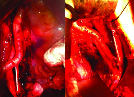
Patient 7: Photo of right and left extended lymph node dissection demonstrating complete removal of lymph node tissue up to the bifurcation of the aorta.
Table 3 summarizes the distribution of nodes between groups. A median of 8 (IQR: 2–12) additional nodes were isolated by the FEP, and almost all of these (189/207, 91.3%) were identified only microscopically. Lymph node metastases were identified in 4 patients (Fig. 8). In 2 patients these were identified by conventional processing only: 1 patient had metastatic nodes discovered after both processing methods, and the other patient had a single positive lymph node only after processing with the FEP. The latter patient had a cystectomy for BCG (bacillus Calmette-Guérin)-refractory carcinoma in situ (CIS). Final pathology confirmed pTisN1M0 urothelial cancer. In this patient, 27 benign nodes were identified conventionally, 10 benign nodes were identified after the FEP, 1 of which contained a 5 × 2-mm foci of carcinoma only seen microscopically (Fig. 9).
Table 3.
Lymph node counts according to method of processing
| Benign lymph nodes | Lymph nodes with metastasis | Final node count | |||||||
|---|---|---|---|---|---|---|---|---|---|
|
| |||||||||
| Median | IQR | n | Median | IQR | n | Median | IQR | n | |
| Conventional processing | 24.5 | 20–32 | 727 | 0 | 0–0 | 22 | 24.5 | 20–40 | 749 |
| FEP | |||||||||
| Macroscopic | 0 | 0–1 | 18 | 0 | 0–0 | 0 | 0 | 0–1 | 186 |
| Microscopic | 5 | 2–11 | 186 | 0 | 0–0 | 3 | 5 | 2–11 | 189 |
| Sum | 5 | 2–12 | 204 | 0 | 0–0 | 3 | 5 | 2–12 | 207 |
| Total nodes | 30 | 24–52 | 931 | 0 | 0–0 | 25 | 30 | 24–52 | 956 |
| p value* | <0.01 | 0.04 | <0.01 | ||||||
IQR: interquartile range; FEP: fat emulsifying protocol.
Students 2 tailed t-test of node count from conventional processing versus the total node count after the FEP.
Fig. 8.

Distribution of node counts.
Fig. 9.

Pelvic lymph node specimen after radical cystectomy and extended lymph node dissection in a patient with BCG (bacillus Calmette-Guérin) refractory disease. This lymph node has been exposed to the fat-emulsifying protocol. Surrounding the lymph node the excess fat and areolar tissue has been dissolved away, bringing the lymph node tissue to the foreground. The metastatic deposit within this lymph node was 5 × 2 mm, occupying most of the node without evidence of extra-nodal extension.
The total number of nodes identified by the conventional method for all patients combined was 749, and this increased to 956 with the FEP (1.27-fold increase). Of the total number of lymph nodes identified conventionally (n = 749), 22 (2.9%) harbored metastases. Of the total 207 lymph nodes identified by FEP alone, 3 (1.5%) were positive. Of the total number of nodes identified (n=956), 25 (2.6%) were metastatic nodes.
The cost of the FEP was $1 174.82 CDN per patient in addition to the conventional fee. Fees included $864.43 dollars for procedural fees, $296.02 for professional fees, and $28.76 for raw supplies. Procedural fees included fees attributed to paraffin processing, sectioning, staining, and use of the microscope. Professional fees included technician fees. Raw supplies included gloves, formalin, cassettes, and scalpel blades.
Our pre-determined sample size to determine a clinically significant 15% increase in node positivity was 91. If we carried the numbers from the first 26 patients forward, we would need to enroll 995 patients to detect the same 15% difference at a power of 0.80. We therefore determined that the study was futile.
Discussion
We have demonstrated that a simple FEP can increase the yield of lymph nodes. However, we found node positive disease was missed in only 1 of 26 patients. The additional nodes identified by FEP were smaller nodes and 91.3% were only identified microscopically. This raises two clinical questions related to lymph node counts.
Firstly, the number of nodes counted is not relevant to the detection of lymph node metastases. Lymph node metastases are mostly found in palpable lymph nodes and only rarely in the additional nodes found after the FEP. Similar results may have been achieved with microscopic evaluation of the residual tissue after removing palpable nodes without the FEP. Metastases are found almost exclusively in palpable lymph nodes, and the dependence on microscopy is evidence for the small size of these additional lymph nodes. Our results suggest that the additional detection of small nodes may not add value. On the other hand, 1 in 4 patients with lymph node metastases were missed by conventional means.
Secondly, the number of nodes clearly varies depending on how the nodes are processed, which has direct implications for the use of node counts as a surrogate measure of the quality of a PLND. Physicians and administrators should not compare between centres without standardized procedures.
The FEP has been applied by Koren’s group in different tumour types, including bladder cancer and was found to dramatically increase (up to twofold) the number of nodes and the number of lymph node metastases detected.15–19 Cancer upstaged in 9.5% to 40% of patients. Specifically in bladder cancer, the FEP discovered 1.95-fold more lymph nodes and upstaged 3 of 12 patients with muscle invasive bladder cancer to node positive.16 Of the 12 patients, 2 were node positive by conventional means. The more dramatic increase in the number of nodes identified could indicate a high-risk population, that the conventional process was less precise, or that the FET was more precise in the Koren study.
This study was powered to require 91 patients, but an interim analysis after 26 patients led to discontinuation. The inclusion of patients post-neoadjuvant chemotherapy and the mix of non-MIBC and MIBC likely reduced the rate of node positivity and further reduced the power of the study. However, it is possible that lower risk patients, including those with BCG refractory NMIBC, have a lower risk of node positivity but could potentially benefit from more precise staging. This latter supposition is supported by the fact that the only patient in our cohort with a positive node only on the FEP had CIS. Even though each patient served as his/her own control and, the extent of PLND was not deemed a critical factor in this analysis, our results would have been strengthened if all patients had an extended PLND.
The FEP is an easy, non-toxic adjunct that should be used if it can improve the identification of node positive patients even just marginally. There is no evidence that a FEP adversely affects routine immunohistochemistry.15 Furthermore, the FEP could reduce the burden on the pathologist in finding individual lymph nodes, thus removing a barrier to careful lymph node counting.22 The use of additional sectioning23 and immunohistochemistry24,25 to enhance lymph node identification is either ineffective or too labour-intensive. The main limitation of the FEP, however, was the additional cost.
Conclusion
A FEP identified more lymph nodes and may be an appropriate method to standardize lymph node processing. However, we were unable to show that such a standardized approach significantly increased the rate of node positivity. The clinical utility of such a protocol is therefore uncertain.
Acknowledgments
We would like to thank Bladder Cancer Canada and the Canadian Urologic Oncology Group for their grant support of this project.
Footnotes
Competing interests: The authors all declare no competing financial or personal interests.
This paper has been peer-reviewed.
References
- 1.Herr H, Lee C, Chang S, et al. Standardization of radical cystectomy and pelvic lymph node dissection for bladder cancer: A collaborative group report. J Urol. 2004;171:1823–8. doi: 10.1097/01.ju.0000120289.78049.0e. discussion 1827–8. [DOI] [PubMed] [Google Scholar]
- 2.Malkowicz SB, van Poppel H, Mickisch G, et al. Muscle-invasive urothelial carcinoma of the bladder. Urology. 2007;69:3–16. doi: 10.1016/j.urology.2006.10.040. [DOI] [PubMed] [Google Scholar]
- 3.Karl A, Carroll PR, Gschwend JE, et al. The impact of lymphadenectomy and lymph node metastasis on the outcomes of radical cystectomy for bladder cancer. Eur Urol. 2009;55:826–35. doi: 10.1016/j.eururo.2009.01.004. [DOI] [PubMed] [Google Scholar]
- 4.Leissner J, Ghoneim MA, Abol-Enein H, et al. Extended radical lymphadenectomy in patients with urothelial bladder cancer: Results of a prospective multicenter study. J Urol. 2004;171:139–144. doi: 10.1097/01.ju.0000102302.26806.fb. [DOI] [PubMed] [Google Scholar]
- 5.Poulsen AL, Horn T, Steven K. Radical cystectomy: Extending the limits of pelvic lymph node dissection improves survival for patients with bladder cancer confined to the bladder wall. J Urol. 1998;160:2015–9. discussion 2020. [PubMed] [Google Scholar]
- 6.Herr HW, Faulkner JR, Grossman HB, et al. Surgical factors influence bladder cancer outcomes: A cooperative group report. J Clin Oncol. 2004;22:2781–9. doi: 10.1200/JCO.2004.11.024. [DOI] [PubMed] [Google Scholar]
- 7.Gilbert SM, Hollenbeck BK. Limitations of lymph node counts as a measure of therapy. J Natl Compr Canc Netw. 2009;7:58–61. doi: 10.6004/jnccn.2009.0005. [DOI] [PubMed] [Google Scholar]
- 8.Dorin R, Daneshmand S, Eisenberg M, et al. Lymph node dissection technique is more important than lymph node count in identifying nodal metastases in radical cystectomy patients. In Society of Urologic Oncology Annual Meeting; Bethesda, MD. 2010. [DOI] [PubMed] [Google Scholar]
- 9.Kassouf W, Leibovici D, Munsell MF, et al. Evaluation of the relevance of lymph node density in a contemporary series of patients undergoing radical cystectomy. J Urol. 2006;176:53–7. doi: 10.1016/S0022-5347(06)00510-6. discussion 57. [DOI] [PubMed] [Google Scholar]
- 10.Kassouf W, Agarwal PK, Herr HW, et al. Lymph node density is superior to TNM nodal status in predicting disease-specific survival after radical cystectomy for bladder cancer: Analysis of pooled data from MDACC and MSKCC. J Clin Oncol. 2008;26:121–6. doi: 10.1200/JCO.2007.12.9247. [DOI] [PubMed] [Google Scholar]
- 11.Wright JL, Lin DW, Porter MP. The association between extent of lymphadenectomy and survival among patients with lymph node metastases undergoing radical cystectomy. Cancer. 2008;112:2401–8. doi: 10.1002/cncr.23474. [DOI] [PubMed] [Google Scholar]
- 12.May M, Herrmann E, Bolenz C, et al. Association Between the Number of Dissected Lymph Nodes During Pelvic Lymphadenectomy and Cancer-Specific Survival in Patients with Lymph Node-Negative Urothelial Carcinoma of the Bladder Undergoing Radical Cystectomy. Ann Surg Oncol. 2011. [DOI] [PubMed]
- 13.Lillie RD, Fullmer HM. Histopathologic Technic and Practical Histochemistry. New York, NY: McGraw-Hill Book Co; 1976. [Google Scholar]
- 14.Newell KJ, Sawka BW, Rudrick BF, et al. GEWF solution. Arch Pathol Lab Med. 2001;125:642–5. doi: 10.5858/2001-125-0642-GS. [DOI] [PubMed] [Google Scholar]
- 15.Koren R, Siegal A, Klein B, et al. Lymph node-revealing solution: Simple new method for detecting minute lymph nodes in colon carcinoma. Dis Colon Rectum. 1997;40:407–10. doi: 10.1007/BF02258384. [DOI] [PubMed] [Google Scholar]
- 16.Koren R, Paz A, Lask D, et al. Lymph-node revealing solution: A new method for detecting minute lymph nodes in cystectomy specimens. Br J Urol. 1997;80:40–3. doi: 10.1046/j.1464-410X.1997.00154.x. [DOI] [PubMed] [Google Scholar]
- 17.Koren R, Shvero J, Yaniv E, et al. Neck-dissection surgical specimens treated by lymph node revealing solution. Otolaryngol Head Neck Surg. 2001;124:72–5. doi: 10.1067/mhn.2001.111290. [DOI] [PubMed] [Google Scholar]
- 18.Koren R, Kyzer S, Paz A, et al. Lymph node revealing solution: A new method for detection of minute axillary lymph nodes in breast cancer specimens. Am J Surg Pathol. 1997;21:1387–90. doi: 10.1097/00000478-199711000-00016. [DOI] [PubMed] [Google Scholar]
- 19.Koren R, Kyzer S, Levin I, et al. Lymph node revealing solution: A new method for lymph node sampling: Results in gastric adenocarcinoma. Oncol Rep. 1998;5:341–4. doi: 10.3892/or.5.2.341. [DOI] [PubMed] [Google Scholar]
- 20.Bochner BH, Cho D, Herr HW, et al. Prospectively packaged lymph node dissections with radical cystectomy: Evaluation of node count variability and node mapping. J Urol. 2004;172:1286–90. doi: 10.1097/01.ju.0000137817.56888.d1. [DOI] [PubMed] [Google Scholar]
- 21.Sanderson KM, Stein JP, Skinner DG. The evolving role of pelvic lymphadenectomy in the treatment of bladder cancer. Urol Oncol. 2004;22:205–11. doi: 10.1016/j.urolonc.2004.04.030. discussion 212–203.22. [DOI] [PubMed] [Google Scholar]
- 22.Wright FC, Law CH, Last LD, et al. Barriers to optimal assessment of lymph nodes in colorectal cancer specimens. Am J Clin Pathol. 2004;121:663–70. doi: 10.1309/17VKM33BFXF9T8WD. [DOI] [PubMed] [Google Scholar]
- 23.Law CH, Wright FC, Rapanos T, et al. Impact of lymph node retrieval and pathological ultra-staging on the prognosis of stage II colon cancer. J Surg Oncol. 2003;84:120–6. doi: 10.1002/jso.10309. [DOI] [PubMed] [Google Scholar]
- 24.Yang XJ, Lecksell K, Epstein JI. Can immunohistochemistry enhance the detection of micrometastases in pelvic lymph nodes from patients with high-grade urothelial carcinoma of the bladder? Am J Clin Pathol. 1999;112:649–53. doi: 10.1093/ajcp/112.5.649. [DOI] [PubMed] [Google Scholar]
- 25.Leissner J, Koeppen C, Wolf HK, et al. Immunohistochemical detection of micrometastases in pelvic lymph nodes from patients with urothelial cancer of the urinary bladder. J Urol. 2002;167:264. [Google Scholar]