
Figure 3. miR-17 is a direct translational repressor of ETV1.
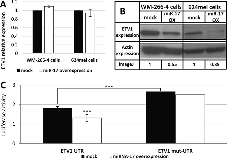
A. The mRNA levels of ETV1 following transfection with scrambled sequence (mock) or miR-17 in the indicated melanoma cell lines was confirmed with qPCR. Figure shows the mean of 3 independent experiments performed; B. ETV1 expression at the protein level was determined by Western blot following transfection with scrambled sequence (mock) or miR-17 in the indicated melanoma cell lines. Actin levels served as control. Normalized densitometry values of ETV1 relative to actin are indicated. A representative blot is shown out of 3 experiments performed; C. UTR and mut-UTR denote ETV1 3′UTR segments containing the reference sequence or mutated sequence in the miR-17-binding site, respectively. Those were cotransfected with miR-17 (miR-17 over-expression) or with an empty vector (mock) to 293T cells. Figure shows the mean of 3 independent experiments performed Y-axis denotes normalized Renilla luciferase activity. ***denotes P < 0.001 (2-tailed t-test).