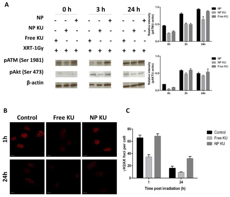
Figure 4.
Effect of KU55933 formulations and radiation on phosphorylation of ATM, Akt and H2AX. H460 cells were pretreated with 10 μM KU or NP KU (containing 10 μM KU) for 3 h and subsequently irradiated (1 Gy). (A) Cell lysates were collected at indicated time points for western blot. The western blot band intensity was quantified after normalized to β-actin based on three independent experiments. (B) Cells were fixed at indicated time points for immunofluorescence of γH2AX. (C) Average numbers of γH2AX foci per cell were presented.

