ABSTRACT
To determine the efficacy of computed tomography (CT) attenuation of cystic lesions measured on an image browsing system to distinguish abscess from hematoma in women with acute abdomen. The medical records of female patients of reproductive age with acute abdomen who were treated over a 7-year period in a single center and who had undergone laparotomy or laparoscopic surgery and preoperative pelvic CT scanning were retrospectively analyzed to identify those with hematoma or abscess cyst formation. Nineteen patients with tubo-ovarian abscess (abscess group) and six patients with hematoma (hematoma group) formation in the pelvis were included in the analysis. The preoperative CT images of the tubo-ovarian cyst were retrospectively investigated on the basis of cyst attenuation. CT attenuation of the cyst measured by both two gynecologists could be used to clearly distinguish inflammatory disease with abscess formation from bleeding disease with hematoma. CT attenuation on a picture archiving and communication system can distinguish hematoma from abscess in women with acute abdomen. This may significantly contribute to making differential diagnosis without interpretation by a medical radiologist.
Key Words: abscess, acute abdomen, CT attenuation, hematoma, differential diagnosis
INTRODUCTION
Diagnosis of acute abdomen in women of reproductive age can be challenging because many symptoms and signs are insensitive and nonspecific. The most common urgent causes of acute lower abdominal pain in these women are pelvic inflammatory disease, ovarian torsion, ruptured ovarian cyst, and appendicitis.1, 2) Among these clinical entities that lead to acute abdomen, it is sometimes difficult to distinguish pelvic inflammatory diseases with tubo-ovarian abscess from hematoma that develops as a result of unexpected bleeding caused by lesions such as hemorrhagic corpus luteum cysts. This is because in most of these entities, cystic lesion formation is observed with retention liquid and reactive abdominal fluid.3) In some cases of hematoma such as cases of hemorrhage as a result of ovarian cyst formation, conservative treatment is possible depending on the amount of bleeding. On the other hand, the emergency surgical intervention of drainage and administration of antibiotics are necessary in cases with abscess formation, as early and accurate diagnosis of acute abdomen with an ovarian cyst is important for the proper management of acute disease and the prevention of sequelae.4) Therefore, it can be helpful for surgeons to know preoperatively whether an ovarian cyst is formed from hematoma or abscess. However, it is sometimes difficult to make an accurate diagnosis using imaging such as ultrasonography or computed tomography (CT). CT is frequently performed as the initial imaging modality in the evaluation of acute lower abdominal pain of unknown etiology. The American College of Radiology has recommended different imaging studies for assessing acute abdomen based on pain location. Ultrasonography is recommended for the assessment of right upper quadrant pain, whereas CT is recommended for right and left lower quadrant pain.5-7)
Prompt differential diagnosis is frequently needed in such patients in an emergency room without interpretation by a medical radiologist. Objective diagnostic methods using an image browsing system at the patient’s bedside will provide significant information concerning the management of women with acute abdomen. Instead of film CT images on the screen of the image viewing system, information is now available to physicians on an electronic medical record system.8, 9) In addition, it is possible to evaluate CT attenuation of an individual point on the image browsing system, and further substantial information can be obtained at the bedside. Although descriptive CT attenuation of abscess and hematoma has been documented in text or review articles,10-14) there are no studies on the diagnostic performance of CT for distinguishing pelvic cystic lesions caused by hemorrhage from inflammatory abscess in an actual clinical setting. Hence, to determine the efficacy of CT attenuation for distinguishing abscess from hematoma, we attempted to compare CT attenuation of the collected fluid in women with acute abdomen on an image browsing system. Such a study would ensure that detailed information of CT attenuation is available to physicians in any emergency.
MATERIALS AND METHOS
Study population
This is a retrospective single-institution study that was reviewed and approved by the Institutional Review Board of the Saitama Medical University, and the need for informed consent was waived. From January 2007 to December 2013, the medical records of menstruate women in reproductive age with acute lower abdomen pain who had undergone laparotomy or laparoscopic surgery and contemporaneous preoperative pelvic CT in our hospital were reviewed to identify those with hematoma or abscess cyst formation in the pelvis. Final diagnosis of these cases had been confirmed via intraoperative findings and pathological diagnosis. Patient age, body mass index (BMI), preoperative white blood cell count (WBC), C-reactive protein (CRP) level, hemoglobin level, and CT attenuation of preoperative plain CT were examined.
CT techniques and image analysis
CT examinations were performed using either Siemens SOMATOM Definition Flash or Siemens SOMATOM Emotion16 (Siemens Medical Solutions, Erlangen, Germany) without contrast agent. The imaging routine for Siemens SOMATOM Definition Flash was as follows: 120 kilovolt (kV); 185 milliampere-second (mAs); detector configuration, 128 × 0.6-mm; pitch, 0.6; rotation time, 0.5 seconds. Siemens SOMATOM Emotion16 was adjusted as follows: 130 kilovolt (kV); 150 milliampere-second (mAs); detector configuration, 16 × 1.5-mm; pitch, 1.0; rotation time, 0.6 seconds. All CT images were retrospectively reviewed independently by two gynecologists. These gynecologists were aware that the purpose of the study was to evaluate imaging findings of ovarian hematoma or abscess in patients with lower abdominal pain. However, they were blinded to all clinical data. One gynecologist (K.S.) was a board-certified gynecologist with 9 years of experience. The other was a third-year gynecology resident (E.H.). All CT images were reviewed on a picture archiving and communication system (PACS; Enterprise-PACS, FUJIFILM Medical Co., Ltd. Tokyo Japan). The CT attenuation values were collected from three round regions of interest (ROI) in the center of cystic lesions, excluding the septum and cyst walls, and the average CT attenuation value were calculated.15)
Statistical analysis
Data are expressed as mean ± standard deviation (SD). Parametric variables were analyzed using the Wilcoxon rank sum test. Statistical analyses were performed using SPSS 15.0 (SPSS, Chicago, IL, USA). P < 0.05 was considered significant.
RESULTS
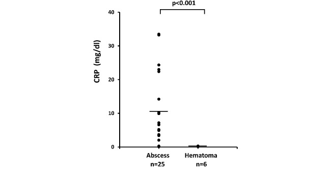
We operated 393 cases during the investigative period for suspecting gynecologic causes of acute abdomen. Among them, we were able to identify nineteen patients as ovarian abscess (abscess group) and six patients as hematoma (hematoma group) in the pelvis whose abdominopelvic CT results were also available for review. Patients’ age, BMI, preoperative WBC, and hemoglobin levels are shown in Table 1. There were no differences between the two groups in mean preoperative WBC or hemoglobin levels. The mean age of the abscess group was significantly higher than that of the hematoma group. Although the mean preoperative CRP level was significantly higher in the abscess group than in the hematoma group, there were three cases without CRP level elevation in the abscess group (Figure 1). The interval between the CT imaging and the surgery for abscess group and hematoma group ranged from 1 to 7 days (mean, 3 days) and from 1 to 2 days (mean, 1 day), respectively.
Table 1.
Comparison of background and hematological examination for ovarian abscess and ovarian hematoma in patients
| Abscess (n = 19) |
Hematoma (n = 6) |
P value | |
|---|---|---|---|
| Age [years; mean ± SD (range)] | 36.8 ± 7.4 (20–47) |
24.3 ± 6.5 (13–30) |
0.001 |
| WBC [/μl; mean ± SD (range)] | 12894 ± 4430 (4550–22630) | 12596 ± 2864 (9560–17480) | N.S. |
| Hb [mg/dl; mean ± SD (range)] | 11.4 ± 2.4 (7.2–14.5) |
10.6 ± 1.1 (9.5–12.5) |
N.S. |
| BMI [mean ± SD (range)] | 22.2 ± 6.1 (15.7–41.1) |
19.9 ± 2.3 (17.4–23.7) |
N.S. |
N.S., not significant
Fig. 1.
The preoperative CRP (mg/ml) level of the abscess group (n = 19) and the hematoma group (n = 6)
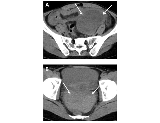
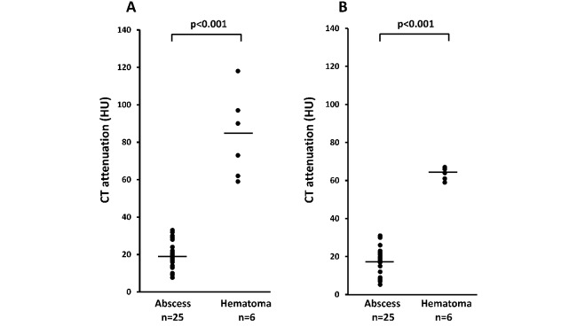
On the other hand, CT attenuation that was measured by both the gynecologists could be used to clearly distinguish inflammatory disease with abscess formation (Figure 2A) from hemorrhagic disease with hematoma (Figure 2B). All cases of CT attenuation in the abscess group were below 40 HU (Hounsfield Number), and those in hematoma were higher than 60 HU (Figure 3A, B).
Fig. 2.
(A) A 38-year-old woman with ovarian abscess. Axial unenhanced CT scan shows a 7-cm unilocular cystic mass (arrows) in the left adnexa. (B) A 20-year-old woman with ovarian hematoma. Axial unenhanced CT scan shows a highly attenuated cystic mass (arrows).
Fig. 3.
CT attenuation of the abscess group (n = 19) and the hematoma group (n = 6) as measured by (A) a board-certified gynecologist and (B) a gynecology resident.
DISCUSSION
The present study demonstrated that CT attenuation of cystic lesions could be used to clearly differentiate hematoma from abscess without interpretation by a medical radiologist. Although the mean preoperative CRP level of the abscess group was significantly higher than that of the hematoma group, there were some cases in which the CRP level of the abscess group overlapped with that of the hematoma group. Therefore, it is not possible to entirely differentiate hematoma from abscess using the preoperative CRP level. Descriptive CT attenuation of abscess and hematoma has previously been documented in text or review articles.10-14) However, there are no studies on the diagnostic performance of CT for distinguishing cystic lesions in the pelvis caused by bleeding from inflammatory abscess. To the best of our knowledge, this is the first report to demonstrate that CT attenuation using PACS on the electronic medical record system in an emergency room may significantly contribute to making differential diagnosis of intrapelvic abscess or hematoma in women with acute lower abdominal pain.
The CT attenuation value of intraperitoneal effusions has been reported to be 0–30 HU for ascites, over 30 HU for blood, and 50–60 HU or above for coagula. CT attenuation of hematoma varies in level as well as with lapse of the time phase.16) It has been reported that there is usually a mixed solid cystic mass with a high-attenuation component (45–100 HU)12, 14) in ovarian hematoma. In the present study, CT attenuation of all hematoma cases was higher than 60 HU. This result is consistent with these previous observations.12, 14) On the other hand, CT attenuation of abscesses has been demonstrated as being near to water attenuation.17, 18) In our study, CT attenuation of all abscess cases was lower than 36 HU. Our series consisted of patients with a relatively short space of time after onset. Therefore, 50 HU set as the boundary line between inflammatory abscess and hematoma may be reasonable to distinguish the early phase of abscess and hematoma.
Magnetic resonance imaging (MRI) is well suited and has obvious advantages over CT. MRI is recognized as a more sensitive tool for discriminating abscess from hematoma, and can eliminate unnecessarily exposure to radiation. However, MRI is not always operable for 24-hour in the majority of clinical settings contrary to CT. As the use of electronic medical records is widespread, PACS is also available to many physicians in Japan. CT images can now be viewed online on the computer screen rather than using the physical film. Therefore, it is generally possible to perform emergency CT as a result of 24-hour accessibility, but prompt interpretation by a radiologist is not always possible. Generally, in Japan, physicians in an emergency room and gynecologists interpret CT images by themselves and diagnose emergency patients with acute abdomen. To establish an objective evaluation of CT images to assist in making differentiated diagnosis may significantly contribute to gynecologic emergencies under such medical circumstances. Therefore, our results provide an index of CT images to distinguish hematoma from abscess; such images may be of great benefit to gynecologic emergencies.
In present study, the mean age of the abscess group was significantly higher than that of the hematoma group. A likely cause of this difference is that abscess group included some relatively elderly women with long time usage of intrauterine device or postoperational infection after endometrial sampling.
Our study had a number of limitations. First, it was retrospective study with a relatively small number of cases. Second, we retrospectively included cases in this study according to pathological findings and excluded some cases that had undergone conservative treatment, especially hematoma cases, from this study. As we could not eliminate inclusion bias completely in this retrospective study, a treatment protocol along with these results will prospectively clarify the efficacy of the classification of CT attenuation. Third, the interval between the CT and surgery of abscess group ranged widely (from 1 to 7 days). However, CT attenuation can clearly distinguish hematoma from abscess in women at any time point in present study.
In conclusion, using PACS on the electronic medical record system, physicians in an emergency room and gynecologists can distinguish hematoma from abscess in women with acute abdomen, which may significantly contribute to making differential diagnosis.
ACKNOWLEDGMENT
The authors appreciate the statistical analysis and helpful discussion provided by Eri Maeda, M.D., from department of Public Health, Graduate School of Medicine, the University of Tokyo, and department of Environmental Health Sciences, Akita University Graduate School of Medicine.
DISCLOSURE
No financial support was received by any of the authors for this study and none of the authors have any relationships with companies that may have a financial interest in the information contained in the manuscript.
REFERENCES
- 1).Kruszka PS, Kruszka SJ. Evaluation of acute pelvic pain in women. Am Fam Physician, 2010; 82: 141–147. [PubMed]
- 2).Jearwattanakanok K, Yamada S, Suntornlimsiri W, Smuthtai W, Patumanond J. Validation of the diagnostic score for acute lower abdominal pain in women of reproductive age. Emerg Med Int, 2014; 2014: 320926. doi: 10.1155/2014/320926. Epub 2014 May 25. [DOI] [PMC free article] [PubMed]
- 3).Allen BC, Barnhart H, Bashir M, Nieman C, Breault S, Jaffe TA. Diagnostic accuracy of intra-abdominal fluid collection characterization in the era of multidetector computed tomography. Am Surg, 2012; 78: 185–189. [PubMed]
- 4).Jung SI, Kim YJ, Park HS, Jeon HJ, Jeong KA. Acute pelvic inflammatory disease: diagnostic performance of CT. J Obstet Gynaecol Res, 2011; 37: 228–235. [DOI] [PubMed]
- 5).Yarmish GM, Smith MP, Rosen MP, Baker ME, Blake MA, Cash BD, Hindman NM, Kamel IR, Kaur H, Nelson RC, Piorkowski RJ, Qayyum A, Tulchinsky M. for the Expert Panel on Gastrointestinal Imaging. American College of Radiology ACR Appropriateness Criteria® Right upper quadrant pain. [Cited 24 Feb 2015]. Available from URL: http://www.acr.org/~/media/ACR/Documents/AppCriteria/Diagnostic/RightUpperQuadrantPain.pdf
- 6).Smith MP, Katz DS, Rosen MP, Lalani T, Carucci LR, Cash BD, Kim DH, Piorkowski RJ, Small WC, Spottswood SE, Tulchinsky M, Yaghmai V, Yee J. for the Expert Panel on Gastrointestinal Imaging. American College of Radiology ACR Appropriateness Criteria® Right lower quadrant pain- Suspected appendicitis. [Cited 24 Feb 2015]. Available from URL: http://www.acr.org/~/media/ACR/Documents/AppCriteria/Diagnostic/RightLowerQuadrantPainSuspectedAppendicitis.pdf
- 7).McNamara MM, Lalani T, Camacho MA, Carucci LR, Cash BD, Feig BW, Fowler KJ, Katz DS, Kim DH, Smith MP, Tulchinsky M, Yaghmai V, Yee J, Rosen MP. for the Expert Panel on Gastrointestinal Imaging. American College of Radiology ACR Appropriateness Criteria® Left lower quadrant pain- Suspected Diverticulitis [Cited 24 Feb 2015]. Available from URL: http://www.acr.org/~/media/ACR/Documents/AppCriteria/Diagnostic/LeftLowerQuadrantPainSuspectedDiverticulitis.pdf
- 8).Carrino JA. Digital imaging overview. Semin Roentgenol, 2003; 38: 200–215. [DOI] [PubMed]
- 9).Tellis WM, Andriole KP, Jovais CS, Avrin DE. RIS minus PACS equals film. J Digit Imaging, 2002; 15 Suppl 1: 20–26. [DOI] [PubMed]
- 10).Ferrucci JT Jr, vanSonnenberg E. Intra-abdominal abscess. Radiological diagnosis and treatment. JAMA, 1981; 246: 2728–2733. [PubMed]
- 11).Yitta S, Hecht EM, Slywotzky CM, Bennett GL. Added value of multiplanar reformation in the multidetector CT evaluation of the female pelvis: a pictorial review. Radiographics, 2009; 29: 1987–2003. [DOI] [PubMed]
- 12).Bennett GL, Slywotzky CM, Giovanniello G. Gynecologic causes of acute pelvic pain: spectrum of CT findings. Radiographics, 2002; 22: 785–801. [DOI] [PubMed]
- 13).Bennett GL, Harvey WB, Slywotzky CM, Birnbaum BA. CT of the acute abdomen: gynecologic etiologies. Abdom Imaging, 2003; 28: 416–432. [DOI] [PubMed]
- 14).Cano Alonso R, Borruel Nacenta S, Díez Martínez P, María NI, Ibáñez Sanz L, Zabía Galíndez E. Role of multidetector CT in the management of acute female pelvic disease. Emerg Radiol, 2009; 16: 453–472. [DOI] [PubMed]
- 15).Choi NJ, Rha SE, Jung SE, Choi BG, Oh SN, Byun JY, Kim MR. Ruptured endometrial cysts as a rare cause of acute pelvic pain: can we differentiate them from ruptured corpus luteal cysts on CT scan? J Comput Assist Tomogr, 2011; 35: 454–458. [DOI] [PubMed]
- 16).Federle MP, Jeffrey RB Jr. Hemoperitoneum studied by computed tomography. Radiology, 1983; 148: 187–192. [DOI] [PubMed]
- 17).Ferrucci JT Jr, vanSonnenberg E. Intra-abdominal abscess. Radiological diagnosis and treatment. JAMA, 1981; 246: 2728–2733. [PubMed]
- 18).Haaga JR, Alfidi RJ, Havrilla TR, Cooperman AM, Seidelmann FE, Reich NE, Weinstein AJ, Meaney TF. CT detection and aspiration of abdominal abscesses. AJR Am J Roentgenol, 1977; 128: 465–474. [DOI] [PubMed]
- 19).McWilliams GD, Hill MJ, Dietrich CS 3rd. Gynecologic emergencies. Surg Clin North Am, 2008; 88: 265–283. [DOI] [PubMed]


